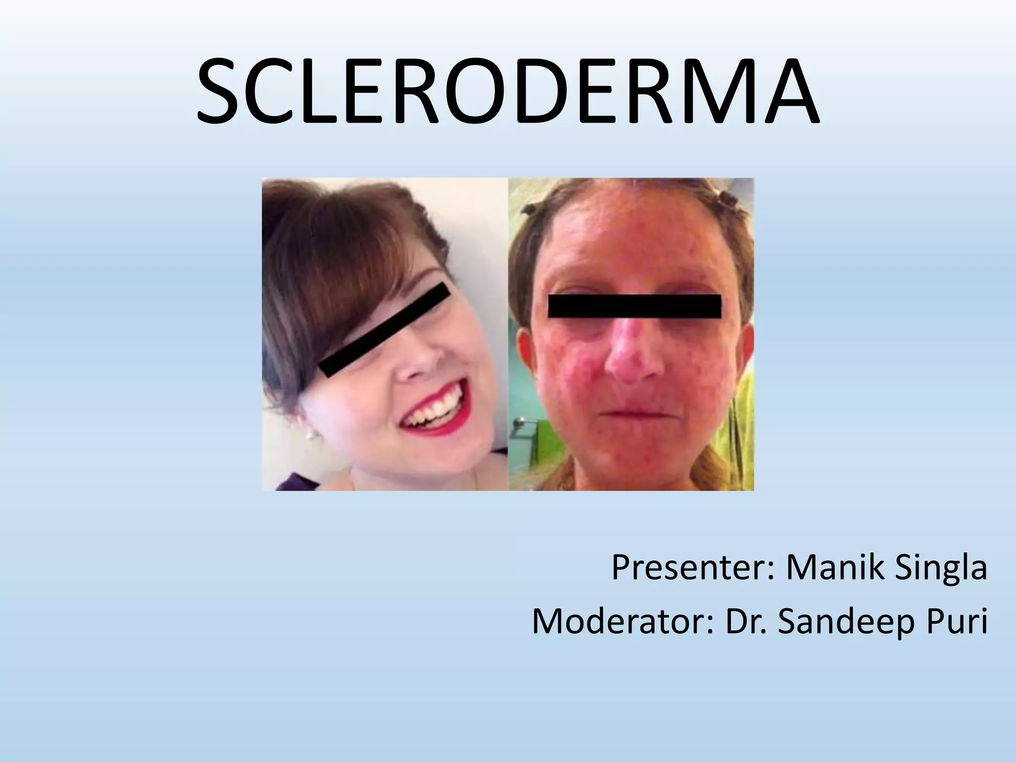
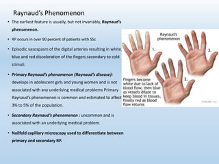
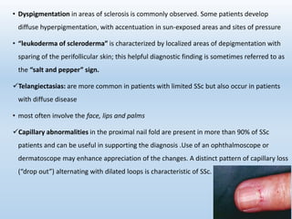
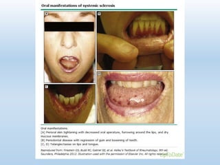
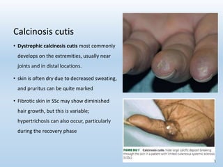
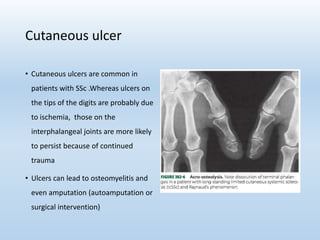
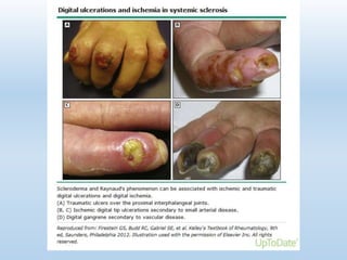
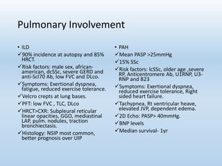
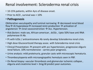
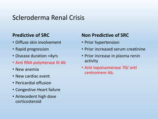
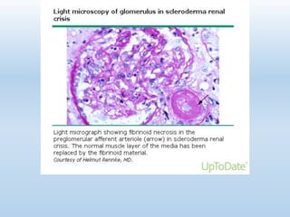
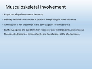
SCLERODERMA
Presenter: Manik Singla
Moderator: Dr. Sandeep Puri
Introduction
•Greek word Sclero meaning hard and derma meaning skin
• Systemic sclerosis (SSc, scleroderma)- autoimmune connective
tissue disease (AI-CTD) of unknown etiology.
• Affects the skin, GI tract, lungs, kidneys and heart.
• Two major clinical subtypes: limited and diffuse cutaneous SSc
History
• The first reported case of SSc (in 1753) written by Carol Curizo was a young
Italian woman who developed progressive induration of her skin.
• Visceral involvement reported by Matsuis in 1924.
• Goetz proposed the name Progressive systemic sclerosis 1945.
Epidemiology
• SSc is a relatively rare acquired sporadic disease with a global
distribution.
• Worldwide estimated incidence of SSc is 2.3-10 people per 1 million
• Women- affected three to four times more
• onset - typically between the ages of 30 and 50 years.
• Approximately 1.5% patients have one or more affected first-degree
relatives, representing a 10- to 15-fold higher risk in family
• SSc - significant mortality rate, 5 yr survival rate ranged from 34-73%.
11.Ashok kumar et al,1990, J Assoc Phsyc India
12. Krishnamurthy et al,1991, J Assoc Phsyc India
13. LeRoy EC,J Rheumatology 1988
Vandana Pradhan et al, 2014,
Indian Journal of Rheumatology
Risk factors and possible causes
• Genetic factors: The risk of SSc in first-degree relatives of individuals
with SSc is markedly increased.
Class II HLA, Fibrillin(Fbn-1), TNFAIP3 (A20) gene, PTPN22 gene
• Chemical Exposure: Petroleum-based products
Vinyl chloride, epoxy resin, silica
Contaminated rapeseed oil – Spain, Toxic oil syndrome
L-tryptophan – Eosinophilia myalgia Syndrome
• Infections
Parvovirus B19, latent CMV infection
• Autoimmunity
Anti-centromere Ab, Anti-topoisomerase Ab
• MICROCHIMERISM
Pathogenesis
Can be discussed under following three headings:
1. Vascular dysregulation
2. Immune dysregulation
3. Fibrosis
Vascular Dysregulation
• Blood vessels effected- smallest capillaries within the proximal nail fold to the
large pulmonary arteries, Renal arteries.
• Endothelial cell injury occurs early-
Anti endothelial antibodies
Perivascular leak & edema
• Surrounding smooth muscle cells - affected & have altered production and
responsiveness to vasoconstrictors (e.g. cold, endothelin) & vasodilators (e.g.
nitric oxide,prostacyclin)
• Intimal proliferation luminal occlusion develop hypoxia, release of
profibrotic cytokines, fibroblast activation & collagen production.
Immune Dysregulation
• Approximately 95 percent of patients with SSc have circulating
autoantibodies directed against one or more of a number of antigens.
• Although not very sensitive, antitopoisomerase-I (Scl-70) antibodies are
highly specific for SSc (98 to 100 percent) and correlate with a higher risk of
interstitial lung disease.
• Complexes containing Topoisomerase I autoantibody, when bound to the
surface of fibroblasts, have been found to stimulate monocyte adhesion and
activation.
Fibrosis
• Fibrosis represents the final common pathway
• Excessive deposition of collagens, proteoglycans, fibronectin, fibrillins and
adhesion molecules (e.g. β1-integrins), which sequester cytokines and
growth factors
• Connective tissue growth factor (CTGF) -responsible for maintenance of
collagen synthesis
• Inherent defect, an autocrine loop, or a hypersensitivity to growth factors in
SSc fibroblasts.
• Accumulation of collagen in SSc seems to be primarily the result of
increased synthesis, rather than decreased degradation
Clinical features
Diagnostic criteria & classification:
• Classification scheme of the American College of Rheumatology And EULAR.
• Two major clinical subtypes: limited and diffuse cutaneous.
• CREST syndrome describes the clinical features in a subset of patients with
limited SSc : calcinosis cutis, Raynaud’s phenomenon, esophageal
involvement, sclerodactyly and telangiectasia
• systemic sclerosis sine scleroderma: Internal organ involvement, Raynaud’s
phenomenon and positive serologies but no cutaneous involvement.
Harrison’s Principles of Internal Medicine 19h Ed.
Skin Involvement
• Early edematous phase, which often feature
pitting edema of the digits
• Subsequently hardens and develops a taut,
shiny appearance (indurated phase)
• Eventually, there may be gradual thinning of
the skin (late atrophic phase)
• Fingers can develop flexion contractures and
ulcers
• Involvement of the face can lead to a beaked
nose, microstomia and a somewhat youthful
appearance
• The earliest feature is usually, but not invariably, Raynaud’s
phenomenon.
• RP occurs in over 90 percent of patients with SSc
• Episodic vasospasm of the digital arteries resulting in white,
blue and red discoloration of the fingers secondary to cold
stimuli.
• Primary Raynaud’s phenomenon (Raynaud’s disease):
develops in adolescent girls and young women and is not
associated with any underlying medical problems Primary
Raynaud’s phenomenon is common and estimated to affect
3% to 5% of the population.
• Secondary Raynaud’s phenomenon : uncommon and is
associated with an underlying medical problem.
• Nailfold capillary microscopy used to differentiate between
primary and secondary RP.
Raynaud’s Phenomenon
• Dyspigmentation in areas of sclerosis is commonly observed. Some patients develop
diffuse hyperpigmentation, with accentuation in sun-exposed areas and sites of pressure
• “leukoderma of scleroderma” is characterized by localized areas of depigmentation with
sparing of the perifollicular skin; this helpful diagnostic finding is sometimes referred to as
the “salt and pepper” sign.
Telangiectasias: are more common in patients with limited SSc but also occur in patients
with diffuse disease
• most often involve the face, lips and palms
Capillary abnormalities in the proximal nail fold are present in more than 90% of SSc
patients and can be useful in supporting the diagnosis .Use of an ophthalmoscope or
dermatoscope may enhance appreciation of the changes. A distinct pattern of capillary loss
(“drop out”) alternating with dilated loops is characteristic of SSc.
• Dystrophic calcinosis cutis most commonly
develops on the extremities, usually near
joints and in distal locations.
• skin is often dry due to decreased sweating,
and pruritus can be quite marked
• Fibrotic skin in SSc may show diminished
hair growth, but this is variable;
hypertrichosis can also occur, particularly
during the recovery phase
Calcinosis cutis
Cutaneous ulcer
• Cutaneous ulcers are common in
patients with SSc .Whereas ulcers on
the tips of the digits are probably due
to ischemia, those on the
interphalangeal joints are more likely
to persist because of continued
trauma
• Ulcers can lead to osteomyelitis and
even amputation (autoamputation or
surgical intervention)
Bone changes
• Phalangeal absorption is associated with calcinosis.
• 70% of patients show absorption, which may be
minimal and only involve one terminal phalanx, or
be gross and involve several phalanges, including
the middle or even proximal phalanges.
• Erosive arthropathy, with ‘pestle and mortar’
deformity of the distal interphalangeal joints,
resembles that seen in psoriatic arthropathy.
• Pain in the temporomandibular area and a
grinding sensation on chewing may be associated
with bone resorption of the angle of the mandible
and zygomatic arches.
Pulmonary Involvement
• Pulmonary involvement leading cause of death.
• Principal forms: ILD and Pulmonary vascular disease.
Less frequent manifestations:
• Asp Pneumonitis complicating chronic Gastroesophageal
reflux
• Pulmonary hemorrhage due to endobronchial
telangiectasia.
• Obliterative bonchiolitis
• Pleural effusion
• Restrictive ventilatory defect due to chest wall fibrosis
• Spontaneous pneumothorax
• Drug induced lung toxicity
• DLco may give an indication of survival; if it is less than 40%
there is a 10% 5-year survival, compared with 75% if it is
greater than 40%
Pulmonary Involvement
• ILD
90% incidence at autopsy and 85%
HRCT.
Risk factors: male sex, african-
american, dcSSc, severe GERD and
anti-Scl70 Ab, low FVC and DLco.
Symptoms: Exertional dyspnea,
fatigue, reduced exercise tolerance.
Velcro crepts at lung bases.
PFT: low FVC , TLC, DLco
HRCT>CXR: Subpleural reticular
linear opacities, GGO, mediastinal
LAP, pulm. nodules, traction
bronchiectasis.
Histology: NSIP most common,
better prognosis over UIP
• PAH
Mean PASP >25mmHg
15% SSc
Risk factors: lcSSc, older age ,severe
RP, Anticentromere Ab, U1RNP, U3-
RNP and B23
Symptoms: Exertional dyspnea,
reduced exercise tolerance, Right
sided heart failure.
Tachypnea, Rt ventricular heave,
elevated JVP, dependent edema.
2D Echo: PASP> 40mmHg.
BNP levels
Median survival- 1yr
Gastro-intestinal tract
1. Macroglosia
2. Oesophagus: involved in approximately 75% of all patients and is the most
frequent part affected.
• Dysphagia & esophageal reflux
• Typical radiological appearance is that of an atonic dilated oesophagus, which
contains air in the resting state.
• Stricture of the lower end of the oesophagus occurs in just over 10% of patients
• Hiatus hernia occurs in approximately 25% of patients
3. Stomach: shows dilatation and lack of peristalsis in approximately 6% of cases
• Systemic sclerosis is one cause of gastric antral vascular ectasia (GAVE) —the so
called ‘watermelon stomach’, because of the striped appearance on endoscopy.
• GI bleeding
4. Intestinal involvement
• Duodenum shows changes of dilatation and lack of peristalsis in approximately one-
third of patients
• Pseudo-obstruction, and death may follow from paralytic ileus.
• Volvulus, bleeding from telangiectasias , bacterial overgrowth & malabsorption
• Steatorrhoea, malabsorption of glucose, calcium, vitamin B12 and folic acid
• Excessive enteric loss of protein
• Pneumatosis cystoides intestinalis may complicate small intestine involvement.
Colon: involved in 43% patients
• Severe constipation
• Colonic telangiectesia – anaemia
• Late stage SSc - presence of wide-mouthed sacculations or diverticula, best demonstrated
on post-evacuation roentgenograms, can cause perforation / bleeding.
• Rectal prolapse and faecal incontinence may result.
Cardiac involvement
• Often silent, usually diagnosed with sensitive
techniques.
• Primary or secondary to PAH/ILD
• dcSSc > lcSSc, within 3yrs of skin thickening.
• Endocardium, myocardium, pericardium may be
involved.
• Abnormalities of rhythm occur and these include
paroxysmal atrial tachycardia, atrial fibrillation
and flutter, heart blocks.
• Recurrent vasospasm and reperfusion  Myocardial
fibrosis  LV dysfunction  overt heart failure.
• Epicardial coronary artery disease not increased.
• TDE,SPECT, Cardiac MRI reveal high prevalence of
abnormal myocardial function/perfusion in SSc
patients.
Renal involvement: Scleroderma renal crisis
• 10-15% patients, within 4yrs of disease onset.
• Prior to ACEI , survival was < 10%
• Pathogenesis
Obliterative vasculopathy and luminal narrowing  decreased renal blood
flow JG hyperplasia increased renin production  activation of
angiotensin  renal vasoconstriction  Acc. Hypertension.
• Risk factors: male sex, African-american , dcSSc , Upto 50% have anti RNA
polymerase III Ab.
• Pt with lcSSc / anticentromere Ab rarely develop Scleroderma renal crisis.
• High dose Glucocorticoid therapy assoc. with Scleroderma renal crisis
• Clinical Presentation: Pt present with acc hypertension, progressive oliguric
renal failure. 10% normotensive- carries poor prognosis.
• Urine analysis: mild proteinuria, granular casts with microscopic hematuria.
• Thrombocytopenia with microangiopathic hemolysis seen in PBF.
• On Renal biopsy: vascular thrombosis and glomerular ischemic collapse ,
oliguria and creatinine level > 3mg/dl predict poor outcomes.
Scleroderma Renal Crisis
Predictive of SRC
• Diffuse skin involvement
• Rapid progression
• Disease duration <4yrs
• Anti RNA polymerase III Ab
• New anemia
• New cardiac event
• Pericardial effusion
• Congestive Heart failure
• Antecedent high dose
corticosteroid
Non Predictive of SRC
• Prior hypertension
• Prior increased serum creatinine
• Prior increase in plasma renin
activity
• Anti topoisomerase 70/ anti
centromere Ab.
Musculoskeletal Involvement
• Carpal tunnel syndrome occurs frequently
• Mobility impaired- Contractures at proximal interphalangeal joints and wrists
• Arthritic pain is not uncommon in the early stages of systemic sclerosis
• Leathery, palpable and audible friction rubs occur over the large joints , due extensive
fibrosis and adhesions of tendon sheaths and fascial planes at the affected joints.
Laboratory Investigations
• CBC - Anemia – Microcytic/normocytic – GAVE
Macrocytic: malabsorption Vit B12, folate
Microangiopathic hemolytic picture with renal crisis
• ESR is normal.
• Urine Routine for Proteinuria, granular casts, hematuria : SRC
• Anti Nuclear Antibodies
• ECG: to detect rhythm & conduction abnormality
• Echocardiography & Right heart catheterization: for pulmonary artery HTN
• Pulmonary function tests: to detect fibrotic changes
• X-rays & HRCT scan: lung involvement
• Esophageal manometry, Endoscopic examination & Barium studies: G.I involvements
• Skin biopsy
• Nail fold capillaroscopy: architectural disorganization, giant capillaries, hemorrhages, loss
of capillaries, angiogenesis, avascular areas
• Modified Rodnan’s skin scores (mRSS): to semi-quantify skin thickness in SSc
Diagnosis
• Primarily on clinical grounds.
• ACR/EULAR criteria high sensitivity and specificity 91 and 92 %.
• Skin induration with visceral organ involvement establishes diagnosis
with high certainty.
• Full thickness skin biopsy is required for establishing the diagnosis of
sclerema, scleromyxedema or nephrogenic systemic fibrosis.
lcSSc: Antecedent RP, Gastroesophageal reflux symptoms ,
sclerodactyly, capillary changes on nailfold capillarascopy, often with
cutaneous telengiectasias and calcinosis, help in establishing diagnosis.
dcSSc: fatigue, swelling ,aching and stiffness initially, with no RP. Upper
extremity edema and puffy fingers, may be diagnosed as RA, SLE,
myositis or MCTD. Within weeks RP with induration of skin and
presence of SSc specific antibodies.
Management
• Due to the wide spectrum of disease manifestations and organ involvement,
the management of the disease is tailored to the individual patient, taking
into account the disease subset and type of internal organ involvement.
• In general, patients with systemic sclerosis (SSc) are treated with organ-
based symptomatic therapy.
As examples,
patients with gastrointestinal reflux are treated with proton-pump
inhibitors,
patients with Raynaud phenomenon are frequently treated with calcium
channel blockers, and
patients with hypertensive scleroderma renal crisis are treated with
angiotensin-converting enzyme (ACE) inhibitors
However, patients with diffuse skin involvement and/or severe inflammatory
organ involvement are usually treated with systemic immunosuppressive
therapy.
Diffuse skin sclerosis
• For patients with progressive and diffuse skin involvement who do
not have visceral involvement, methotrexate (MTX)
or mycophenolate mofetil (MMF) is ideally initiated within three
years of disease onset.
• Although cyclophosphamide is generally reserved for dcSSc patients
with interstitial lung disease, several studies have also reported
improvements in skin involvement.
• For refractory cases: IVIG and Rituximab may be used as adjuvant.
• Investigations are ongoing to evaluate the possible benefit of the
anti-interleukin (IL)-6 receptor antibody, toclizumab, for the
treatment of both skin and lung disease in patients with diffuse SSc.
Raynaud’s Phenomenon
• First line therapy: Behavioural
-Involves educating patients to keep warm and avoid smoking to minimize the frequency
and severity of attacks
• Second-line therapy : Pharmacologic: starting with vasodilators
- Calcium channel blockers- Nifedepine
- Angiotensin II Rs blockers- Losartan
- Phosphodiesterase type 5 inhibitors (e.g. sildenafil, tadalafil) that target the nitric
oxide-mediated vasodilation pathway have been employed.
• SSRI (fluoxetine), topical nitroglycerin and intermittent IV prostaglandins.
• Oral antiplatelet agents (e.g. low-dose aspirin) and/or pentoxifylline may theoretically
provide additional blood flow to compromised distal sites.
Cutaneous ulcers
• Digital ulcers on the fingers are difficult to treat and a source of great morbidity
• Bosentan, an oral endothelin receptor antagonist, has been approved
• Oral iloprost (a prostacyclin analogue)- result in a reduction in digital ulcers and improved
healing.
• Subcutaneous prostacyclin analogue treprostinil suggest that it may also be effective
• In refractory cases, nerve blocks and sympathectomies may be considered
• Moist hydrocolloid dressings provide a better wound healing environment than dry or
wet-to-dry dressings
• Enzymatic debriding agents (e.g. collagenase) and topically applied growth factors (e.g.
PDGF) have been utilized.
• Skin equivalents or grafts can also be used to stimulate the wound bed and decrease
wound pain
Scleroderma Renal crisis
• It’s a medical emergency
• High risk SSc patients monitor blood pressure daily.
• Nephrotoxic drugs avoided, Glucocorticoids to be used at very low dose if
absolutely necessary.
• Short acting ACE inhibitors with goal of achieving normal blood pressures
• ARB’s and CCB’s or Direct renin inhibitors if blood pressure not controlled.
• Upto 2/3rd of patients go on to dialysis
• Outcome worse with Anti topoisomerase Ab than Anti RNA polymerase III
Ab.
• 30-50% patients recover and dialysis can be discontinued.
• Kidney transplantation for those unable to discontinue dialysis after 2yrs.
Antifibrotic therapy
• The treatment of established fibrosis is a very difficult therapeutic problem,
since there is no safe and effective method of removing excess insoluble
cross-linked collagen fibers without damaging the structural framework of
the body and individual organs.
• D-Penicillamine - standard dose (750mg/day) vs low dose (125mg
EOD) - no difference
• Interferons- Apha and gamma
• Iloprost
• Relaxin
• Anticytokine therapies: Against TGFB,CTGF
• Investigational approaches: tyrosine kinase inhibitors- Imatinib, nilotinib.
Nintenib and pirfenidone have been suggested for lung fibrosis but still under
evaluation in SSc.
Pulmonary Manifestation - ILD
• In the vast majority of patients with SSc-ILD, the lung injury is
characterized by a pattern termed nonspecific interstitial pneumonia
(NSIP).
• Histopathologically, NSIP is characterized by varying degrees of
pulmonary inflammation and fibrosis, with some forms being
primarily inflammatory (cellular NSIP) and others primarily fibrotic
(fibrotic NSIP)
• Patients with a more fibrosing form of NSIP have worse outcomes
compared with those with cellular NSIP.
• Few patients with SSc-ILD have the histopathologic pattern of UIP, the
pattern that is associated with idiopathic pulmonary fibrosis (IPF).
Indication
Pulmonary Manifestation-ILD
• Cyclophosphamide pulse therapy : has a definitive role in SSc with
alveolitis.
Three trials have shown improvement in lung function with
cyclophosphamide as compared to D-Penicillamine or Prednisone.
Largest series with 103 patients followed upto 13 months showed
median survival of 89% in those treated with cyclophosphamide
compared to 71% in untreated group.
White B et al, Ann Int Med 2000;132:947-54
Pulmonary Manifestation-ILD
• Scleroderma Lung Study II (SLS II) included 142 patients with SSc-ILD,
characterized by a forced vital capacity (FVC) <80 percent, but >45
percent, of predicted, exertional dyspnea grade 2 or higher, ground
glass opacities on high resolution computed tomography (HRCT) with
or without reticular opacities, and onset of the first non-Raynaud
symptom of SSc within the period 7 years.
• Mycophenolate mofetil is preferred over Cyclophosphamide due to a
better safety profile and comparable efficacy
• Participants were randomly assigned to MMF 1500 mg twice daily for
24 months or oral cyclophosphamide titrated up to a maximum daily
dose of 1.8 to 2.3 mg/kg for 12 months.
• No significant difference was noted between groups. Dyspnea
improved in both groups. MMF was better tolerated than
cyclophosphamide based on a longer time to patient withdrawal and
a lower incidence of leukopenia and thrombocytopenia.
Lancet Respir Med. 2016;4(9):708. Epub 2016 Jul 25
Pulmonary Artery Hypertension
• Advances in the treatment of pulmonary artery hypertension:
- Endothelin receptor antagonists (bosentan, sitaxsentan, ambrisentan)
- Phosphodiesterase type 5 inhibitors (sildenafil, tadalafil)
- Prostacyclin analogues iloprost [inhaled], epoprostenol [intravenous],
treprostinil [subcutaneous] are approved for pulmonary arterial hypertension
• Immunosuppressive agents: mycophenolate mofetil, azathioprine, chlorambucil,
5-fluorouracil, cyclosporine.
• Autologous hematopoietic stem cell transplantation is currently being studied.
Prognosis
• Substantial increase in risk of premature death.
• dcSSc: 5 and 10yr survival rate are 70% and 55% respectively.
• lcSSc: 5 and 10yr survival rate are 90% and 75% respectively.
• Cause of death: PAH, Pulmonary fibrosis, GI involvement, cardiac
disease.
• SRC assoc with 30% 3yr mortality. ACE inhibitors increased survival
rate from <10% at 1 yr to >70% 3yr survival a present time.
• 10yr survival rate in SSc has improved from <60% in 1970 to >66-78%
in 90’s
Thank you

Systemic Sclerosis 2017

  • 1.
  • 2.
    Introduction •Greek word Scleromeaning hard and derma meaning skin • Systemic sclerosis (SSc, scleroderma)- autoimmune connective tissue disease (AI-CTD) of unknown etiology. • Affects the skin, GI tract, lungs, kidneys and heart. • Two major clinical subtypes: limited and diffuse cutaneous SSc
  • 3.
    History • The firstreported case of SSc (in 1753) written by Carol Curizo was a young Italian woman who developed progressive induration of her skin. • Visceral involvement reported by Matsuis in 1924. • Goetz proposed the name Progressive systemic sclerosis 1945.
  • 4.
    Epidemiology • SSc isa relatively rare acquired sporadic disease with a global distribution. • Worldwide estimated incidence of SSc is 2.3-10 people per 1 million • Women- affected three to four times more • onset - typically between the ages of 30 and 50 years. • Approximately 1.5% patients have one or more affected first-degree relatives, representing a 10- to 15-fold higher risk in family • SSc - significant mortality rate, 5 yr survival rate ranged from 34-73%.
  • 5.
    11.Ashok kumar etal,1990, J Assoc Phsyc India 12. Krishnamurthy et al,1991, J Assoc Phsyc India 13. LeRoy EC,J Rheumatology 1988
  • 6.
    Vandana Pradhan etal, 2014, Indian Journal of Rheumatology
  • 7.
    Risk factors andpossible causes • Genetic factors: The risk of SSc in first-degree relatives of individuals with SSc is markedly increased. Class II HLA, Fibrillin(Fbn-1), TNFAIP3 (A20) gene, PTPN22 gene • Chemical Exposure: Petroleum-based products Vinyl chloride, epoxy resin, silica Contaminated rapeseed oil – Spain, Toxic oil syndrome L-tryptophan – Eosinophilia myalgia Syndrome • Infections Parvovirus B19, latent CMV infection • Autoimmunity Anti-centromere Ab, Anti-topoisomerase Ab • MICROCHIMERISM
  • 8.
    Pathogenesis Can be discussedunder following three headings: 1. Vascular dysregulation 2. Immune dysregulation 3. Fibrosis
  • 9.
    Vascular Dysregulation • Bloodvessels effected- smallest capillaries within the proximal nail fold to the large pulmonary arteries, Renal arteries. • Endothelial cell injury occurs early- Anti endothelial antibodies Perivascular leak & edema • Surrounding smooth muscle cells - affected & have altered production and responsiveness to vasoconstrictors (e.g. cold, endothelin) & vasodilators (e.g. nitric oxide,prostacyclin) • Intimal proliferation luminal occlusion develop hypoxia, release of profibrotic cytokines, fibroblast activation & collagen production.
  • 10.
    Immune Dysregulation • Approximately95 percent of patients with SSc have circulating autoantibodies directed against one or more of a number of antigens. • Although not very sensitive, antitopoisomerase-I (Scl-70) antibodies are highly specific for SSc (98 to 100 percent) and correlate with a higher risk of interstitial lung disease. • Complexes containing Topoisomerase I autoantibody, when bound to the surface of fibroblasts, have been found to stimulate monocyte adhesion and activation.
  • 11.
    Fibrosis • Fibrosis representsthe final common pathway • Excessive deposition of collagens, proteoglycans, fibronectin, fibrillins and adhesion molecules (e.g. β1-integrins), which sequester cytokines and growth factors • Connective tissue growth factor (CTGF) -responsible for maintenance of collagen synthesis • Inherent defect, an autocrine loop, or a hypersensitivity to growth factors in SSc fibroblasts. • Accumulation of collagen in SSc seems to be primarily the result of increased synthesis, rather than decreased degradation
  • 14.
    Clinical features Diagnostic criteria& classification: • Classification scheme of the American College of Rheumatology And EULAR. • Two major clinical subtypes: limited and diffuse cutaneous. • CREST syndrome describes the clinical features in a subset of patients with limited SSc : calcinosis cutis, Raynaud’s phenomenon, esophageal involvement, sclerodactyly and telangiectasia • systemic sclerosis sine scleroderma: Internal organ involvement, Raynaud’s phenomenon and positive serologies but no cutaneous involvement.
  • 15.
    Harrison’s Principles ofInternal Medicine 19h Ed.
  • 17.
    Skin Involvement • Earlyedematous phase, which often feature pitting edema of the digits • Subsequently hardens and develops a taut, shiny appearance (indurated phase) • Eventually, there may be gradual thinning of the skin (late atrophic phase) • Fingers can develop flexion contractures and ulcers • Involvement of the face can lead to a beaked nose, microstomia and a somewhat youthful appearance
  • 18.
    • The earliestfeature is usually, but not invariably, Raynaud’s phenomenon. • RP occurs in over 90 percent of patients with SSc • Episodic vasospasm of the digital arteries resulting in white, blue and red discoloration of the fingers secondary to cold stimuli. • Primary Raynaud’s phenomenon (Raynaud’s disease): develops in adolescent girls and young women and is not associated with any underlying medical problems Primary Raynaud’s phenomenon is common and estimated to affect 3% to 5% of the population. • Secondary Raynaud’s phenomenon : uncommon and is associated with an underlying medical problem. • Nailfold capillary microscopy used to differentiate between primary and secondary RP. Raynaud’s Phenomenon
  • 19.
    • Dyspigmentation inareas of sclerosis is commonly observed. Some patients develop diffuse hyperpigmentation, with accentuation in sun-exposed areas and sites of pressure • “leukoderma of scleroderma” is characterized by localized areas of depigmentation with sparing of the perifollicular skin; this helpful diagnostic finding is sometimes referred to as the “salt and pepper” sign. Telangiectasias: are more common in patients with limited SSc but also occur in patients with diffuse disease • most often involve the face, lips and palms Capillary abnormalities in the proximal nail fold are present in more than 90% of SSc patients and can be useful in supporting the diagnosis .Use of an ophthalmoscope or dermatoscope may enhance appreciation of the changes. A distinct pattern of capillary loss (“drop out”) alternating with dilated loops is characteristic of SSc.
  • 21.
    • Dystrophic calcinosiscutis most commonly develops on the extremities, usually near joints and in distal locations. • skin is often dry due to decreased sweating, and pruritus can be quite marked • Fibrotic skin in SSc may show diminished hair growth, but this is variable; hypertrichosis can also occur, particularly during the recovery phase Calcinosis cutis
  • 22.
    Cutaneous ulcer • Cutaneousulcers are common in patients with SSc .Whereas ulcers on the tips of the digits are probably due to ischemia, those on the interphalangeal joints are more likely to persist because of continued trauma • Ulcers can lead to osteomyelitis and even amputation (autoamputation or surgical intervention)
  • 24.
    Bone changes • Phalangealabsorption is associated with calcinosis. • 70% of patients show absorption, which may be minimal and only involve one terminal phalanx, or be gross and involve several phalanges, including the middle or even proximal phalanges. • Erosive arthropathy, with ‘pestle and mortar’ deformity of the distal interphalangeal joints, resembles that seen in psoriatic arthropathy. • Pain in the temporomandibular area and a grinding sensation on chewing may be associated with bone resorption of the angle of the mandible and zygomatic arches.
  • 25.
    Pulmonary Involvement • Pulmonaryinvolvement leading cause of death. • Principal forms: ILD and Pulmonary vascular disease. Less frequent manifestations: • Asp Pneumonitis complicating chronic Gastroesophageal reflux • Pulmonary hemorrhage due to endobronchial telangiectasia. • Obliterative bonchiolitis • Pleural effusion • Restrictive ventilatory defect due to chest wall fibrosis • Spontaneous pneumothorax • Drug induced lung toxicity • DLco may give an indication of survival; if it is less than 40% there is a 10% 5-year survival, compared with 75% if it is greater than 40%
  • 26.
    Pulmonary Involvement • ILD 90%incidence at autopsy and 85% HRCT. Risk factors: male sex, african- american, dcSSc, severe GERD and anti-Scl70 Ab, low FVC and DLco. Symptoms: Exertional dyspnea, fatigue, reduced exercise tolerance. Velcro crepts at lung bases. PFT: low FVC , TLC, DLco HRCT>CXR: Subpleural reticular linear opacities, GGO, mediastinal LAP, pulm. nodules, traction bronchiectasis. Histology: NSIP most common, better prognosis over UIP • PAH Mean PASP >25mmHg 15% SSc Risk factors: lcSSc, older age ,severe RP, Anticentromere Ab, U1RNP, U3- RNP and B23 Symptoms: Exertional dyspnea, reduced exercise tolerance, Right sided heart failure. Tachypnea, Rt ventricular heave, elevated JVP, dependent edema. 2D Echo: PASP> 40mmHg. BNP levels Median survival- 1yr
  • 27.
    Gastro-intestinal tract 1. Macroglosia 2.Oesophagus: involved in approximately 75% of all patients and is the most frequent part affected. • Dysphagia & esophageal reflux • Typical radiological appearance is that of an atonic dilated oesophagus, which contains air in the resting state. • Stricture of the lower end of the oesophagus occurs in just over 10% of patients • Hiatus hernia occurs in approximately 25% of patients 3. Stomach: shows dilatation and lack of peristalsis in approximately 6% of cases • Systemic sclerosis is one cause of gastric antral vascular ectasia (GAVE) —the so called ‘watermelon stomach’, because of the striped appearance on endoscopy. • GI bleeding 4. Intestinal involvement • Duodenum shows changes of dilatation and lack of peristalsis in approximately one- third of patients • Pseudo-obstruction, and death may follow from paralytic ileus. • Volvulus, bleeding from telangiectasias , bacterial overgrowth & malabsorption
  • 28.
    • Steatorrhoea, malabsorptionof glucose, calcium, vitamin B12 and folic acid • Excessive enteric loss of protein • Pneumatosis cystoides intestinalis may complicate small intestine involvement. Colon: involved in 43% patients • Severe constipation • Colonic telangiectesia – anaemia • Late stage SSc - presence of wide-mouthed sacculations or diverticula, best demonstrated on post-evacuation roentgenograms, can cause perforation / bleeding. • Rectal prolapse and faecal incontinence may result.
  • 29.
    Cardiac involvement • Oftensilent, usually diagnosed with sensitive techniques. • Primary or secondary to PAH/ILD • dcSSc > lcSSc, within 3yrs of skin thickening. • Endocardium, myocardium, pericardium may be involved. • Abnormalities of rhythm occur and these include paroxysmal atrial tachycardia, atrial fibrillation and flutter, heart blocks. • Recurrent vasospasm and reperfusion  Myocardial fibrosis  LV dysfunction  overt heart failure. • Epicardial coronary artery disease not increased. • TDE,SPECT, Cardiac MRI reveal high prevalence of abnormal myocardial function/perfusion in SSc patients.
  • 30.
    Renal involvement: Sclerodermarenal crisis • 10-15% patients, within 4yrs of disease onset. • Prior to ACEI , survival was < 10% • Pathogenesis Obliterative vasculopathy and luminal narrowing  decreased renal blood flow JG hyperplasia increased renin production  activation of angiotensin  renal vasoconstriction  Acc. Hypertension. • Risk factors: male sex, African-american , dcSSc , Upto 50% have anti RNA polymerase III Ab. • Pt with lcSSc / anticentromere Ab rarely develop Scleroderma renal crisis. • High dose Glucocorticoid therapy assoc. with Scleroderma renal crisis • Clinical Presentation: Pt present with acc hypertension, progressive oliguric renal failure. 10% normotensive- carries poor prognosis. • Urine analysis: mild proteinuria, granular casts with microscopic hematuria. • Thrombocytopenia with microangiopathic hemolysis seen in PBF. • On Renal biopsy: vascular thrombosis and glomerular ischemic collapse , oliguria and creatinine level > 3mg/dl predict poor outcomes.
  • 31.
    Scleroderma Renal Crisis Predictiveof SRC • Diffuse skin involvement • Rapid progression • Disease duration <4yrs • Anti RNA polymerase III Ab • New anemia • New cardiac event • Pericardial effusion • Congestive Heart failure • Antecedent high dose corticosteroid Non Predictive of SRC • Prior hypertension • Prior increased serum creatinine • Prior increase in plasma renin activity • Anti topoisomerase 70/ anti centromere Ab.
  • 33.
    Musculoskeletal Involvement • Carpaltunnel syndrome occurs frequently • Mobility impaired- Contractures at proximal interphalangeal joints and wrists • Arthritic pain is not uncommon in the early stages of systemic sclerosis • Leathery, palpable and audible friction rubs occur over the large joints , due extensive fibrosis and adhesions of tendon sheaths and fascial planes at the affected joints.
  • 34.
    Laboratory Investigations • CBC- Anemia – Microcytic/normocytic – GAVE Macrocytic: malabsorption Vit B12, folate Microangiopathic hemolytic picture with renal crisis • ESR is normal. • Urine Routine for Proteinuria, granular casts, hematuria : SRC • Anti Nuclear Antibodies • ECG: to detect rhythm & conduction abnormality • Echocardiography & Right heart catheterization: for pulmonary artery HTN • Pulmonary function tests: to detect fibrotic changes • X-rays & HRCT scan: lung involvement • Esophageal manometry, Endoscopic examination & Barium studies: G.I involvements • Skin biopsy • Nail fold capillaroscopy: architectural disorganization, giant capillaries, hemorrhages, loss of capillaries, angiogenesis, avascular areas • Modified Rodnan’s skin scores (mRSS): to semi-quantify skin thickness in SSc
  • 40.
    Diagnosis • Primarily onclinical grounds. • ACR/EULAR criteria high sensitivity and specificity 91 and 92 %. • Skin induration with visceral organ involvement establishes diagnosis with high certainty. • Full thickness skin biopsy is required for establishing the diagnosis of sclerema, scleromyxedema or nephrogenic systemic fibrosis. lcSSc: Antecedent RP, Gastroesophageal reflux symptoms , sclerodactyly, capillary changes on nailfold capillarascopy, often with cutaneous telengiectasias and calcinosis, help in establishing diagnosis. dcSSc: fatigue, swelling ,aching and stiffness initially, with no RP. Upper extremity edema and puffy fingers, may be diagnosed as RA, SLE, myositis or MCTD. Within weeks RP with induration of skin and presence of SSc specific antibodies.
  • 41.
    Management • Due tothe wide spectrum of disease manifestations and organ involvement, the management of the disease is tailored to the individual patient, taking into account the disease subset and type of internal organ involvement. • In general, patients with systemic sclerosis (SSc) are treated with organ- based symptomatic therapy. As examples, patients with gastrointestinal reflux are treated with proton-pump inhibitors, patients with Raynaud phenomenon are frequently treated with calcium channel blockers, and patients with hypertensive scleroderma renal crisis are treated with angiotensin-converting enzyme (ACE) inhibitors However, patients with diffuse skin involvement and/or severe inflammatory organ involvement are usually treated with systemic immunosuppressive therapy.
  • 42.
    Diffuse skin sclerosis •For patients with progressive and diffuse skin involvement who do not have visceral involvement, methotrexate (MTX) or mycophenolate mofetil (MMF) is ideally initiated within three years of disease onset. • Although cyclophosphamide is generally reserved for dcSSc patients with interstitial lung disease, several studies have also reported improvements in skin involvement. • For refractory cases: IVIG and Rituximab may be used as adjuvant. • Investigations are ongoing to evaluate the possible benefit of the anti-interleukin (IL)-6 receptor antibody, toclizumab, for the treatment of both skin and lung disease in patients with diffuse SSc.
  • 43.
    Raynaud’s Phenomenon • Firstline therapy: Behavioural -Involves educating patients to keep warm and avoid smoking to minimize the frequency and severity of attacks • Second-line therapy : Pharmacologic: starting with vasodilators - Calcium channel blockers- Nifedepine - Angiotensin II Rs blockers- Losartan - Phosphodiesterase type 5 inhibitors (e.g. sildenafil, tadalafil) that target the nitric oxide-mediated vasodilation pathway have been employed. • SSRI (fluoxetine), topical nitroglycerin and intermittent IV prostaglandins. • Oral antiplatelet agents (e.g. low-dose aspirin) and/or pentoxifylline may theoretically provide additional blood flow to compromised distal sites.
  • 44.
    Cutaneous ulcers • Digitalulcers on the fingers are difficult to treat and a source of great morbidity • Bosentan, an oral endothelin receptor antagonist, has been approved • Oral iloprost (a prostacyclin analogue)- result in a reduction in digital ulcers and improved healing. • Subcutaneous prostacyclin analogue treprostinil suggest that it may also be effective • In refractory cases, nerve blocks and sympathectomies may be considered • Moist hydrocolloid dressings provide a better wound healing environment than dry or wet-to-dry dressings • Enzymatic debriding agents (e.g. collagenase) and topically applied growth factors (e.g. PDGF) have been utilized. • Skin equivalents or grafts can also be used to stimulate the wound bed and decrease wound pain
  • 46.
    Scleroderma Renal crisis •It’s a medical emergency • High risk SSc patients monitor blood pressure daily. • Nephrotoxic drugs avoided, Glucocorticoids to be used at very low dose if absolutely necessary. • Short acting ACE inhibitors with goal of achieving normal blood pressures • ARB’s and CCB’s or Direct renin inhibitors if blood pressure not controlled. • Upto 2/3rd of patients go on to dialysis • Outcome worse with Anti topoisomerase Ab than Anti RNA polymerase III Ab. • 30-50% patients recover and dialysis can be discontinued. • Kidney transplantation for those unable to discontinue dialysis after 2yrs.
  • 48.
    Antifibrotic therapy • Thetreatment of established fibrosis is a very difficult therapeutic problem, since there is no safe and effective method of removing excess insoluble cross-linked collagen fibers without damaging the structural framework of the body and individual organs. • D-Penicillamine - standard dose (750mg/day) vs low dose (125mg EOD) - no difference • Interferons- Apha and gamma • Iloprost • Relaxin • Anticytokine therapies: Against TGFB,CTGF • Investigational approaches: tyrosine kinase inhibitors- Imatinib, nilotinib. Nintenib and pirfenidone have been suggested for lung fibrosis but still under evaluation in SSc.
  • 49.
    Pulmonary Manifestation -ILD • In the vast majority of patients with SSc-ILD, the lung injury is characterized by a pattern termed nonspecific interstitial pneumonia (NSIP). • Histopathologically, NSIP is characterized by varying degrees of pulmonary inflammation and fibrosis, with some forms being primarily inflammatory (cellular NSIP) and others primarily fibrotic (fibrotic NSIP) • Patients with a more fibrosing form of NSIP have worse outcomes compared with those with cellular NSIP. • Few patients with SSc-ILD have the histopathologic pattern of UIP, the pattern that is associated with idiopathic pulmonary fibrosis (IPF).
  • 50.
  • 51.
    Pulmonary Manifestation-ILD • Cyclophosphamidepulse therapy : has a definitive role in SSc with alveolitis. Three trials have shown improvement in lung function with cyclophosphamide as compared to D-Penicillamine or Prednisone. Largest series with 103 patients followed upto 13 months showed median survival of 89% in those treated with cyclophosphamide compared to 71% in untreated group. White B et al, Ann Int Med 2000;132:947-54
  • 52.
    Pulmonary Manifestation-ILD • SclerodermaLung Study II (SLS II) included 142 patients with SSc-ILD, characterized by a forced vital capacity (FVC) <80 percent, but >45 percent, of predicted, exertional dyspnea grade 2 or higher, ground glass opacities on high resolution computed tomography (HRCT) with or without reticular opacities, and onset of the first non-Raynaud symptom of SSc within the period 7 years. • Mycophenolate mofetil is preferred over Cyclophosphamide due to a better safety profile and comparable efficacy • Participants were randomly assigned to MMF 1500 mg twice daily for 24 months or oral cyclophosphamide titrated up to a maximum daily dose of 1.8 to 2.3 mg/kg for 12 months. • No significant difference was noted between groups. Dyspnea improved in both groups. MMF was better tolerated than cyclophosphamide based on a longer time to patient withdrawal and a lower incidence of leukopenia and thrombocytopenia. Lancet Respir Med. 2016;4(9):708. Epub 2016 Jul 25
  • 53.
    Pulmonary Artery Hypertension •Advances in the treatment of pulmonary artery hypertension: - Endothelin receptor antagonists (bosentan, sitaxsentan, ambrisentan) - Phosphodiesterase type 5 inhibitors (sildenafil, tadalafil) - Prostacyclin analogues iloprost [inhaled], epoprostenol [intravenous], treprostinil [subcutaneous] are approved for pulmonary arterial hypertension • Immunosuppressive agents: mycophenolate mofetil, azathioprine, chlorambucil, 5-fluorouracil, cyclosporine. • Autologous hematopoietic stem cell transplantation is currently being studied.
  • 54.
    Prognosis • Substantial increasein risk of premature death. • dcSSc: 5 and 10yr survival rate are 70% and 55% respectively. • lcSSc: 5 and 10yr survival rate are 90% and 75% respectively. • Cause of death: PAH, Pulmonary fibrosis, GI involvement, cardiac disease. • SRC assoc with 30% 3yr mortality. ACE inhibitors increased survival rate from <10% at 1 yr to >70% 3yr survival a present time. • 10yr survival rate in SSc has improved from <60% in 1970 to >66-78% in 90’s
  • 55.

Editor's Notes

  • #8 MICROCHIMERISM — Normal women harbor viable immunologic stem cells of fetal origin many years following pregnancy [76]. The number of fetal cells present in women with systemic sclerosis (SSc) who have been pregnant is elevated compared with healthy control women with a history of pregnancy [77,78]. Male cells were found in at least one organ obtained at autopsy from women with SSc who had male offspring [79]. The persistence of fetal cells may be linked to the pathogenesis of SSc via several potential mechanisms: ●Fetal (the graft) cells may mount an immune response against the mother (the host) in a graft-versus-host-like reaction. ●A maternal response to the fetal cells may subsequently be redirected against “self,” leading to autoimmunity. It is noteworthy that patients with SSc are more likely to be closely human leukocyte antigen (HLA)-matched to the paternal HLA carried by their children [79,80]. ●Maternal cells that cross the placenta and that are carried by the fetus may play an immunomodulatory role in men with SSc or in women with SSc who have never been pregnant