Downloaded 171 times
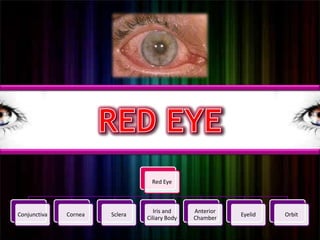

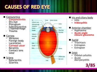


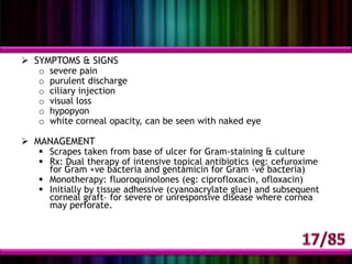


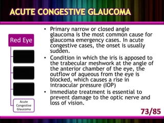


This document provides an overview of causes and symptoms of red eye. It discusses various conditions that can cause redness of the eye including conjunctivitis, pterygium, subconjunctival hemorrhage, corneal abrasion, keratitis, iritis, episcleritis, trichiasis, entropion, orbital cellulitis, acute dacryocystitis, hyphaema, and corneal ulcer. Signs and symptoms of red eye such as pain, discharge, photophobia, and visual changes are described for different conditions. Evaluation of red eye involves characterizing symptoms and performing an examination of the conjunctiva, cornea, anterior chamber, eyelids, and orbit.


