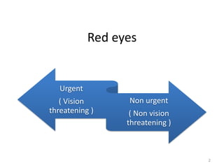
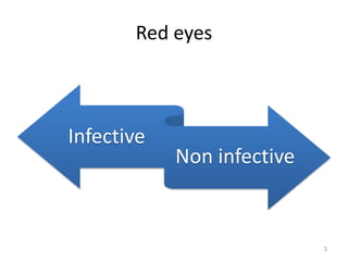
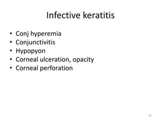
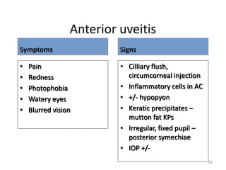
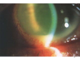
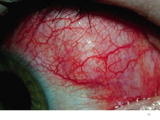
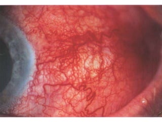
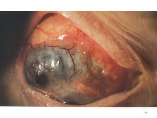
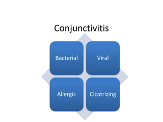
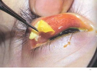
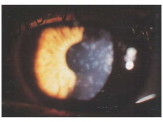
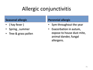
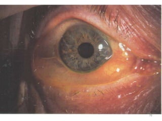
This document provides information on evaluating and differentiating between urgent and non-urgent cases of red eye. It discusses various conditions that can cause red eye including infectious causes like bacterial and viral conjunctivitis, as well as non-infectious causes like allergic conjunctivitis. Clinical features, differential diagnoses, management strategies are outlined for conditions like anterior uveitis, episcleritis, scleritis, blepharitis and subconjunctival hemorrhage. The document emphasizes the importance of identifying vision-threatening conditions like corneal ulcers that require urgent referral and treatment.
This document provides an outline of a lecture on the red eye, summarizing various potential causes organized by anatomical structure. Key structures discussed include the lids, conjunctiva, episclera, sclera, cornea, anterior chamber, and orbit. Common conditions are summarized such as conjunctivitis (bacterial, viral, allergic), subconjunctival hemorrhage, episcleritis, keratitis, uveitis, glaucoma. Diagnostic features and treatments are briefly mentioned for many conditions. The document concludes with reminding the importance of thorough history and eye examination to determine the cause and guide management of the red eye.
This document provides an overview of causes and management of red eye. It discusses various red eye disorders including non-vision threatening conditions like allergic conjunctivitis, dry eye, and corneal abrasions. Vision threatening conditions like orbital cellulitis, uveitis, and acute glaucoma are also reviewed. For each condition, the document outlines symptoms, signs, investigations, and treatment approach. Common etiologies of red eye include bacterial and viral conjunctivitis, subconjunctival hemorrhage, and dry eye syndrome. Evaluation of red eye involves thorough history taking and physical examination of the eye and surrounding structures.
This document discusses red eye conditions with a high risk of vision loss, including infectious keratitis/corneal ulcer, anterior uveitis, and acute angle closure glaucoma. It provides details on the causes, signs, symptoms, investigations, and treatment approaches for each condition. Infectious keratitis can be caused by bacteria, viruses, fungi or parasites and results in eye pain, discharge and corneal infiltration. Anterior uveitis commonly presents with eye pain and redness, and can be caused by infections, autoimmune disorders or injuries. Acute angle closure glaucoma presents as eye pain, blurred vision and rainbow halos, and requires prompt medical or surgical treatment to lower intraocular pressure.
Red Eye - Common Causes, Diagnosis and Treatment.pptxMedinfopedia Blog
Red eye is a non-specific term that is used to describe an eye that appears red due to intraocular or extra-ocular pathologies which can be as a result of infections, inflammations, allergies or trauma.
It is usually as a result of vasodilation in the anterior portion of the eye. It is a sign of an underlying disease, not a diagnosis.
This document discusses various systemic diseases that can manifest ocularly and be detected on eye examination. It covers 10 categories of disease including congenital, traumatic, vascular, neoplastic, autoimmune, idiopathic, infectious, metabolic/endocrine, and drugs/toxins. For each category, specific diseases are described along with their characteristic ocular signs and symptoms. The importance of comprehensive eye exams for evaluating systemic health is emphasized.
Bacterial eyelid infections and blepharitis.SristiThakur
This document discusses bacterial infections and blepharitis that can affect the eyelids. It begins by covering the anatomy of the eyelid and then describes several specific bacterial infections including external hordeolum, impetigo, erysipelas, necrotizing fasciitis, anthrax, and syphilis. It then discusses blepharitis in detail, describing the different types (staphylococcal, seborrheic, posterior), associated conditions like meibomian gland dysfunction, symptoms, signs, treatment, and complications.
This document provides information on various types of uveitis (inflammation of the uveal tract of the eye) including:
- Types of uveitis based on location such as iritis, cyclitis, choroiditis, etc.
- Causes of uveitis including infectious, autoimmune, neoplastic, and idiopathic.
- Presentation of uveitis depending on factors such as onset (sudden or insidious), duration (limited or persistent), activity level, and presence of complications.
- Investigations, treatment approaches including medications, injections and surgery, and management considerations for different types of uveitis.
This document discusses keratitis, an infection or inflammation of the cornea caused by various microorganisms or other factors. It can involve just the cornea or both the cornea and conjunctiva. Causes include bacterial, viral, fungal infections, contact lenses, vitamin A deficiency, cosmetics. Symptoms are eye redness, pain, tearing, blurred vision, photophobia. Diagnosis involves examination with a slit lamp and corneal smears. Treatment consists of antibiotic eye drops, antiviral medications, antifungal drops, cycloplegic drops, and phototherapeutic keratectomy using laser treatment for diseased corneal tissue.
The uveal tract disorders include uveitis, uveal melanoma, aniridia, and albinism. Uveitis is the inflammation of the uveal tract and is a common cause of blindness. Symptoms include eye redness, blurred vision, eye pain, and photophobia. Uveitis can be classified anatomically by the area of inflammation (anterior, intermediate, posterior, or pan) and clinically or pathologically. Treatment involves medications like corticosteroids and lifestyle changes. Complications can include cataracts, glaucoma, synechiae, and retinal issues if not properly treated and monitored.
This document discusses a case of a 65-year-old man presenting with decreased vision, pain, photophobia, and whiteness and discharge from his eye due to a suspected dust injury. Upon examination, he was found to have lid swelling, conjunctival congestion, ciliary congestion, a poorly-defined ulcer with slough and hypopyon in the floor. Testing found pseudomonas pyocyanea infection. The stages and complications of corneal ulcers are outlined. Treatment involves antibiotic drops, cycloplegics, and managing complications like perforation which may require gluing, grafting or surgery. Causes of non-healing ulcers and fungal and acanthamoeba infections are also
This document discusses anterior uveitis, which is inflammation of the iris, ciliary body, and anterior chamber of the eye. It can be caused by infection, autoimmune disorders, trauma, or idiopathic factors. Symptoms include eye pain, redness, photophobia, and blurred vision. Signs include cells in the anterior chamber, flare, hypopyon, keratic precipitates, iris nodules, and synechiae. Treatment involves cycloplegic eye drops, topical corticosteroids, and sometimes systemic corticosteroids or immunosuppressants depending on the severity and cause of inflammation. Complications like glaucoma and cataracts may require additional treatment.
2. This document presents an overview of red eye conditions and their management. It identifies signs requiring urgent referral, such as absent pupil response or corneal damage. Common differentials discussed include
Red eye, or ocular hyperemia, is caused by enlarged, dilated blood vessels leading to redness of the eye's surface. It can be caused by various conditions affecting the conjunctiva, cornea, iris, anterior chamber, eyelids, or orbit. Common causes include conjunctivitis (bacterial, viral, allergic), corneal abrasions, iritis, acute glaucoma, eyelid disorders like entropion or ectropion, and orbital cellulitis. A thorough history and examination is needed to determine the underlying cause and guide appropriate treatment.
This document discusses recurrent ocular herpes, which occurs when the herpes virus lies dormant in the trigeminal ganglion and periodically reactivates, causing recurrent infections of the trigeminal nerve. The main manifestations include epithelial keratitis, stromal keratitis, herpetic endothelitis, trophic ulceration, herpetic uveitis, and herpetic trabeculitis. Treatment involves antiviral medications like acyclovir applied topically or taken systemically, as well as corticosteroids depending on the specific manifestation.
This document provides an overview of common causes of a red eye and guidelines for evaluation and management. It discusses various conditions that can affect the eyelids, conjunctiva, sclera, cornea, iris and anterior chamber. Conditions range from self-limiting issues like blepharitis, styes and viral conjunctivitis to more serious conditions like bacterial ulcers, iritis, uveitis and acute angle closure glaucoma which require prompt referral. A basic history of symptoms and external exam can help form a differential diagnosis, while fluorescein staining aids examination of the cornea. Early referral is advised for decreased vision, severe pain, poor response to initial treatment or unilateral red eye.
This document provides information on various conditions related to the eye. It begins by discussing unilateral red eye in a 25-year-old female, noting that ciliary injection is a characteristic of intraocular pathology. It then covers topics like uveitis and its classification by anatomical site of involvement. Other sections discuss keratic precipitates, aqueous flare, conjunctival reactions including follicles and papillae, as well as conditions like chalazion, pterygium, and various forms of keratitis. Complications of conditions like ulcerative keratitis and scleritis are also outlined. Differential diagnoses are provided for clinical cases.
This document discusses various disorders and conditions of the eye. It begins by covering conditions of the eyelids and conjunctiva, such as blepharitis, hordeolum, entropion, ectropion, and ptosis. It then discusses disorders of the cornea and uveal tract, focusing on keratitis (inflammation and ulceration of the cornea), which can be caused by viruses, bacteria, fungi, or trauma. The document also covers conjunctivitis, pterygium, trachoma, and errors of refraction like myopia, hyperopia, and astigmatism as well as other disorders such as cataracts, glaucoma, retinal problems, and eye
This document describes various eye conditions including:
1) Conjunctivitis which can be caused by allergies, viruses, bacteria, herpes, gonorrhea, and Kawasaki disease.
2) Blepharitis which causes redness and swelling of the eyelid margins.
3) Preseptal and postseptal cellulitis which are infections anterior or posterior to the orbital septum respectively.
4) Other conditions described include chalazion, insect bites, pterygium, allergic shiners, raccoon eyes from basilar skull fractures, and scleral epithelial melanosis. Causes, symptoms, signs, time courses and treatments are provided for
This document discusses ocular involvement in leprosy. It notes that 70-75% of leprosy patients experience ocular involvement, with 10-50% experiencing severe symptoms and 5% becoming blind. The major parts of the eye affected include the eyelids, cornea, iris, ciliary body, conjunctiva, and nerves. Common complications include blepharochalasis, madarosis, trichiasis, lagophthalmos, dacryocystitis, punctate keratitis, pannus, iridocyclitis, glaucoma, and cataract. Modes of infection include direct invasion via blood or nerves. Management involves antibiotics, steroids, surgery, and
The document discusses signs of intraocular inflammation and distinguishes between granulomatous and non-granulomatous uveitis. It describes key signs of acute anterior uveitis including redness, pain, photophobia, aqueous cells and flare, hypopyon, iris signs like synechiae, and lens complications. Granulomatous uveitis is characterized by mutton fat KPs, iris nodules, and thick synechiae, while non-granulomatous uveitis presents with fine KPs, no iris nodules, and fine synechiae with more cells and flare. The document also notes the distinction between acute versus chronic conditions as causes of red eye
This document discusses several common complications that can arise from contact lens wear, including dry eye, papillary conjunctivitis, corneal abrasion, corneal hypoxia/edema, neovascularization, keratitis, and corneal ulcer. Dry eye is the most common problem and results from a lack of sufficient lubrication and moisture on the eye surface. Papillary conjunctivitis appears as localized swelling or papillae on the tarsal conjunctiva. Corneal abrasion is a scratch or staining on the cornea surface caused by a poorly fitted lens or lens deposits. Prolonged lens wear can lead to corneal hypoxia/edema from oxygen deprivation. Neovascularization is the growth of new blood
Cataract Surgery and LASIK Update 2013 - Dr. Jeff Martin of North Shore Eye C...Jeff Martin, MD, FACS
This document provides case studies and information about cataract surgery, LASIK, and corneal conditions from Dr. Jeffrey Martin of North Shore Eye Care. It includes summaries of two case studies, one involving a post-PRK patient with reduced vision and discomfort, and another involving a post-LASIK patient with pain, redness and photophobia. It also discusses techniques for cataract surgery and LASIK, medications used, and conditions like bacterial keratitis, herpes keratitis, and various forms of peripheral corneal thinning.
This document describes the clinical signs and symptoms of various types of conjunctivitis including bacterial, viral, allergic, and other causes. Bacterial conjunctivitis presents with redness, discharge, and irritation with symptoms ranging from mild to severe depending on if it is acute or chronic. Viral conjunctivitis often causes redness, watering, discomfort and photophobia. Allergic conjunctivitis commonly presents with intense itching, burning, watery discharge and photophobia. The document outlines several specific types of allergic conjunctivitis and their characteristics. Other types discussed include giant papillary conjunctivitis, phlyctenular keratoconjunctivitis, and contact der
This document discusses the causes and differential diagnosis of red eye. It lists various etiologies of red eye including adnexal causes like blepharitis, conjunctival causes like conjunctivitis, corneal causes like infectious keratitis, and other causes like dry eye syndrome. Key conditions are described in more detail such as subconjunctival hemorrhage, dry eye syndrome, corneal abrasion, corneal ulcer, episcleritis, and scleritis. The document concludes with a table comparing the signs and symptoms of subconjunctival hemorrhage, conjunctivitis, iritis, and acute glaucoma to aid in differential diagnosis of red eye.
This document discusses the causes and differential diagnosis of red eye. It lists various etiologies of red eye including adnexal causes like blepharitis, conjunctival causes like conjunctivitis, corneal causes like infectious keratitis, and other causes like dry eye syndrome. Key conditions are described in more detail such as subconjunctival hemorrhage, dry eye syndrome, corneal abrasion, corneal ulcer, episcleritis, and scleritis. The document concludes by providing a differential diagnosis table to distinguish between conditions like subconjunctival hemorrhage, conjunctivitis, iritis, and acute glaucoma based on conjunctival, pupil, corneal, anterior chamber, and intraocular
Red eye can be caused by conditions affecting the conjunctiva, cornea, sclera, iris, ciliary body, anterior chamber, eyelid, or orbit. Common causes include conjunctivitis, keratitis, scleritis, iritis, acute glaucoma, corneal ulcer, eyelid abnormalities like entropion or ectropion, and orbital cellulitis. Symptoms vary depending on the underlying condition but may include pain, visual loss, eye discharge, photophobia, and gritty or itchy sensation. Examination findings can provide clues to diagnose the specific cause and guide treatment.
Optic Neuritis, Papilledema document discusses optic nerve conditions. It defines optic neuritis as inflammation of the optic nerve impairing nerve conduction that can be caused by demyelination, infection, or autoimmunity. Papilledema is defined as bilateral, non-inflammatory swelling of the optic disc due to increased intracranial pressure. The document covers causes, signs, symptoms, diagnostic tests, treatment, and prognosis for both conditions.
Walmart Business+ and Spark Good for Nonprofits.pdfTechSoup
"Learn about all the ways Walmart supports nonprofit organizations.
You will hear from Liz Willett, the Head of Nonprofits, and hear about what Walmart is doing to help nonprofits, including Walmart Business and Spark Good. Walmart Business+ is a new offer for nonprofits that offers discounts and also streamlines nonprofits order and expense tracking, saving time and money.
The webinar may also give some examples on how nonprofits can best leverage Walmart Business+.
The event will cover the following::
Walmart Business + (https://business.walmart.com/plus) is a new shopping experience for nonprofits, schools, and local business customers that connects an exclusive online shopping experience to stores. Benefits include free delivery and shipping, a 'Spend Analytics” feature, special discounts, deals and tax-exempt shopping.
Special TechSoup offer for a free 180 days membership, and up to $150 in discounts on eligible orders.
Spark Good (walmart.com/sparkgood) is a charitable platform that enables nonprofits to receive donations directly from customers and associates.
Answers about how you can do more with Walmart!"
How to Make a Field Mandatory in Odoo 17Celine George
In Odoo, making a field required can be done through both Python code and XML views. When you set the required attribute to True in Python code, it makes the field required across all views where it's used. Conversely, when you set the required attribute in XML views, it makes the field required only in the context of that particular view.
বাংলাদেশের অর্থনৈতিক সমীক্ষা ২০২৪ [Bangladesh Economic Review 2024 Bangla.pdf] কম্পিউটার , ট্যাব ও স্মার্ট ফোন ভার্সন সহ সম্পূর্ণ বাংলা ই-বুক বা pdf বই " সুচিপত্র ...বুকমার্ক মেনু 🔖 ও হাইপার লিংক মেনু 📝👆 যুক্ত ..
আমাদের সবার জন্য খুব খুব গুরুত্বপূর্ণ একটি বই ..বিসিএস, ব্যাংক, ইউনিভার্সিটি ভর্তি ও যে কোন প্রতিযোগিতা মূলক পরীক্ষার জন্য এর খুব ইম্পরট্যান্ট একটি বিষয় ...তাছাড়া বাংলাদেশের সাম্প্রতিক যে কোন ডাটা বা তথ্য এই বইতে পাবেন ...
তাই একজন নাগরিক হিসাবে এই তথ্য গুলো আপনার জানা প্রয়োজন ...।
বিসিএস ও ব্যাংক এর লিখিত পরীক্ষা ...+এছাড়া মাধ্যমিক ও উচ্চমাধ্যমিকের স্টুডেন্টদের জন্য অনেক কাজে আসবে ...
This document provides an overview of wound healing, its functions, stages, mechanisms, factors affecting it, and complications.
A wound is a break in the integrity of the skin or tissues, which may be associated with disruption of the structure and function.
Healing is the body’s response to injury in an attempt to restore normal structure and functions.
Healing can occur in two ways: Regeneration and Repair
There are 4 phases of wound healing: hemostasis, inflammation, proliferation, and remodeling. This document also describes the mechanism of wound healing. Factors that affect healing include infection, uncontrolled diabetes, poor nutrition, age, anemia, the presence of foreign bodies, etc.
Complications of wound healing like infection, hyperpigmentation of scar, contractures, and keloid formation.
it describes the bony anatomy including the femoral head , acetabulum, labrum . also discusses the capsule , ligaments . muscle that act on the hip joint and the range of motion are outlined. factors affecting hip joint stability and weight transmission through the joint are summarized.
6. Laterality
• Infection may be unilateral initially but later
bilateral
• Allergy – usually bilateral, young
• Iritis / acute glaucoma – usually unilateral
6
7. Vision
• Transient blurring due to watery eyes /
discharge
• Persistent significant visual loss indicates more
serious disease
• Haloes – urgent!
7
8. General medical
• Headache, vomiting, eye pain – high IOP
• Hx of allergy, atopy, bronchial asthma, eczema
• Systemic assoc : iritis with sarcoidosis,
Ankylosing spondylitis, scleritis, CTD
• Acne rosacea assoc with blepharoconjunctivitis
• Hx of trauma to the eye – iritis, subconj
hemorrhage, glaucoma
8
9. General medical
• URTI accompanies infective conjunctivitis
• Preauricular lymphnode enlargement
9
32. Episcleritis
• Non – infective inflammation involving a
tissue layer superficial to the sclera and deep
to the conjunctiva.
• Extremely common
• Pain +/-
• Hyperemia : diffuse or segmental ( simple /
nodular )
39
36. Scleritis
• Uncommon
• More serious than episcleritis
• Oedema & cellular infiltration of the entire
thickness of sclera.
• Spectrum of disease – from self limiting to
blindness. ( necrotising & non-necrotising )
• Systemic associations – RA (most common
), Wegener’s granulomatosis, Relapsing
polychondritis, PAN ( polyarteritis nodosa ),
SLE.
43
37. Scleritis
Symptom
• Severe eye pain
• Red eyes
• Reduce vision
Signs
• Vascular congestion and
dilatation associated
with oedema. ( violet
hues )
• Eye is tender to touch
44
50. Management
• Self limiting, most no need Ix
• Broad spectrum eyedrops – CMC, gentamycin,
fucidic acid
• If x resolved – conj swab
• Atypical cases need referal – x respond to rx,
chronic
57
56. Chlamydial conjunctivitis
• Auto inoculation from genital secretions
although eye to eye spread may occur in
about 10% of cases.
• Urogenital symptoms
• Symptom – subacute, watery discharge.
Untreated may persis for several months.
63
68. Allergic conjunctivitis
Seasonal allergic
• ( hay fever )
• Spring , summer
• Tree & grass pollen
Perennial allergic
• Sym throughout the year
• Exacerbation in autum,
expose to house dust mite,
animal dander, fungal
allergens.
75
84. • Examine the hemorhage, try to see it’s border.
• If minimal – non-urgent
• Hx of penetrating, sharpnel injury must not be
taken lightly.
• If spontaneous, quite safe to refer the next
day.
• Bleeding tendency?
91
85. • If has hx of
definite trauma,
examine properly.
• Look for signs of
laceration.
• Refer eye
accordingly.
92
90. Blepharitis
Staphylococcal
• Infxn of the lid margins by
staphylococcal bacteria.
• Lid margin inflammed and
coated with scales.
• Lashes stuck together,
matted with crusts.
Seborrhoeic
• Assoc with seborrhoeic
dermatitis.
• Foamy tear film
• Can give rise to secondary
conjunctivitis
91. Rx
• Lid hygiene – reduce to bacterial load
• Antibiotics – topical ointments cmc.
• Systemic antibiotics – severe persistent cases
• Tetracycline, doxycycline
• Artificial tears
97. Pterygium
• Triangular fibrovascular
subepithelial ingrowth of
degenerative bulbar
conjunctival tissue over the
limbus to the cornea.
• Living in hot climates
• Response to chronic
dryness and UV exposure.
• Encroaches visual axis -
astigmatisme