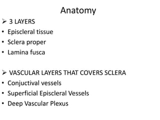
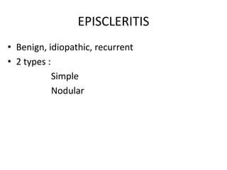
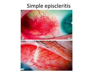
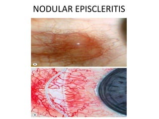
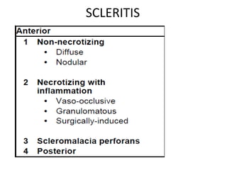
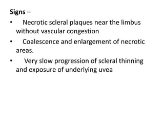
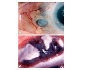
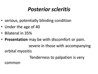
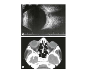
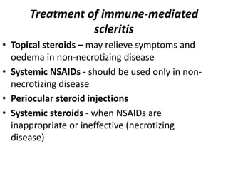
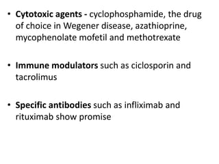
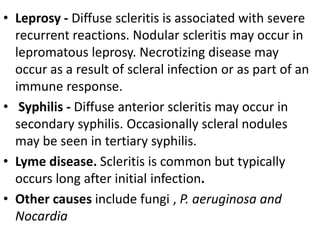
Scleritis and episcleritis affect the layers of the sclera and surrounding tissues. Episcleritis is usually benign and recurrent, occurring as either simple or nodular types. Scleritis can be anterior and non-necrotizing, nodular, or necrotizing. Complications of scleritis include keratitis, uveitis, glaucoma, hypotony, and perforation. Treatment involves topical steroids, NSAIDs, immunosuppressants, or antibiotics depending on the type and cause.
This presentation describes the background of the cornea and the corneal diseases in general, also it describes in detailed manner how to manage the corneal ulcer with its different causes
Dry eye occurs when the eye does not produce
tears properly, or when the tears are not of the correct consistency and
evaporate too quickly.
In addition, inflammation of the surface of
the eye may occur along with dry eye. If left untreated, this condition can
lead to pain, ulcers, or scars on the cornea, and some loss of vision. However,
permanent loss of vision from dry eye is uncommon.
Dry eye can make it more difficult to perform
some activities, such as using a computer or reading for an extended period of
time, and it can decrease tolerance for dry environments, such as the air
inside an airplane.
Other names for dry eye include dry eye syndrome,
keratoconjunctivitis sicca (KCS), dysfunctional tear syndrome, lacrimal
keratoconjunctivitis, evaporative tear deficiency, aqueous tear deficiency, and
LASIK-induced neurotrophic epitheliopathy (LNE).
you will get information about the layers of sclera and its diseases such as episcleritis and scleritis.
types of scleritis and episcleritis are also eplained in these slides. such as diffuse and nodular types of episclera, necrotizing and non-necrotizing types of anterior scleritis, posterior sleritis.
there etiologies. complications, investigations and treatment are also explained in detail.
Diseases of sclera
2. anatomy • Sclera posterior 5/6th opaque part of the external fibrous tunic of the eyeball.
3. • outer surface }covered by Tenon's capsule. • anterior part } covered by bulbar conjunctiva.
4. Its inner surface lies in contact with choroid with a potential suprachoroidal space in between
5. Thickness of sclera. • thinner }children and in females Sclera • thickest} posteriorly (1mm) • gradually becomes thin when traced anteriorly. • thinnest } insertion of extraocular muscles (0.3 mm). • Lamina cribrosa is a sieve-like sclera from which fibres of optic nerve pass.
6. Apertures of sclera • Anterior • Anterior ciliary vessels • Middle • four vortex veins (vena verticosae) • Posterior • Optic nerve • Long & short ciliary nerves
7. Layers of sclera sclera episclera Sclera proper Lamina fusca thin, dense vascularised layer of connective tissue fibroblasts, macrophages and lymphocytes avascular structure dense bundles of collagen fibres. innermost blends with suprachoroidal and supraciliary laminae of the uveal tract. brownish in colour presence of pigmented cells.
Lung Cancer: Artificial Intelligence, Synergetics, Complex System Analysis, S...Oleg Kshivets
RESULTS: Overall life span (LS) was 2252.1±1742.5 days and cumulative 5-year survival (5YS) reached 73.2%, 10 years – 64.8%, 20 years – 42.5%. 513 LCP lived more than 5 years (LS=3124.6±1525.6 days), 148 LCP – more than 10 years (LS=5054.4±1504.1 days).199 LCP died because of LC (LS=562.7±374.5 days). 5YS of LCP after bi/lobectomies was significantly superior in comparison with LCP after pneumonectomies (78.1% vs.63.7%, P=0.00001 by log-rank test). AT significantly improved 5YS (66.3% vs. 34.8%) (P=0.00000 by log-rank test) only for LCP with N1-2. Cox modeling displayed that 5YS of LCP significantly depended on: phase transition (PT) early-invasive LC in terms of synergetics, PT N0—N12, cell ratio factors (ratio between cancer cells- CC and blood cells subpopulations), G1-3, histology, glucose, AT, blood cell circuit, prothrombin index, heparin tolerance, recalcification time (P=0.000-0.038). Neural networks, genetic algorithm selection and bootstrap simulation revealed relationships between 5YS and PT early-invasive LC (rank=1), PT N0—N12 (rank=2), thrombocytes/CC (3), erythrocytes/CC (4), eosinophils/CC (5), healthy cells/CC (6), lymphocytes/CC (7), segmented neutrophils/CC (8), stick neutrophils/CC (9), monocytes/CC (10); leucocytes/CC (11). Correct prediction of 5YS was 100% by neural networks computing (area under ROC curve=1.0; error=0.0).
CONCLUSIONS: 5YS of LCP after radical procedures significantly depended on: 1) PT early-invasive cancer; 2) PT N0--N12; 3) cell ratio factors; 4) blood cell circuit; 5) biochemical factors; 6) hemostasis system; 7) AT; 8) LC characteristics; 9) LC cell dynamics; 10) surgery type: lobectomy/pneumonectomy; 11) anthropometric data. Optimal diagnosis and treatment strategies for LC are: 1) screening and early detection of LC; 2) availability of experienced thoracic surgeons because of complexity of radical procedures; 3) aggressive en block surgery and adequate lymph node dissection for completeness; 4) precise prediction; 5) adjuvant chemoimmunoradiotherapy for LCP with unfavorable prognosis.
HOT NEW PRODUCT! BIG SALES FAST SHIPPING NOW FROM CHINA!! EU KU DB BK substit...GL Anaacs
Contact us if you are interested:
Email / Skype : kefaya1771@gmail.com
Threema: PXHY5PDH
New BATCH Ku !!! MUCH IN DEMAND FAST SALE EVERY BATCH HAPPY GOOD EFFECT BIG BATCH !
Contact me on Threema or skype to start big business!!
Hot-sale products:
NEW HOT EUTYLONE WHITE CRYSTAL!!
5cl-adba precursor (semi finished )
5cl-adba raw materials
ADBB precursor (semi finished )
ADBB raw materials
APVP powder
5fadb/4f-adb
Jwh018 / Jwh210
Eutylone crystal
Protonitazene (hydrochloride) CAS: 119276-01-6
Flubrotizolam CAS: 57801-95-3
Metonitazene CAS: 14680-51-4
Payment terms: Western Union,MoneyGram,Bitcoin or USDT.
Deliver Time: Usually 7-15days
Shipping method: FedEx, TNT, DHL,UPS etc.Our deliveries are 100% safe, fast, reliable and discreet.
Samples will be sent for your evaluation!If you are interested in, please contact me, let's talk details.
We specializes in exporting high quality Research chemical, medical intermediate, Pharmaceutical chemicals and so on. Products are exported to USA, Canada, France, Korea, Japan,Russia, Southeast Asia and other countries.
Title: Sense of Taste
Presenter: Dr. Faiza, Assistant Professor of Physiology
Qualifications:
MBBS (Best Graduate, AIMC Lahore)
FCPS Physiology
ICMT, CHPE, DHPE (STMU)
MPH (GC University, Faisalabad)
MBA (Virtual University of Pakistan)
Learning Objectives:
Describe the structure and function of taste buds.
Describe the relationship between the taste threshold and taste index of common substances.
Explain the chemical basis and signal transduction of taste perception for each type of primary taste sensation.
Recognize different abnormalities of taste perception and their causes.
Key Topics:
Significance of Taste Sensation:
Differentiation between pleasant and harmful food
Influence on behavior
Selection of food based on metabolic needs
Receptors of Taste:
Taste buds on the tongue
Influence of sense of smell, texture of food, and pain stimulation (e.g., by pepper)
Primary and Secondary Taste Sensations:
Primary taste sensations: Sweet, Sour, Salty, Bitter, Umami
Chemical basis and signal transduction mechanisms for each taste
Taste Threshold and Index:
Taste threshold values for Sweet (sucrose), Salty (NaCl), Sour (HCl), and Bitter (Quinine)
Taste index relationship: Inversely proportional to taste threshold
Taste Blindness:
Inability to taste certain substances, particularly thiourea compounds
Example: Phenylthiocarbamide
Structure and Function of Taste Buds:
Composition: Epithelial cells, Sustentacular/Supporting cells, Taste cells, Basal cells
Features: Taste pores, Taste hairs/microvilli, and Taste nerve fibers
Location of Taste Buds:
Found in papillae of the tongue (Fungiform, Circumvallate, Foliate)
Also present on the palate, tonsillar pillars, epiglottis, and proximal esophagus
Mechanism of Taste Stimulation:
Interaction of taste substances with receptors on microvilli
Signal transduction pathways for Umami, Sweet, Bitter, Sour, and Salty tastes
Taste Sensitivity and Adaptation:
Decrease in sensitivity with age
Rapid adaptation of taste sensation
Role of Saliva in Taste:
Dissolution of tastants to reach receptors
Washing away the stimulus
Taste Preferences and Aversions:
Mechanisms behind taste preference and aversion
Influence of receptors and neural pathways
Impact of Sensory Nerve Damage:
Degeneration of taste buds if the sensory nerve fiber is cut
Abnormalities of Taste Detection:
Conditions: Ageusia, Hypogeusia, Dysgeusia (parageusia)
Causes: Nerve damage, neurological disorders, infections, poor oral hygiene, adverse drug effects, deficiencies, aging, tobacco use, altered neurotransmitter levels
Neurotransmitters and Taste Threshold:
Effects of serotonin (5-HT) and norepinephrine (NE) on taste sensitivity
Supertasters:
25% of the population with heightened sensitivity to taste, especially bitterness
Increased number of fungiform papillae
TEST BANK for Operations Management, 14th Edition by William J. Stevenson, Ve...kevinkariuki227
TEST BANK for Operations Management, 14th Edition by William J. Stevenson, Verified Chapters 1 - 19, Complete Newest Version.pdf
- Video recording of this lecture in English language: https://youtu.be/lK81BzxMqdo
- Video recording of this lecture in Arabic language: https://youtu.be/Ve4P0COk9OI
- Link to download the book free: https://nephrotube.blogspot.com/p/nephrotube-nephrology-books.html
- Link to NephroTube website: www.NephroTube.com
- Link to NephroTube social media accounts: https://nephrotube.blogspot.com/p/join-nephrotube-on-social-media.html
These lecture slides, by Dr Sidra Arshad, offer a quick overview of physiological basis of a normal electrocardiogram.
Learning objectives:
1. Define an electrocardiogram (ECG) and electrocardiography
2. Describe how dipoles generated by the heart produce the waveforms of the ECG
3. Describe the components of a normal electrocardiogram of a typical bipolar leads (limb II)
4. Differentiate between intervals and segments
5. Enlist some common indications for obtaining an ECG
Study Resources:
1. Chapter 11, Guyton and Hall Textbook of Medical Physiology, 14th edition
2. Chapter 9, Human Physiology - From Cells to Systems, Lauralee Sherwood, 9th edition
3. Chapter 29, Ganong’s Review of Medical Physiology, 26th edition
4. Electrocardiogram, StatPearls - https://www.ncbi.nlm.nih.gov/books/NBK549803/
5. ECG in Medical Practice by ABM Abdullah, 4th edition
6. ECG Basics, http://www.nataliescasebook.com/tag/e-c-g-basics
Title: Sense of Smell
Presenter: Dr. Faiza, Assistant Professor of Physiology
Qualifications:
MBBS (Best Graduate, AIMC Lahore)
FCPS Physiology
ICMT, CHPE, DHPE (STMU)
MPH (GC University, Faisalabad)
MBA (Virtual University of Pakistan)
Learning Objectives:
Describe the primary categories of smells and the concept of odor blindness.
Explain the structure and location of the olfactory membrane and mucosa, including the types and roles of cells involved in olfaction.
Describe the pathway and mechanisms of olfactory signal transmission from the olfactory receptors to the brain.
Illustrate the biochemical cascade triggered by odorant binding to olfactory receptors, including the role of G-proteins and second messengers in generating an action potential.
Identify different types of olfactory disorders such as anosmia, hyposmia, hyperosmia, and dysosmia, including their potential causes.
Key Topics:
Olfactory Genes:
3% of the human genome accounts for olfactory genes.
400 genes for odorant receptors.
Olfactory Membrane:
Located in the superior part of the nasal cavity.
Medially: Folds downward along the superior septum.
Laterally: Folds over the superior turbinate and upper surface of the middle turbinate.
Total surface area: 5-10 square centimeters.
Olfactory Mucosa:
Olfactory Cells: Bipolar nerve cells derived from the CNS (100 million), with 4-25 olfactory cilia per cell.
Sustentacular Cells: Produce mucus and maintain ionic and molecular environment.
Basal Cells: Replace worn-out olfactory cells with an average lifespan of 1-2 months.
Bowman’s Gland: Secretes mucus.
Stimulation of Olfactory Cells:
Odorant dissolves in mucus and attaches to receptors on olfactory cilia.
Involves a cascade effect through G-proteins and second messengers, leading to depolarization and action potential generation in the olfactory nerve.
Quality of a Good Odorant:
Small (3-20 Carbon atoms), volatile, water-soluble, and lipid-soluble.
Facilitated by odorant-binding proteins in mucus.
Membrane Potential and Action Potential:
Resting membrane potential: -55mV.
Action potential frequency in the olfactory nerve increases with odorant strength.
Adaptation Towards the Sense of Smell:
Rapid adaptation within the first second, with further slow adaptation.
Psychological adaptation greater than receptor adaptation, involving feedback inhibition from the central nervous system.
Primary Sensations of Smell:
Camphoraceous, Musky, Floral, Pepperminty, Ethereal, Pungent, Putrid.
Odor Detection Threshold:
Examples: Hydrogen sulfide (0.0005 ppm), Methyl-mercaptan (0.002 ppm).
Some toxic substances are odorless at lethal concentrations.
Characteristics of Smell:
Odor blindness for single substances due to lack of appropriate receptor protein.
Behavioral and emotional influences of smell.
Transmission of Olfactory Signals:
From olfactory cells to glomeruli in the olfactory bulb, involving lateral inhibition.
Primitive, less old, and new olfactory systems with different path
Factory Supply Best Quality Pmk Oil CAS 28578–16–7 PMK Powder in Stockrebeccabio
Factory Supply Best Quality Pmk Oil CAS 28578–16–7 PMK Powder in Stock
Telegram: bmksupplier
signal: +85264872720
threema: TUD4A6YC
You can contact me on Telegram or Threema
Communicate promptly and reply
Free of customs clearance, Double Clearance 100% pass delivery to USA, Canada, Spain, Germany, Netherland, Poland, Italy, Sweden, UK, Czech Republic, Australia, Mexico, Russia, Ukraine, Kazakhstan.Door to door service
Hot Selling Organic intermediates
Prix Galien International 2024 Forum ProgramLevi Shapiro
June 20, 2024, Prix Galien International and Jerusalem Ethics Forum in ROME. Detailed agenda including panels:
- ADVANCES IN CARDIOLOGY: A NEW PARADIGM IS COMING
- WOMEN’S HEALTH: FERTILITY PRESERVATION
- WHAT’S NEW IN THE TREATMENT OF INFECTIOUS,
ONCOLOGICAL AND INFLAMMATORY SKIN DISEASES?
- ARTIFICIAL INTELLIGENCE AND ETHICS
- GENE THERAPY
- BEYOND BORDERS: GLOBAL INITIATIVES FOR DEMOCRATIZING LIFE SCIENCE TECHNOLOGIES AND PROMOTING ACCESS TO HEALTHCARE
- ETHICAL CHALLENGES IN LIFE SCIENCES
- Prix Galien International Awards Ceremony
ARTIFICIAL INTELLIGENCE IN HEALTHCARE.pdfAnujkumaranit
Artificial intelligence (AI) refers to the simulation of human intelligence processes by machines, especially computer systems. It encompasses tasks such as learning, reasoning, problem-solving, perception, and language understanding. AI technologies are revolutionizing various fields, from healthcare to finance, by enabling machines to perform tasks that typically require human intelligence.
Report Back from SGO 2024: What’s the Latest in Cervical Cancer?bkling
Are you curious about what’s new in cervical cancer research or unsure what the findings mean? Join Dr. Emily Ko, a gynecologic oncologist at Penn Medicine, to learn about the latest updates from the Society of Gynecologic Oncology (SGO) 2024 Annual Meeting on Women’s Cancer. Dr. Ko will discuss what the research presented at the conference means for you and answer your questions about the new developments.
8. Anterior non-necrotizing scleritis
• DIFFUSE
5th decade, women
ocular redness followed a few days later by
aching
may spread to the face and temple
Vascular congestion and dilatation associated
with oedema
Recurrences at the same location are common
9.
10. NODULAR
• Previous attack of herpes zoster ophthalmicus
• Fifth decade
• C/F - pain followed by increasing redness,
tenderness of the globe and the appearance
of a scleral nodule
• Scleral nodule may be single or multiple and
mostly 3–4 mm away from the limbus
11. • Multiple nodules may coalesce, become confluent
• 10% of patients with nodular scleritis develop
necrotizing
12. Anterior necrotizing scleritis with
inflammation
• the aggressive form of scleritis
• 60 years
• bilateral in 60%
• may result in severe visual morbidity and
sometimes loss of the eye
• Symptoms - gradual onset of pain which becomes
severe and persistent
radiates to the temple, brow or jaw
13. Signs
• three types of necrotizing disease
a) Vaso-occlusive - often associated with rheumatoid
arthritis
Isolated patches of scleral oedema
with overlying non-perfused episclera & conjunctiva
The patches coalesce, and if
unchecked rapidly proceed to progressive scleral
necrosis
14.
15. b) Granulomatous
• often associated with Wegener
granulomatosis and polyarteritis nodosa
• starts with injection adjacent to the limbus
and then extends posteriorly
• Within 24 hours, the sclera, episclera,
conjunctiva and adjacent cornea become
irregularly raised and oedematous
16.
17. c) Surgically-induced scleritis
• Within 3 weeks of the surgical procedure
• strabismus repair, trabeculectomy , scleral buckling ,
treatment of pterygium with adjunctive mitomycin
C
• necrotizing process starts at the site of surgery and
then extends outwards
• But it tends to remain localized to one segment
18.
19. Investigations
• Laboratory –
RF, ANA, ANCA (cANCA, pANCA) and
antiphospholipid antibodies
• FA –
Most ptients - vascular non-perfusion
Systemic vasculitis - transudation, localized areas of
vasculitis and new vessel formation
20. Complications
1 Acute infiltrative stromal keratitis may be localized or
diffuse.
2 Sclerosing keratitis, characterized by chronic thinning
and opacification in which the peripheral cornea
adjacent to the site of scleritis resembles sclera
3 Peripheral ulcerative keratitis is characterized by
progressive melting and ulceration. In granulomatous
scleritis the destruction extends directly from the sclera
into the limbus and cornea. This characteristic pattern
is seen in Wegener granulomatosis, polyarteritis
nodosa and relapsing polychondritis.
21. 4 Uveitis, if severe, usually denotes aggressive
scleritis.
5 Glaucoma is the most common cause of eventual
loss of vision. The intraocular pressure is very
difficult to control in the presence of active
scleritis.
6 Hypotony may be the result of ciliary body
detachment, inflammatory damage or ischaemia.
7 Perforation of the sclera as a result of the
inflammatory process alone is extremely rare
22.
23. Scleromalacia perforans
• necrotizing scleritis without inflammation
• typically affects elderly women with longstanding
rheumatoid arthritis
• perforation of the globe is extremely rare
• Presentation - slight non-specific irritation and
keratoconjunctivitis sicca may be
suspected; pain is absent and vision unaffected.
24. Signs –
• Necrotic scleral plaques near the limbus
without vascular congestion
• Coalescence and enlargement of necrotic
areas.
• Very slow progression of scleral thinning
and exposure of underlying uvea
25.
26. Treatment
• Effective in patients with very early disease
• Repair of scleral perforation must be attempted
otherwise phthisis bulbi ensues
• Differential diagnosis - scleral hyaline plaque
oval, dark-greyish area located close to the
insertion of the horizontal rectus muscles.
27. Posterior scleritis
• serious, potentially blinding condition
• Under the age of 40
• Bilateral in 35%
• Presentation may be with discomfort or pain.
severe in those with accompanying
orbital myositis
Tenderness to palpation is very
common
28. Signs
a Exudative retinal detachment occurs in almost
25% of cases
b Uveal effusion characterized by exudative retinal
detachment and choroidal detachment
c Choroidal folds represent an anterior
displacement of the choroid. They are usually
confined to the posterior pole and run
horizontally.
d Subretinal mass characterized by a yellowish-
brown elevation which may be mistaken for a
choroidal tumour.
29. e Disc oedema with an accompanying slight
reduction of vision is common. It iscaused by
spread of the granulomatous process into the
orbital tissue and the optic nerve.
f Myositis is common and gives rise to diplopia,
pain on eye movement, tenderness to touch and
redness around a muscle insertion.
g Proptosis is usually mild and frequently
associated with ptosis.
h Other features - glaucoma, periorbital oedema,
chemosis and conjunctival injection
30.
31. Ultrasound :
• Increased scleral thickness, scleral nodules and
separation of Tenon capsule from the sclera.
• Fluid in Tenon's space gives rise to the
characteristic ‘T’ sign in which the stem of the T is
formed by the optic nerve on its side and the cross
bar by the gap containing fluid
• Disc oedema, choroidal folds or retinal
detachment.
MR and CT may show scleral thickening and proptosis
32.
33. Differential Diagnosis
1 Subretinal mass - granuloma associated with some other
pathology, amelanotic choroidal melanoma, choroidal
metastasis and choroidal hemangioma.
2 Choroidal folds, retinal striae and disc oedema - orbital
tumours, orbital inflammatory disease, thyroid eye
disease, papilloedema and hypotony.
3 Exudative retinal detachment - Vogt–Koyanagi–Harada
(VKH) syndrome and central serous retinopathy.
4 Orbital cellulitis
34. Treatment of immune-mediated
scleritis
• Topical steroids – may relieve symptoms and
oedema in non-necrotizing disease
• Systemic NSAIDs - should be used only in non-
necrotizing disease
• Periocular steroid injections
• Systemic steroids - when NSAIDs are
inappropriate or ineffective (necrotizing
disease)
35. • Cytotoxic agents - cyclophosphamide, the drug
of choice in Wegener disease, azathioprine,
mycophenolate mofetil and methotrexate
• Immune modulators such as ciclosporin and
tacrolimus
• Specific antibodies such as infliximab and
rituximab show promise
36. Infectious scleritis
CAUSES
• Herpes zoster – extremely resistant to treatment
and may result in a punched-out area or a
thinned scleral patch
• Tuberculous scleritis – by direct spread from a
local conjunctival or choroidal lesion, or more
commonly by haematogenous spread. Clinically
involvement may be nodular or necrotizing
37.
38. • Leprosy - Diffuse scleritis is associated with severe
recurrent reactions. Nodular scleritis may occur in
lepromatous leprosy. Necrotizing disease may
occur as a result of scleral infection or as part of an
immune response.
• Syphilis - Diffuse anterior scleritis may occur in
secondary syphilis. Occasionally scleral nodules
may be seen in tertiary syphilis.
• Lyme disease. Scleritis is common but typically
occurs long after initial infection.
• Other causes include fungi , P. aeruginosa and
Nocardia
39. Treatment
• specific antimicrobial therapy
• Topical and systemic steroids
• surgical debridement not only facilitates the
penetration of antibiotics but also debulks the
infected scleral tissue