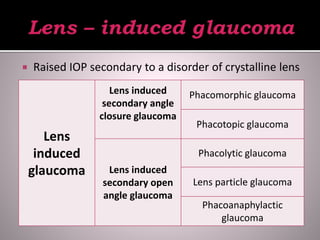
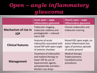
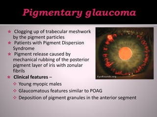
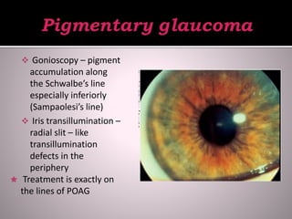
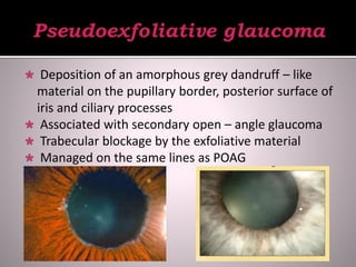
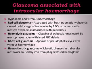
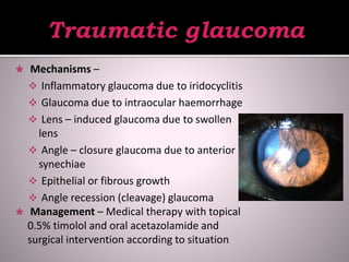
 Glaucoma - Group of disorders characterized by a
progressive optic neuropathy resulting in
characteristic appearance of optic disc and a specific
pattern of irreversible visual field defects associated
with raised intraocular pressure.
 Secondary Glaucoma – Group of disorders in which
the raised IOP is associated with a primary ocular or
systemic disease.
 Depending on the mechanism of rise in IOP –
 Secondary open angle glaucoma
 Secondary angle closure glaucoma
 Depending on the causative primary disease –
 Lens - induced glaucoma
 Inflammatory glaucoma
 Pigmentary glaucoma
 Neovascular glaucoma
 Glaucomas associated with irido – corneal
endothelial syndromes
 Pseudoexfoliative glaucoma
 Glaucomas associated with intraocular
haemorrhage
 Steroid-induced glaucoma
 Traumatic glaucoma
 Glaucoma-in-aphakia
 Glaucoma associated with intraocular tumours
 Ciliary block glaucoma
 Raised IOP secondary to a disorder of crystalline lens
Lens
induced
glaucoma
Lens induced
secondary angle
closure glaucoma
Phacomorphic glaucoma
Phacotopic glaucoma
Lens induced
secondary open
angle glaucoma
Phacolytic glaucoma
Lens particle glaucoma
Phacoanaphylactic
glaucoma
 Causes -
 Intumescent lens
Anterior subluxation or dislocation of the lens
and spherophakia
 Pathogenesis – Swollen lens pushes iris forwards,
obliterating the angle
 Presentation – Acute congestive glaucoma and
shows features of acute primary angle closure
glaucoma
 Treatment –
 Medical treatment –
Control of IOP by iv
mannitol, systemic
acetazolamide and
topical beta blockers
 Laser iridotomy
 Cataract extraction
with implantation of
PCIOL
 Trabecular meshwork is clogged by the lens
proteins, macrophages which phagocytose
the lens protein and inflammatory debris
 Deep anterior chamber and aqueous may
contain fine white protein particles, which
settle down as pseudohypopyon
 Treatment includes medical therapy to
lower IOP followed by extraction of
hypermature cataractous lens with PCIOL
implantation
 Trabecular meshwork is blocked by the lens
particles floating in aqueous humour.
 Symptoms of acute rise in IOP associated with lens
particles in the anterior chamber
 Medical therapy to lower IOP and irrigation –
aspiration of the lens particles from the anterior
chamber
 Fulminating acute inflammatory reaction due to
antigen – antibody reaction
 Granulomatous inflammation in the involved eye
 Preceding disruption of lens capsule by
extracapsular cataract extraction, penetrating injury
of leak of proteins from the capsule
 IOP is raised due to inflammatory reaction of the
uveal tissue excited by the lens matter.
 Management includes
medical therapy to
lower IOP, treatment
of iridocyclitis with
steroids and
cycloplegics and
irrigation – aspiration
of lens matter from
anterior chamber ( if
required).
Non specific
inflammatory glaucoma
Open – angle
inflammatory glaucoma
Angle – closure
inflammatory glaucoma
Specific hypertensive
uveitis syndrome
Fuchs’ uveitis syndrome
Glaucomatocyclitic crisis
Acute open – angle
inflammatory glaucoma
Chronic open – angle
inflammatory glaucoma
Mechanism of rise in
IOP
Trabecular clogging ,
trabecular oedema and
prostaglandin – induced
rise in IOP
Chronic trabeculitis and
trabecular scarring
Clinical features
Features of acute
iridocyclitis associated with
raised IOP with open-angle
of anterior chamber
Raised IOP, open angle, no
active inflammation but
signs of previous episode
of uveitis present
Management
Treatment of iridocyclitis
and medical therapy to
lower IOP by use of
hyperosmotic agents,
acetazolamide and beta –
blockers eye drops
Medical therapy
Trabeculectomy
Cyclodestructive
procedures
 Mechanism of rise in IOP –
 Secondary angle – closure with pupil block
 Secondary angle – closure without pupil block
 Clinical features – Raised IOP, seclusio papillae,
shallow anterior chamber
 Management –
 Prophylaxis – Local steroids and atropine to
prevent formation of synechiae
 Curative treatment – Medical therapy, surgical or
laser iridotomy and filtration surgery
 Clogging up of trabecular meshwork
by the pigment particles
 Patients with Pigment Dispersion
Syndrome
 Pigment release caused by
mechanical rubbing of the posterior
pigment layer of iris with zonular
fibrils
 Clinical features –
 Young myopic males
 Glaucomatous features similar to POAG
 Deposition of pigment granules in the anterior segment
 Gonioscopy – pigment
accumulation along
the Schwalbe’s line
especially inferiorly
(Sampaolesi’s line)
 Iris transillumination –
radial slit – like
transillumination
defects in the
periphery
 Treatment is exactly on
the lines of POAG
 Intractable glaucoma results due to formation of
neovascular membrane involving the angle of
anterior chamber
 Etiology – Neovascularization of iris following retinal
ischaemia, feature of
 Proliferative diabetic retinopathy
 Central retinal vein occlusion
 Sickle – cell retinopathy
 Rare causes (intraocular tumours and long
standing retinal detachment)
 Clinical profile –
 Pre – glaucomatous stage
 Open – angle glaucoma stage
 Secondary angle closure
glaucoma
 Treatment –
 Panretinal photocoagulation
 Medical therapy not
effective
 Artificial filtration shunt
(Seton operation)
 3 entities –
 Progressive iris atrophy
 Chandler’s syndrome
 Cogan – Reese syndrome
 Presence of abnormal corneal endothelial cells
which proliferate to form an endothelial membrane
in the angle of anterior chamber
 Clinical features – Affects middle – aged women
 Progressive iris atrophy – iris features predominates with
corectopia, atrophy and hole formation
 Chandler’s syndrome – Mild iris changes and corneal
oedema predominates
 Cogan – Reese syndrome – nodular and diffuse pigmented
lesions of iris, may or may not be associated with corneal
changes
 Treatment –
 Medical treatment
 Trabeculectomy
 Artificial filtration
 Deposition of an amorphous grey dandruff – like
material on the pupillary border, posterior surface of
iris and ciliary processes
 Associated with secondary open – angle glaucoma
 Trabecular blockage by the exfoliative material
 Managed on the same lines as POAG
 Hyphaema and vitreous haemorrhage
 Red cell glaucoma – Associated with fresh traumatic hyphaema;
caused by blockage of trabeculae by RBCs in patients with
massive hyphaema; associated with pupil block
 Haemolytic glaucoma – Clogging of trabecular meshwork by
macrophages laden with lysed RBC debris
 Ghost cell glaucoma – Aphakic or pseudophakic eyes with
vitreous haemorrhage
 Hemosiderotic glaucoma – Sclerotic changes in trabecular
meshwork caused by iron from phagocytosed hemoglobin
 Type of secondary open – angle glaucoma which develops
following topical or systemic steroid therapy
 Etiopathogenesis –
 Glycosaminoglycans (GAG) theory
 Endothelial cell theory
 Prostaglandin theory
 Symptoms similar to POAG
 Prevented by judicious use of steroids and regular
monitoring of IOP
 Treatment –
 Discontinuation of steroids
 Medical therapy by 0.5% timolol maleate
 Filtration surgery
 Mechanisms –
 Inflammatory glaucoma due to iridocyclitis
 Glaucoma due to intraocular haemorrhage
 Lens – induced glaucoma due to swollen
lens
 Angle – closure glaucoma due to anterior
synechiae
 Epithelial or fibrous growth
 Angle recession (cleavage) glaucoma
 Management – Medical therapy with topical
0.5% timolol and oral acetazolamide and
surgical intervention according to situation
 Raised IOP with deep anterior chamber in early
postoperative period
 Secondary angle – closure glaucoma due to flat
anterior chamber
 Secondary angle – closure glaucoma due to pupil
block
 Undiagnosed pre – existing primary open – angle
glaucoma
 Steroid – induced glaucoma
 Epithelial ingrowth
 Malignant glaucoma
 Malignant melanoma, retinoblastoma
 Mechanisms –
 Trabecular block due to blockage by tumour cells
 Neovascularization of the angle
 Venous stasis
 Angle closure due to forward displacement of iris – lens
diaphragm
 Treatment – Enucleation of the
eyeball
 Rare condition occurring as
complication of any intraocular
operation
 Patients with primary angle –
closure glaucoma operated for
peripheral iridectomy or
trabeculectomy
 Markedly raised IOP associated
with shallow or absent anterior
chamber
 Clinical features includes severe
pain and blurring of vision
following any intraocular operation
 On examination,
 Persistent flat anterior chamber
 Markedly raised IOP
 Unresponsiveness or even aggravation by
miotics
 Phakic, aphakic or pseudophakic
 Treatment –
 Medical therapy – 1% atropine drops,
acetazolamide, 0.5% timolol maleate eye
drops and iv mannitol
 YAG laser hyaloidotomy
 Surgical therapy
Secondary glaucoma

Secondary glaucoma

  • 2.
     Glaucoma -Group of disorders characterized by a progressive optic neuropathy resulting in characteristic appearance of optic disc and a specific pattern of irreversible visual field defects associated with raised intraocular pressure.  Secondary Glaucoma – Group of disorders in which the raised IOP is associated with a primary ocular or systemic disease.
  • 3.
     Depending onthe mechanism of rise in IOP –  Secondary open angle glaucoma  Secondary angle closure glaucoma  Depending on the causative primary disease –  Lens - induced glaucoma  Inflammatory glaucoma  Pigmentary glaucoma  Neovascular glaucoma
  • 4.
     Glaucomas associatedwith irido – corneal endothelial syndromes  Pseudoexfoliative glaucoma  Glaucomas associated with intraocular haemorrhage  Steroid-induced glaucoma  Traumatic glaucoma  Glaucoma-in-aphakia  Glaucoma associated with intraocular tumours  Ciliary block glaucoma
  • 5.
     Raised IOPsecondary to a disorder of crystalline lens Lens induced glaucoma Lens induced secondary angle closure glaucoma Phacomorphic glaucoma Phacotopic glaucoma Lens induced secondary open angle glaucoma Phacolytic glaucoma Lens particle glaucoma Phacoanaphylactic glaucoma
  • 6.
     Causes - Intumescent lens Anterior subluxation or dislocation of the lens and spherophakia  Pathogenesis – Swollen lens pushes iris forwards, obliterating the angle  Presentation – Acute congestive glaucoma and shows features of acute primary angle closure glaucoma
  • 7.
     Treatment – Medical treatment – Control of IOP by iv mannitol, systemic acetazolamide and topical beta blockers  Laser iridotomy  Cataract extraction with implantation of PCIOL
  • 8.
     Trabecular meshworkis clogged by the lens proteins, macrophages which phagocytose the lens protein and inflammatory debris  Deep anterior chamber and aqueous may contain fine white protein particles, which settle down as pseudohypopyon  Treatment includes medical therapy to lower IOP followed by extraction of hypermature cataractous lens with PCIOL implantation
  • 9.
     Trabecular meshworkis blocked by the lens particles floating in aqueous humour.  Symptoms of acute rise in IOP associated with lens particles in the anterior chamber  Medical therapy to lower IOP and irrigation – aspiration of the lens particles from the anterior chamber
  • 10.
     Fulminating acuteinflammatory reaction due to antigen – antibody reaction  Granulomatous inflammation in the involved eye  Preceding disruption of lens capsule by extracapsular cataract extraction, penetrating injury of leak of proteins from the capsule  IOP is raised due to inflammatory reaction of the uveal tissue excited by the lens matter.
  • 11.
     Management includes medicaltherapy to lower IOP, treatment of iridocyclitis with steroids and cycloplegics and irrigation – aspiration of lens matter from anterior chamber ( if required).
  • 12.
    Non specific inflammatory glaucoma Open– angle inflammatory glaucoma Angle – closure inflammatory glaucoma Specific hypertensive uveitis syndrome Fuchs’ uveitis syndrome Glaucomatocyclitic crisis
  • 13.
    Acute open –angle inflammatory glaucoma Chronic open – angle inflammatory glaucoma Mechanism of rise in IOP Trabecular clogging , trabecular oedema and prostaglandin – induced rise in IOP Chronic trabeculitis and trabecular scarring Clinical features Features of acute iridocyclitis associated with raised IOP with open-angle of anterior chamber Raised IOP, open angle, no active inflammation but signs of previous episode of uveitis present Management Treatment of iridocyclitis and medical therapy to lower IOP by use of hyperosmotic agents, acetazolamide and beta – blockers eye drops Medical therapy Trabeculectomy Cyclodestructive procedures
  • 14.
     Mechanism ofrise in IOP –  Secondary angle – closure with pupil block  Secondary angle – closure without pupil block  Clinical features – Raised IOP, seclusio papillae, shallow anterior chamber  Management –  Prophylaxis – Local steroids and atropine to prevent formation of synechiae  Curative treatment – Medical therapy, surgical or laser iridotomy and filtration surgery
  • 15.
     Clogging upof trabecular meshwork by the pigment particles  Patients with Pigment Dispersion Syndrome  Pigment release caused by mechanical rubbing of the posterior pigment layer of iris with zonular fibrils  Clinical features –  Young myopic males  Glaucomatous features similar to POAG  Deposition of pigment granules in the anterior segment
  • 16.
     Gonioscopy –pigment accumulation along the Schwalbe’s line especially inferiorly (Sampaolesi’s line)  Iris transillumination – radial slit – like transillumination defects in the periphery  Treatment is exactly on the lines of POAG
  • 17.
     Intractable glaucomaresults due to formation of neovascular membrane involving the angle of anterior chamber  Etiology – Neovascularization of iris following retinal ischaemia, feature of  Proliferative diabetic retinopathy  Central retinal vein occlusion  Sickle – cell retinopathy  Rare causes (intraocular tumours and long standing retinal detachment)
  • 18.
     Clinical profile–  Pre – glaucomatous stage  Open – angle glaucoma stage  Secondary angle closure glaucoma  Treatment –  Panretinal photocoagulation  Medical therapy not effective  Artificial filtration shunt (Seton operation)
  • 19.
     3 entities–  Progressive iris atrophy  Chandler’s syndrome  Cogan – Reese syndrome  Presence of abnormal corneal endothelial cells which proliferate to form an endothelial membrane in the angle of anterior chamber
  • 20.
     Clinical features– Affects middle – aged women  Progressive iris atrophy – iris features predominates with corectopia, atrophy and hole formation  Chandler’s syndrome – Mild iris changes and corneal oedema predominates  Cogan – Reese syndrome – nodular and diffuse pigmented lesions of iris, may or may not be associated with corneal changes  Treatment –  Medical treatment  Trabeculectomy  Artificial filtration
  • 21.
     Deposition ofan amorphous grey dandruff – like material on the pupillary border, posterior surface of iris and ciliary processes  Associated with secondary open – angle glaucoma  Trabecular blockage by the exfoliative material  Managed on the same lines as POAG
  • 22.
     Hyphaema andvitreous haemorrhage  Red cell glaucoma – Associated with fresh traumatic hyphaema; caused by blockage of trabeculae by RBCs in patients with massive hyphaema; associated with pupil block  Haemolytic glaucoma – Clogging of trabecular meshwork by macrophages laden with lysed RBC debris  Ghost cell glaucoma – Aphakic or pseudophakic eyes with vitreous haemorrhage  Hemosiderotic glaucoma – Sclerotic changes in trabecular meshwork caused by iron from phagocytosed hemoglobin
  • 23.
     Type ofsecondary open – angle glaucoma which develops following topical or systemic steroid therapy  Etiopathogenesis –  Glycosaminoglycans (GAG) theory  Endothelial cell theory  Prostaglandin theory  Symptoms similar to POAG  Prevented by judicious use of steroids and regular monitoring of IOP  Treatment –  Discontinuation of steroids  Medical therapy by 0.5% timolol maleate  Filtration surgery
  • 24.
     Mechanisms – Inflammatory glaucoma due to iridocyclitis  Glaucoma due to intraocular haemorrhage  Lens – induced glaucoma due to swollen lens  Angle – closure glaucoma due to anterior synechiae  Epithelial or fibrous growth  Angle recession (cleavage) glaucoma  Management – Medical therapy with topical 0.5% timolol and oral acetazolamide and surgical intervention according to situation
  • 25.
     Raised IOPwith deep anterior chamber in early postoperative period  Secondary angle – closure glaucoma due to flat anterior chamber  Secondary angle – closure glaucoma due to pupil block  Undiagnosed pre – existing primary open – angle glaucoma  Steroid – induced glaucoma  Epithelial ingrowth  Malignant glaucoma
  • 26.
     Malignant melanoma,retinoblastoma  Mechanisms –  Trabecular block due to blockage by tumour cells  Neovascularization of the angle  Venous stasis  Angle closure due to forward displacement of iris – lens diaphragm  Treatment – Enucleation of the eyeball
  • 27.
     Rare conditionoccurring as complication of any intraocular operation  Patients with primary angle – closure glaucoma operated for peripheral iridectomy or trabeculectomy  Markedly raised IOP associated with shallow or absent anterior chamber  Clinical features includes severe pain and blurring of vision following any intraocular operation
  • 28.
     On examination, Persistent flat anterior chamber  Markedly raised IOP  Unresponsiveness or even aggravation by miotics  Phakic, aphakic or pseudophakic  Treatment –  Medical therapy – 1% atropine drops, acetazolamide, 0.5% timolol maleate eye drops and iv mannitol  YAG laser hyaloidotomy  Surgical therapy