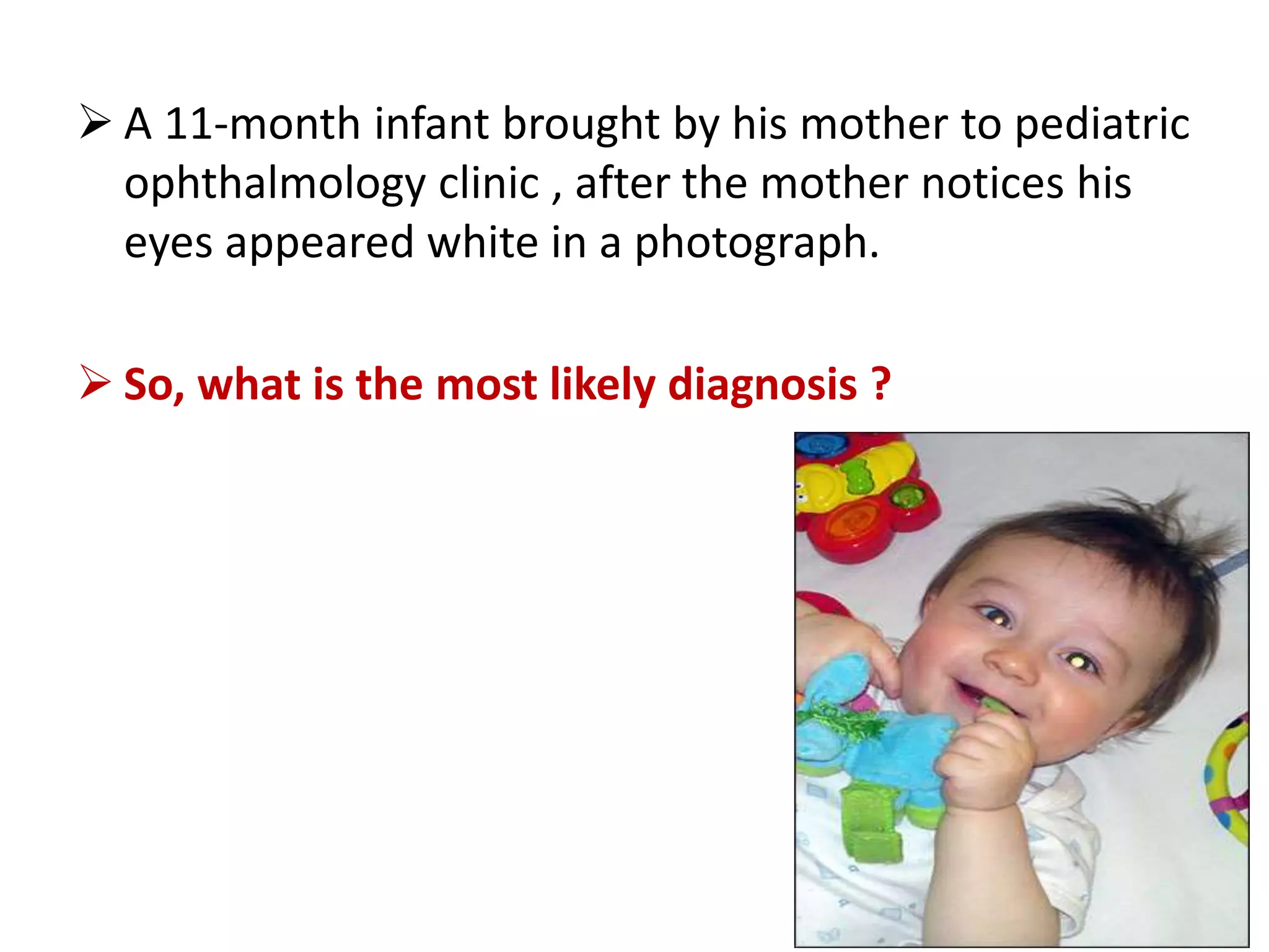
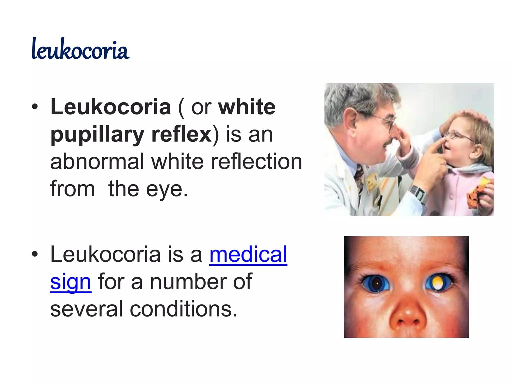
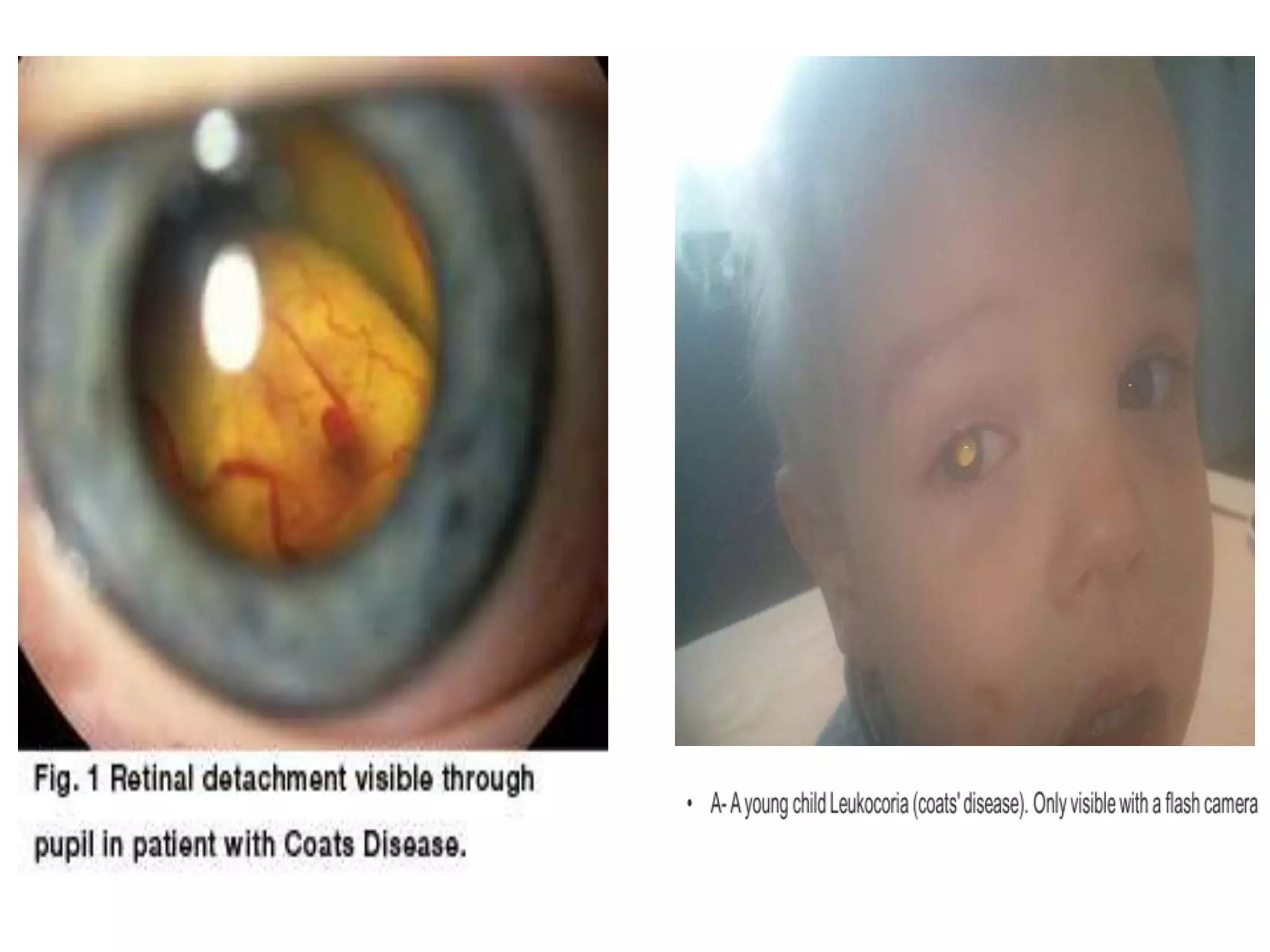
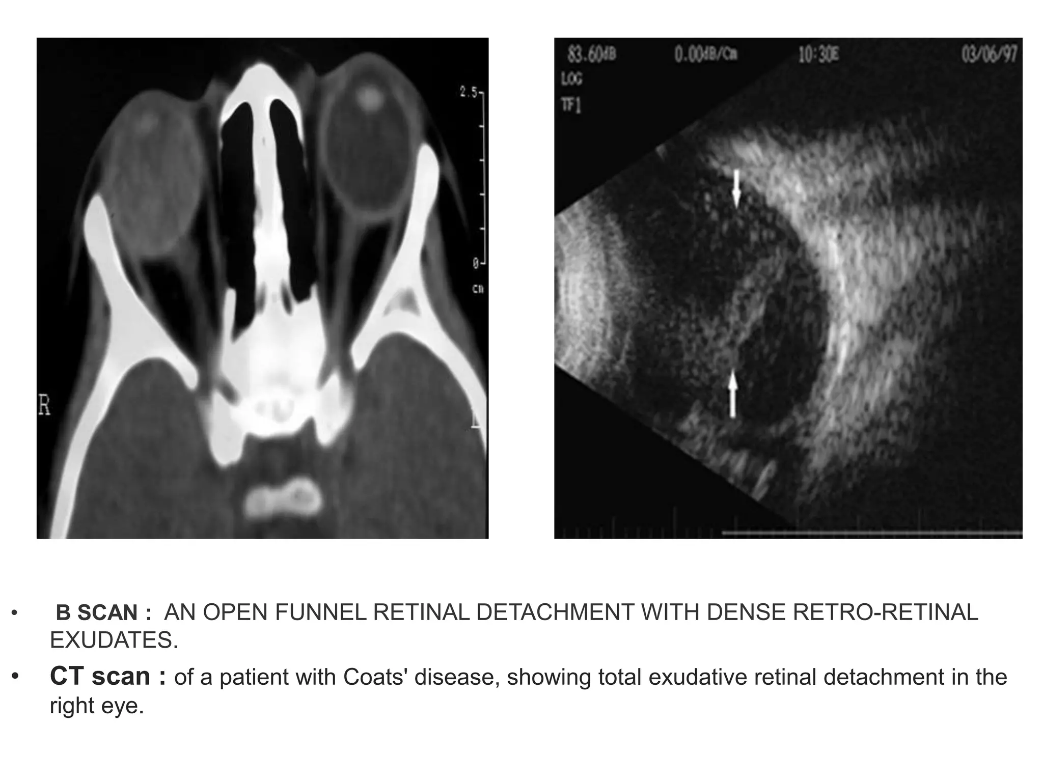
leukocoria
Prof. Dr. Abdelmongy Elsayed Ali
professor of ophthalmology
Al azhar faculty of medicine
2017
• designed by :
Mohamed Abdelmongy
Misr University for Science and Technology
MBBCh Candidate
 A 11-month infant brought by his mother to pediatric
ophthalmology clinic , after the mother notices his
eyes appeared white in a photograph.
 So, what is the most likely diagnosis ?
leukocoria
• Leukocoria ( or white
pupillary reflex) is an
abnormal white reflection
from the eye.
• Leukocoria is a medical
sign for a number of
several conditions.
DIFFERENTIAL DIAGNOSIS OF
LEUKOCORIA
• Congenital Cataract
• Retinoblastoma
• PHPV
• Toxocariasis
• Coat´s disease
• ROP
• Coloboma of optic
disc &retina
• Retinal detachment
• Norrie´s disease
Clinical work-up for child with
leukocoria
 the differential diagnosis can be narrowed through :
• History :
- age of presention ----
at birth ……. PHPV , cong. cataract
1-3 Y………… RB , cong.cataract
Pre school age …….. toxocariases , Coats
- birth history
pre-mature , low birth wight ……. ROP
• Ocular examination : EUA, fumdus ex.
• Investgation :
B-scan
CT-scan, MRI
Persistent hyperplastic
primary vitreous
Persistent hyperplastic primary vitreous
- It’s a congenital anomaly of the eye that
results from failure of embryological primary
vitreous and hyaloid vasculature to regress.
- It is characterized by persistence of various
portions of the primary vitreous , associated
with microphthalmia, cataract and glaucoma.
PHPV is usually divided into three types:
1- Anterior PHPV:
• occurs when the remnant
vascular stalk is seen
attached to the back of
the lens but not extended
back to the optic nerve.
• This form is typically
associated with cataract,
glaucoma and
retrolenticular
membrane.
2- Posterior PHPV:
• The remnant vascular
stalk is seen arising off
the optic nerve but not
reaching the lens, and
usually not causing
cataract.
• Posterior PHPV may be
associated with
abnormal development
of the retina, optic nerve
and macula, vitreal
membranes.
3- A combination of anterior and posterior PHPV:
• Is the most commonly
seen type.
(a band extending from
retina to posterior lens
capsule).
• COLOR DOPPLER
SHOWING ARTERIALIZED
BLOOD FLOW IN THE
ECHOGENIC BAND
Treatment
- Anterior form is most often treated with
observation, lensectomy, I/A with ant. vitrectomy
and glaucoma management, whether medical or
surgical.
- Posterior form is usually associated with a
poor visual outcome regardless of intervention due
to retinal and optic nerve abnormalities.
- combined type is also typically associated
with a poor outcome due to the high prevalence of
posterior segment abnormalities.
Ocular Toxocariasis
- Ocular toxocariasis is an uncommon disease that affects mostly
children and young adults, resulting in significant vision loss.
a-Toxocara canis (a common parasite of dogs)
b-Toxocara cati (a common parasite of cats).
- Humans acquire the infection as accidental hosts by ingesting soil
contaminated with Toxocara eggs ingestion of contaminated food, or
the oral-fecal route.
Clinical presentation of Ocular Toxocariasis
• Chronic
endophthalmitis (2-9
years)
• Posterior pole
granuloma (6-14
years)
• Peripheral granuloma
(6-40 years)
TREATMENT
• Medical therapy
• 5-day course of albendazole (10 mg/kg/day)
• Consider systemic or periocular corticosteroids to
suppress the immune response .
• Surgical therapy (If inflammation persists)
• Pars plana vitrectomy
• Perfluorocarbon liquids injection
• Indicated to facilitate removal of epiretinal membranes (ERMs)
and the posterior hyaloid in cases of tractional retinal detachment.
• Cryotherapy
• Applied directly at the areas of exudation at the pars plana with a
double freeze-thaw technique.
• Endolaser
• Indicated for treatment of ocular granulomas
COATS' DISEASE
• Coats’ disease is caused by( telangiectasia )a problem of
blood vessels inside the eye that provide blood and oxygen to
the retina.
• In Coats’ disease, the blood vessels are dilated, abnormally
twisted and leaky.
• This prevents the normal flow of blood, and allows fluid to leak
out of the blood vessels (exudate) builds up, it can cause a
detachment of the retina and loss of vision.
• B SCAN : AN OPEN FUNNEL RETINAL DETACHMENT WITH DENSE RETRO-RETINAL
EXUDATES.
• CT scan : of a patient with Coats' disease, showing total exudative retinal detachment in the
right eye.
stages of Coats’ disease :
• Stage I: There is presence of
telangiectasia (abnormal blood
vessels ) only.
• Stage II: both telangiectasia as
well as exudates.
• Stage III: Quite an advanced
stage of Coats disease and is
characterized by subtotal
retinal detachment.
• Stage IV: Severely advanced
and there is total detachment
of the retina.
• Stage V: Completely advanced
and the affected individual
loses vision completely with
secondary glaucoma and
painful eye.
Treatment
• For mild form of Coats Disease , Laser
photocoagulation with cryotherapy may
be useful for treatment.
• Advanced stage : vitreoretinal surgery.
• If severe pain in the eye, enucleation is
required as a method of treatment.
Retinopathy of prematurity
(ROP)
• Disease of retinal vasculature in
immature retina of a premature
neonate.
- Results from interruption of normal
vascularization.
• Characterized by vaso-obliteration/ vaso
cessation followed by abnormal
neovascularization and ultimately
cicatrisation.
• Risk factor :
 Birth before 32 weeks' of gestation.
 Birth weight of less than 1500 g.
 supplemental oxygen, hypoxemia.
Stages (1-5)
Stage 1:
Demarcation line -a flat, thin, whitish, clear-cut demarcation
between vascularised and avascular retina
Stage 2:
Elevated ridge - demarcation line has ”3D” appearnce & extends
anteriorly from the retinal plane as a ridge into the vitreous
Stage 3:
Neovascularisation - Extraretinal fibrovascular tissue begins
to grow on the top of the ridge or posterior to the ridge and
extends into the vitreous.
“Pop-corn lesion
STAGE 4 (Partial RD)
4A: Extra foveal
4B: Foveal
STAGE 5 (TOTAL RD)
Plus Disease
 When the blood vessels of the retina have become enlarged and twisted,
indicating a worsening of the disease.
 poor pupil dilatation.
 vitreous haze.
 vascular engorgement of the iris with extension onto anterior lens surface.
Coloboma
• Congenital coloboma is
developmental defects.
• either retinal coloboma
• Or optic nerve
coloboma can cause
leukocoria.
• Other optic disc
abnormalities such as a
(morning glory disc)
myelinated nerve fibers.
Leukocoria (White Pupillary Reflex)

Leukocoria (White Pupillary Reflex)

  • 1.
    leukocoria Prof. Dr. AbdelmongyElsayed Ali professor of ophthalmology Al azhar faculty of medicine 2017 • designed by : Mohamed Abdelmongy Misr University for Science and Technology MBBCh Candidate
  • 2.
     A 11-monthinfant brought by his mother to pediatric ophthalmology clinic , after the mother notices his eyes appeared white in a photograph.  So, what is the most likely diagnosis ?
  • 3.
    leukocoria • Leukocoria (or white pupillary reflex) is an abnormal white reflection from the eye. • Leukocoria is a medical sign for a number of several conditions.
  • 4.
    DIFFERENTIAL DIAGNOSIS OF LEUKOCORIA •Congenital Cataract • Retinoblastoma • PHPV • Toxocariasis • Coat´s disease • ROP • Coloboma of optic disc &retina • Retinal detachment • Norrie´s disease
  • 5.
    Clinical work-up forchild with leukocoria  the differential diagnosis can be narrowed through : • History : - age of presention ---- at birth ……. PHPV , cong. cataract 1-3 Y………… RB , cong.cataract Pre school age …….. toxocariases , Coats - birth history pre-mature , low birth wight ……. ROP • Ocular examination : EUA, fumdus ex. • Investgation : B-scan CT-scan, MRI
  • 6.
  • 7.
    Persistent hyperplastic primaryvitreous - It’s a congenital anomaly of the eye that results from failure of embryological primary vitreous and hyaloid vasculature to regress. - It is characterized by persistence of various portions of the primary vitreous , associated with microphthalmia, cataract and glaucoma.
  • 8.
    PHPV is usuallydivided into three types: 1- Anterior PHPV: • occurs when the remnant vascular stalk is seen attached to the back of the lens but not extended back to the optic nerve. • This form is typically associated with cataract, glaucoma and retrolenticular membrane.
  • 9.
    2- Posterior PHPV: •The remnant vascular stalk is seen arising off the optic nerve but not reaching the lens, and usually not causing cataract. • Posterior PHPV may be associated with abnormal development of the retina, optic nerve and macula, vitreal membranes.
  • 10.
    3- A combinationof anterior and posterior PHPV: • Is the most commonly seen type. (a band extending from retina to posterior lens capsule). • COLOR DOPPLER SHOWING ARTERIALIZED BLOOD FLOW IN THE ECHOGENIC BAND
  • 11.
    Treatment - Anterior formis most often treated with observation, lensectomy, I/A with ant. vitrectomy and glaucoma management, whether medical or surgical. - Posterior form is usually associated with a poor visual outcome regardless of intervention due to retinal and optic nerve abnormalities. - combined type is also typically associated with a poor outcome due to the high prevalence of posterior segment abnormalities.
  • 12.
  • 13.
    - Ocular toxocariasisis an uncommon disease that affects mostly children and young adults, resulting in significant vision loss. a-Toxocara canis (a common parasite of dogs) b-Toxocara cati (a common parasite of cats). - Humans acquire the infection as accidental hosts by ingesting soil contaminated with Toxocara eggs ingestion of contaminated food, or the oral-fecal route.
  • 14.
    Clinical presentation ofOcular Toxocariasis • Chronic endophthalmitis (2-9 years) • Posterior pole granuloma (6-14 years) • Peripheral granuloma (6-40 years)
  • 15.
    TREATMENT • Medical therapy •5-day course of albendazole (10 mg/kg/day) • Consider systemic or periocular corticosteroids to suppress the immune response . • Surgical therapy (If inflammation persists) • Pars plana vitrectomy • Perfluorocarbon liquids injection • Indicated to facilitate removal of epiretinal membranes (ERMs) and the posterior hyaloid in cases of tractional retinal detachment. • Cryotherapy • Applied directly at the areas of exudation at the pars plana with a double freeze-thaw technique. • Endolaser • Indicated for treatment of ocular granulomas
  • 16.
  • 17.
    • Coats’ diseaseis caused by( telangiectasia )a problem of blood vessels inside the eye that provide blood and oxygen to the retina. • In Coats’ disease, the blood vessels are dilated, abnormally twisted and leaky. • This prevents the normal flow of blood, and allows fluid to leak out of the blood vessels (exudate) builds up, it can cause a detachment of the retina and loss of vision.
  • 19.
    • B SCAN: AN OPEN FUNNEL RETINAL DETACHMENT WITH DENSE RETRO-RETINAL EXUDATES. • CT scan : of a patient with Coats' disease, showing total exudative retinal detachment in the right eye.
  • 20.
    stages of Coats’disease : • Stage I: There is presence of telangiectasia (abnormal blood vessels ) only. • Stage II: both telangiectasia as well as exudates. • Stage III: Quite an advanced stage of Coats disease and is characterized by subtotal retinal detachment. • Stage IV: Severely advanced and there is total detachment of the retina. • Stage V: Completely advanced and the affected individual loses vision completely with secondary glaucoma and painful eye.
  • 21.
    Treatment • For mildform of Coats Disease , Laser photocoagulation with cryotherapy may be useful for treatment. • Advanced stage : vitreoretinal surgery. • If severe pain in the eye, enucleation is required as a method of treatment.
  • 22.
  • 23.
    • Disease ofretinal vasculature in immature retina of a premature neonate. - Results from interruption of normal vascularization. • Characterized by vaso-obliteration/ vaso cessation followed by abnormal neovascularization and ultimately cicatrisation. • Risk factor :  Birth before 32 weeks' of gestation.  Birth weight of less than 1500 g.  supplemental oxygen, hypoxemia.
  • 24.
    Stages (1-5) Stage 1: Demarcationline -a flat, thin, whitish, clear-cut demarcation between vascularised and avascular retina
  • 25.
    Stage 2: Elevated ridge- demarcation line has ”3D” appearnce & extends anteriorly from the retinal plane as a ridge into the vitreous
  • 26.
    Stage 3: Neovascularisation -Extraretinal fibrovascular tissue begins to grow on the top of the ridge or posterior to the ridge and extends into the vitreous. “Pop-corn lesion
  • 27.
    STAGE 4 (PartialRD) 4A: Extra foveal 4B: Foveal
  • 28.
  • 29.
    Plus Disease  Whenthe blood vessels of the retina have become enlarged and twisted, indicating a worsening of the disease.  poor pupil dilatation.  vitreous haze.  vascular engorgement of the iris with extension onto anterior lens surface.
  • 31.
  • 32.
    • Congenital colobomais developmental defects. • either retinal coloboma • Or optic nerve coloboma can cause leukocoria. • Other optic disc abnormalities such as a (morning glory disc) myelinated nerve fibers.