Downloaded 153 times


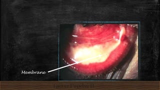
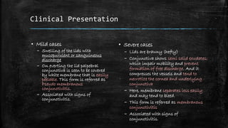
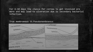

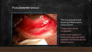
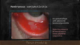


Membranous conjunctivitis is characterized by the presence of a pseudo membrane or fibrinous membrane on the palpebral or bulbar conjunctiva. It can be caused by bacteria like Corynebacterium diphtheriae or viruses. Clinically, it presents as swelling of the eyelids with mucopurulent discharge and a white membrane covering the palpebral conjunctiva that can easily peel off (pseudo membranous) or be semi-solid and not peel off easily (membranous). Treatment involves topical antibiotics and systemic antibiotics if bacteria are causing it. For diphtheritic cases, diphtheria antitoxin is also given to prevent complications like symblepharon


