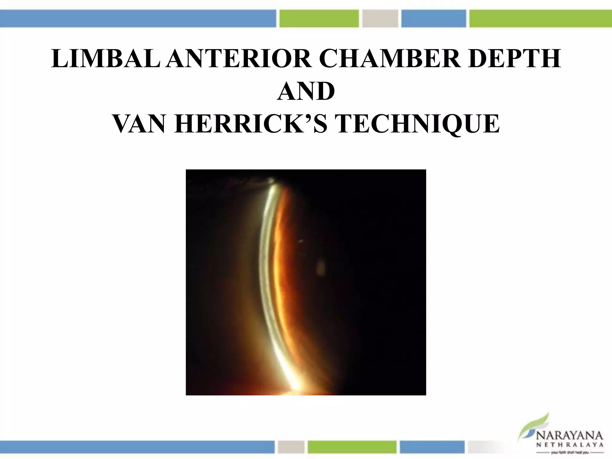
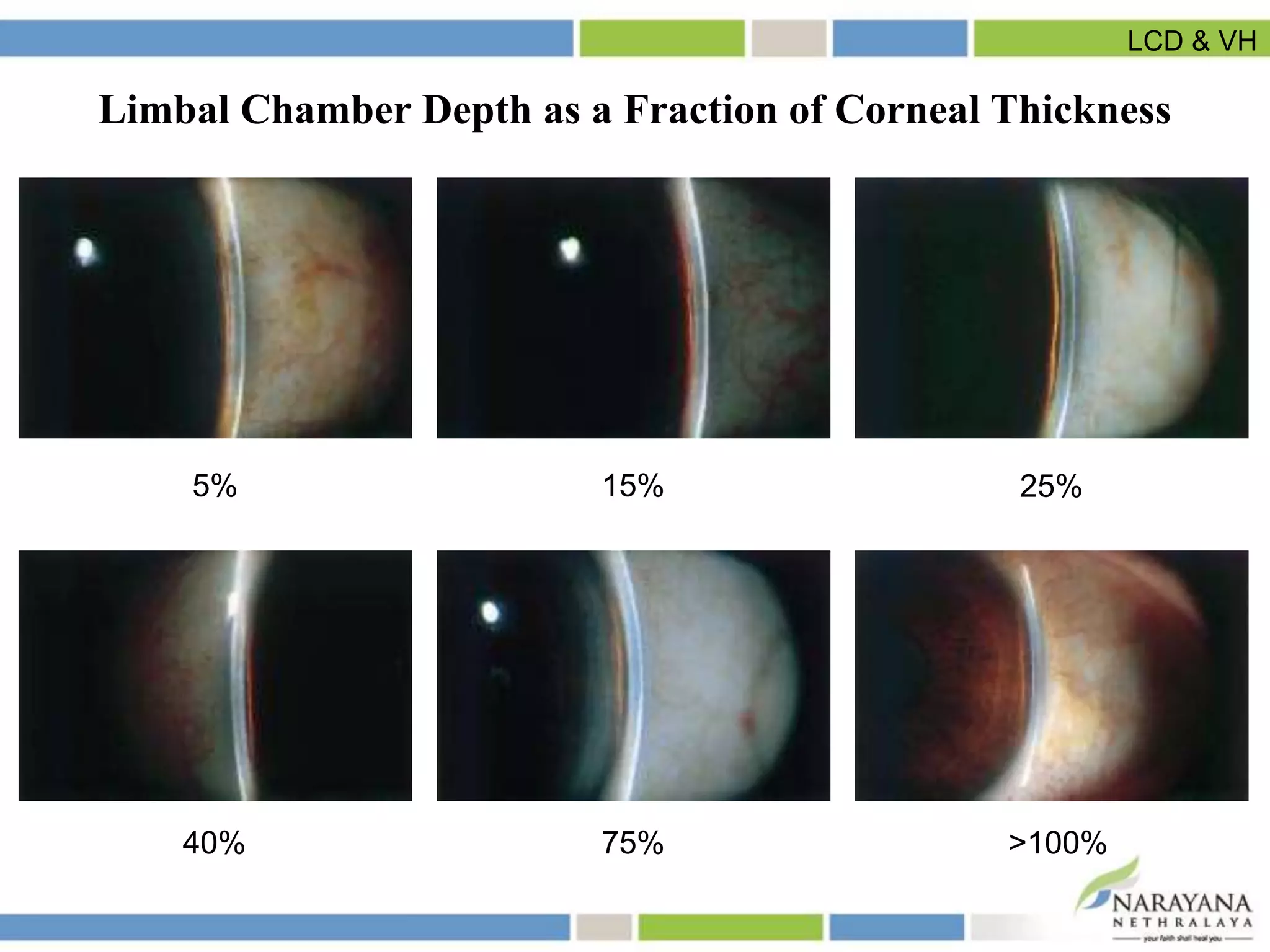
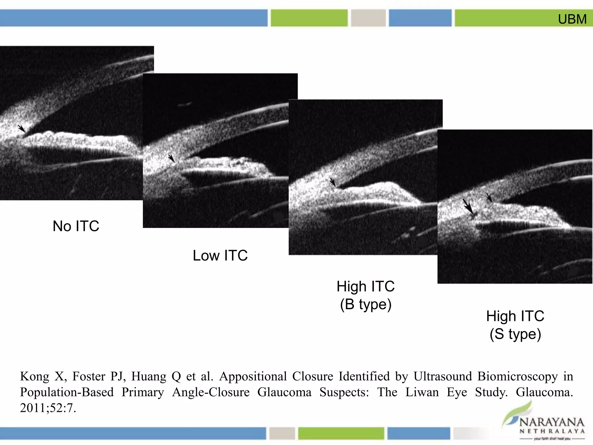
SlideShare a Scribd company logo
Sujay Chauhan
ASSESSMENT OF
ANTERIOR CHAMBER ANGLE
INDICATIONS FOR ASSESSMENT OF
ANTERIOR CHAMBER ANGLE
All patients on the first visit
• Glaucoma suspects
• Open angle glaucoma
• Ocular hypertension
• Angle closure post-iridotomy
• Retinal vein occlusion
• Blunt trauma
Indications
METHODS FOR ASSESSMENT OF
ANTERIOR CHAMBER ANGLE
SUBJECTIVE ASSESSMENT
• Oblique flashlight test
• Van Herrick’s technique
• Gonioscopy
OBLIQUE FLASHLIGHT TEST
GRADE I GRADE II GRADE III GRADE IV
< 1/3
illuminated
1/3-2/3
illuminated
>2/3
illuminated
Fully
illuminated
Oblique flashlight test
• Good inter-observer agreement (p = 0.04)
• Good reproducibility (weighted kappa value = 0.74)
• Sensitivity: 75-97%
• Specificity: 67-99.4%
• Gracitelli et al. Ability of non-ophthalmologist doctors to detect eyes with occludable angles using the
flashlight test. Int Ophthalmol. 2014 Jun;34(3):557-561
• Congdon NG et al. Screening techniques for angle-closure glaucoma in rural Taiwan. Acta Ophthalmol
Scand 1996;74:113–119
• He M et al. Angle-closure glaucoma in East Asian and European people. Different diseases? Eye
2006;20:3–12
• Thomas R et al. The flashlight test and van Herick’s test are poor predictors for occludable angles.
Aust N Z J Ophthalmol 1996;24:251–256
• Vargas E, Drance SM Anterior chamber depth in angle-closure glaucoma. Clinical methods of depth
determination in people with and without the disease. Arch Ophthalmol 1973;90:438–439
Oblique flashlight test
• Simple
• No sophisticated equipment required
Advantages
Oblique flashlight test
• Lack of standardization
• Arbitrary cut-offs
Limitations
Oblique flashlight test
LIMBALANTERIOR CHAMBER DEPTH
AND
VAN HERRICK’S TECHNIQUE
5% 15% 25%
40% 75% >100%
Limbal Chamber Depth as a Fraction of Corneal Thickness
LCD & VH
All Occludable Angles PAC PACG
LCD VH Sensitivity Specificity Sensitivity Specificity Sensitivity Specificity
0% NA 17.8% 99.6% 29.3% 99.2% 52.2% 98.9%
≥5% NA 60.5% 96.1% 70.7% 93.7% 91.3% 92.6%
≥15% 1 83.7% 85.7% 84.5% 82.4% 91.3% 81.1%
≥25% 2 99.2% 65.5% 100% 62.4% 100% 61.1%
≥40% 3 100% 40.3% 100% 38.3% 100% 37.5%
Performance Indices of Limbal Chamber Depth Estimation in
Detection of Gonioscopically Occludable Angles, PAC and PACG
Foster PJ et al. Detection of gonioscopically occludable angles and primary angle closure glaucoma by
estimation of limbal chamber depth in Asians: modified grading scheme. Br J Ophthalmol.
2000;84:186-192.
LCD & VH
• Good interobserver agreement (weighted kappa value = 0.73-0.76)
• High intraobserver repeatability
• Sensitivity = 99%
• Thomas R et al. The flashlight test and van Herick’s test are poor predictors for occludable angles.
Aust N Z J Ophthalmol 1996;24:251–256
• Foster PJ et al. Detection of gonioscopically occludable angles and primary angle closure glaucoma
by estimation of limbal chamber depth in Asians: modified grading scheme. Br J Ophthalmol
2000;84:186–192
• Cockburn DM. Slitlamp estimate of anterior chamber depth as a predictor of the gonioscopic
visibility of angle structures. Am J Optom Physiol Opt 1982;59:904–908
LCD & VH
Advantages
• Quick
• Non-invasive
• No auxiliary equipment
LCD & VH
• Subjective
• Overestimation of angle depth (if slit beam moved too far on to
the cornea, nasal limbus)
• Little or no information regarding angle morphology
• Affected by corneal degenerations such as arcus senilis
• Horizontal quadrants only
Limitations
LCD & VH
GONIOSCOPY
• Indirect gonioscopy – most common, reference standard
• Grading of anterior chamber angle depth
• Iris configuration
• Insertion of iris root
• Peripheral anterior synechiae
• Pigment deposition
• Neovascularisation
• Uveitis (nodules)
• Blunt trauma (angle recession, cyclodialysis cleft)
High interobserver agreement (kappa value = 0.62-0.82)
• Foster PJ et al. Detection of gonioscopically occludable angles and primary angle closure glaucoma by
estimation of limbal chamber depth in Asians: modified grading scheme. Br J Ophthalmol
2000;84:186–192
• Jacob JT et al. Evaluation of interobserver agreement in gonioscopy. Kerala J Ophthalmol.
2008;XX:253–255
• Lavanya R et al. Screening for narrow angles in the Singapore population: evaluation of new
noncontact screening methods. Ophthalmology 2008:115:1720–1727
• Thomas R et al. The flashlight test and van Herick’s test are poor predictors for occludable angles. Aust
N Z J Ophthalmol. 1996;24:251–256
Gonioscopy
Advantages
• Easily and quickly performed
• Not expensive
Gonioscopy
• Invasive
• Subjective, requires skill and experience
• Variables – inadvertent pressure on cornea, light exposure onto
pupil, patient cooperation, type of lens used, direction of gaze
• Does not provide quantitative evaluation of the angle
• Can not visualize structures posterior to iris
Limitations
Gonioscopy
OBJECTIVE MEASURES
• Ultrasound biomicroscopy (UBM)
• Anterior segment optical coherence tomography (AS-OCT)
Gonioscopy
ULTRASOUND BIOMICROSCOPY (UBM)
• Two-dimensional high-resolution gray-scale images of ocular
structures anterior to pars plana
• Quantitative and qualitative evaluation
• Principle similar to that of B-scan (10 MHz)
• Frequency – 50 MHz – More frequency – less penetration (5 mm)
and more resolution
UBM
Interpretation of anterior chamber angle configuration is
dependent on determining the location of scleral spur (inward
protrusion of sclera)
UBM
Kong X, Foster PJ, Huang Q et al. Appositional Closure Identified by Ultrasound Biomicroscopy in
Population-Based Primary Angle-Closure Glaucoma Suspects: The Liwan Eye Study. Glaucoma.
2011;52:7.
No ITC
High ITC
(S type)
High ITC
(B type)
Low ITC
UBM
• Angle opening distance (AOD500): Length of a perpendicular
from TM to iris at a point 500 µm from SS
• Trabecular-iris angle (Ø1): Apex in the iris sulcus and arms
passing through the point 500 µm from SS and the point
perpendicularly opposite on the iris
UBM
AOD500 :
• Sensitivity = 82%
• Specificity = 96%
Trabecular iris angle:
• Sensitivity = 83%
• Specificity = 93%
Henzan, IM et al. Comparison of Ultrasound Biomicroscopic Configurations Among Primary Angle
Closure, Its Suspects, and Nonoccludable Angles: The Kumejima Study. Am J Ophthalmol.
2011;151(6):1065-1073
UBM
Advantages
• High resolution of 50 µm at a frequency of 50 MHz
• Cross-sections of angle can be captured without lighting –
changes in iris shape with or without light can be ascertained
• Independent of corneal opacity
• Dynamic changes can be recorded
• Images can be automatically quantified
UBM
Assessment of areas not possible by gonioscopy:
• Ciliary body
• Inside the iris
• Posterior chamber
• Inside filtering blebs
• Outflow tract for aqueous humour inside sclera
UBM
Limitations
• Supine position – iris diaphragm falls back – angle opens
• Only 1 quadrant can be imaged at a time
• Angle region measured is located subjectively as superior, nasal,
inferior and temporal
• Skilled examiner
• Longer image acquisition time
• Use of anaesthesia
• Contact with the eye using a cup with a coupling medium or a
probe – corneal abrasion, infection
UBM
Can not detect:
• Pigmentation in the angle
• Inflammatory exudate in the angle
• Neovascularization in the angle
UBM
ANTERIOR SEGMENT OPTICAL
COHERENCE TOMOGRAPHY (AS-OCT)
POSTERIOR SEGMENT OCT ANTERIOR SEGMENT OCT
830 nm 1310 nm
Unable to penetrate sclera Penetrates sclera and iris
More time for image acquisition Reduced time
Compromised image quality Reduced motion artefacts – better
image quality
AS-OCT
• Low coherence interferometry to obtain cross-sectional images of
the ocular structures
• Similar to UBM, interpretation of anterior chamber angle
configuration is dependent on determining the location of scleral
spur
AS-OCT
Angle closure in more than one quadrant was detected by:
• AS-OCT in 71% of patients
• Gonioscopy in 49.5% of patients
When performed in dark conditions,
AS-OCT identified angle closure in:
• 98% of subjects found to have angle closure on gonioscopy
• 44.6% of subjects found to have open angles on gonioscopy
Specificity of AS-OCT to detect angle closure: 55.4%
Nolan et al. Detection of primary angle closure using anterior segment optical coherence tomography in
Asian eyes. Ophthalmology. 2007 Jan;114(1):33-39.
AS-OCT
Intraobserver variability = 9.4% to 12.5%
Interobserver variability = 10.7%
High reproducibility: 0.875-0.942
Tan AN et al. Reproducibility of anterior chamber angle measurements with anterior segment optical
coherence tomography. Invest Ophthalmol Vis Sci, 52(5), 2095-2099
AS-OCT
AS-OCT scanning protocol Sensitivity Specificity
Nasal-temporal quadrants 47% 88%
Inferior quadrant 84% 69%
Superior-inferior quadrants 92% 54%
Khor WB et al. Evaluation of scanning protocols for imaging the anterior chamber angle with anterior
segment-optical coherence tomography. J Glaucoma. 2010 Aug;19(6):365-368.
AS-OCT
Advantages
• Higher resolution than UBM (18 µm vs 25 µm)
• Non-contact method
• All 4 quadrants are imaged at once
• Entire cross-section of anterior segment is imaged into one frame
– permits assessment and documentation of iris profile and its
relationship to other anatomical parameters of eye
• Patient in sitting position – no alteration in anterior chamber
depth
• Does not require a skilled operator
AS-OCT
Limitations
• Infrared light blocked by iris pigment – precludes assessment of
structures located behind iris
• Degradation of light by sclera – ciliary body not imaged fully
• Can not obtain clear images through opaque media
• Obstruction by eyelids – difficulty in imaging superior and
inferior angles
AS-OCT
OCT and UBM had similar
• mean values of angle opening distance, angle recess area and
trabecular-iris space area
• reproducibility, and
• sensitivity-specificity profiles
in identifying eyes with narrow angles
Radhakrishnan S et al. Comparision of optical coherence tomography and ultrasound biomicroscopy for
detection of narrow anterior chamber angles. Arch Ophthalmol. 2005 Aug:123(8):1053-1059.
AS-OCT
CONCLUSION
• UBM and AS-OCT provide real-time cross-sectional images of
anterior chamber angle – accuracy and reproducibility yet to be
proved
• Gonioscopy – continues to remain essential for glaucoma
evaluation and management
Assessment of Anterior Chamber Angle

More Related Content

What's hot

A scan ultrasonography
A scan ultrasonographyA scan ultrasonography
A scan ultrasonography
Samuel Ponraj
 
Subjective refraction
Subjective refractionSubjective refraction
Subjective refraction
Azizul Islam
 
Slit lamp biomicroscope
Slit lamp biomicroscopeSlit lamp biomicroscope
Slit lamp biomicroscope
Md. Nurul Islam
 
FUNDUS FLUORESCEIN ANGIOGRAPHY
FUNDUS FLUORESCEIN ANGIOGRAPHYFUNDUS FLUORESCEIN ANGIOGRAPHY
FUNDUS FLUORESCEIN ANGIOGRAPHY
Anuraag Singh
 
Slit lamp biomicroscopy
Slit lamp biomicroscopySlit lamp biomicroscopy
Slit lamp biomicroscopy
Hira Dahal
 
Pachymetry
PachymetryPachymetry
Pachymetry
SSSIHMS-PG
 
Soft Contact Lens Fitting
Soft Contact Lens FittingSoft Contact Lens Fitting
Soft Contact Lens FittingVishakh Nair
 
Indirect ophthalmoscopy
Indirect ophthalmoscopy Indirect ophthalmoscopy
Indirect ophthalmoscopy
Shruti Laddha
 
Maddox rod n wing
Maddox rod n wingMaddox rod n wing
Maddox rod n wing
Rizwan Sajjad Chachar
 
Slit lamp techniques.pptx
Slit lamp techniques.pptxSlit lamp techniques.pptx
Slit lamp techniques.pptx
Raju Kaiti
 
Pentacam
PentacamPentacam
Pentacam
SimiAfroz2
 
Gonioscopy: gonioscopic lenses, principle and clinical aspects
Gonioscopy: gonioscopic lenses, principle and clinical aspectsGonioscopy: gonioscopic lenses, principle and clinical aspects
Gonioscopy: gonioscopic lenses, principle and clinical aspects
Dr Samarth Mishra
 
AMBLYOPIA AND ITS MANAGEMENT
AMBLYOPIA AND ITS MANAGEMENTAMBLYOPIA AND ITS MANAGEMENT
AMBLYOPIA AND ITS MANAGEMENT
SSSIHMS-PG
 
Clinical examination of squint
Clinical examination of squintClinical examination of squint
Clinical examination of squint
Reshma Peter
 
ultrasound biomicroscopy
ultrasound biomicroscopyultrasound biomicroscopy
ultrasound biomicroscopy
SSSIHMS-PG
 
Lensometers
LensometersLensometers
Lensometers
Sahil Rockzz
 
Slit Lamp Illumination Techniques
Slit Lamp Illumination TechniquesSlit Lamp Illumination Techniques
Slit Lamp Illumination Techniques
Irina Kezik
 
Pentacam
Pentacam Pentacam
Pentacam
Mehdi Khanlari
 
pentacam
pentacampentacam
pentacam
nrvdad
 

What's hot (20)

A scan ultrasonography
A scan ultrasonographyA scan ultrasonography
A scan ultrasonography
 
Subjective refraction
Subjective refractionSubjective refraction
Subjective refraction
 
Maddox rod
Maddox rodMaddox rod
Maddox rod
 
Slit lamp biomicroscope
Slit lamp biomicroscopeSlit lamp biomicroscope
Slit lamp biomicroscope
 
FUNDUS FLUORESCEIN ANGIOGRAPHY
FUNDUS FLUORESCEIN ANGIOGRAPHYFUNDUS FLUORESCEIN ANGIOGRAPHY
FUNDUS FLUORESCEIN ANGIOGRAPHY
 
Slit lamp biomicroscopy
Slit lamp biomicroscopySlit lamp biomicroscopy
Slit lamp biomicroscopy
 
Pachymetry
PachymetryPachymetry
Pachymetry
 
Soft Contact Lens Fitting
Soft Contact Lens FittingSoft Contact Lens Fitting
Soft Contact Lens Fitting
 
Indirect ophthalmoscopy
Indirect ophthalmoscopy Indirect ophthalmoscopy
Indirect ophthalmoscopy
 
Maddox rod n wing
Maddox rod n wingMaddox rod n wing
Maddox rod n wing
 
Slit lamp techniques.pptx
Slit lamp techniques.pptxSlit lamp techniques.pptx
Slit lamp techniques.pptx
 
Pentacam
PentacamPentacam
Pentacam
 
Gonioscopy: gonioscopic lenses, principle and clinical aspects
Gonioscopy: gonioscopic lenses, principle and clinical aspectsGonioscopy: gonioscopic lenses, principle and clinical aspects
Gonioscopy: gonioscopic lenses, principle and clinical aspects
 
AMBLYOPIA AND ITS MANAGEMENT
AMBLYOPIA AND ITS MANAGEMENTAMBLYOPIA AND ITS MANAGEMENT
AMBLYOPIA AND ITS MANAGEMENT
 
Clinical examination of squint
Clinical examination of squintClinical examination of squint
Clinical examination of squint
 
ultrasound biomicroscopy
ultrasound biomicroscopyultrasound biomicroscopy
ultrasound biomicroscopy
 
Lensometers
LensometersLensometers
Lensometers
 
Slit Lamp Illumination Techniques
Slit Lamp Illumination TechniquesSlit Lamp Illumination Techniques
Slit Lamp Illumination Techniques
 
Pentacam
Pentacam Pentacam
Pentacam
 
pentacam
pentacampentacam
pentacam
 

Viewers also liked

Assessment of anterior chamber
Assessment of anterior chamberAssessment of anterior chamber
Assessment of anterior chamber
LABISHETTY CHARAN
 
Anatomy of anterior chamber
Anatomy of anterior chamberAnatomy of anterior chamber
Anatomy of anterior chamber
Dr.Prathibha S
 
Mechanisms of angle closure glaucoma
Mechanisms of angle closure glaucomaMechanisms of angle closure glaucoma
Mechanisms of angle closure glaucoma
SSSIHMS-PG
 
Angle closure glaucoma
Angle  closure  glaucomaAngle  closure  glaucoma
Angle closure glaucomaSamuel Ponraj
 
Bio microscopy
Bio microscopyBio microscopy
Bio microscopy
Niv09
 
Primary Glaucoma
Primary GlaucomaPrimary Glaucoma
Primary Glaucoma
John Paul Taday
 
Gonioscopy and optic nerve head evaluation
Gonioscopy and optic nerve head evaluationGonioscopy and optic nerve head evaluation
Gonioscopy and optic nerve head evaluation
Ahmedfaik
 
Steroid induced glaucoma
Steroid induced glaucomaSteroid induced glaucoma
Steroid induced glaucoma
Farukhzad Hafizyar
 
Review of angle closure glaucomas, By Fritz Allen, MD
Review of angle closure glaucomas, By Fritz Allen, MDReview of angle closure glaucomas, By Fritz Allen, MD
Review of angle closure glaucomas, By Fritz Allen, MDVisionary Ophthamology
 
Glaucoma 2 primary angle closure glaucoma,dr.k.n.jha,02.11.16
Glaucoma 2 primary angle closure glaucoma,dr.k.n.jha,02.11.16Glaucoma 2 primary angle closure glaucoma,dr.k.n.jha,02.11.16
Glaucoma 2 primary angle closure glaucoma,dr.k.n.jha,02.11.16
ophthalmgmcri
 
Role of oct in glaucoma
Role of oct in glaucomaRole of oct in glaucoma
Role of oct in glaucoma
Dr Samarth Mishra
 
Angle closure glaucoma
Angle closure glaucomaAngle closure glaucoma
Angle closure glaucoma
Arushi Prakash
 
Lecture1 anatomy of the eye
Lecture1   anatomy of the eyeLecture1   anatomy of the eye
Lecture1 anatomy of the eyespecialclass
 
Bohomolets 4th year Surgery Complication of Appendicitis
Bohomolets 4th year Surgery Complication of AppendicitisBohomolets 4th year Surgery Complication of Appendicitis
Bohomolets 4th year Surgery Complication of Appendicitis
Dr. Rubz
 
Glaucoma for undergraduates
Glaucoma for undergraduates Glaucoma for undergraduates
Glaucoma for undergraduates
KafrELShiekh University
 
Ang.Closure Glaucoma
Ang.Closure GlaucomaAng.Closure Glaucoma
Ang.Closure Glaucoma
Zaitsev
 
Ocular physiology_2014
Ocular physiology_2014Ocular physiology_2014
Ocular physiology_2014Subrata Roy
 

Viewers also liked (20)

Assessment of anterior chamber
Assessment of anterior chamberAssessment of anterior chamber
Assessment of anterior chamber
 
Anatomy of anterior chamber
Anatomy of anterior chamberAnatomy of anterior chamber
Anatomy of anterior chamber
 
Mechanisms of angle closure glaucoma
Mechanisms of angle closure glaucomaMechanisms of angle closure glaucoma
Mechanisms of angle closure glaucoma
 
Angle closure glaucoma
Angle  closure  glaucomaAngle  closure  glaucoma
Angle closure glaucoma
 
Bio microscopy
Bio microscopyBio microscopy
Bio microscopy
 
Gonioscopy
GonioscopyGonioscopy
Gonioscopy
 
Primary Glaucoma
Primary GlaucomaPrimary Glaucoma
Primary Glaucoma
 
Gonioscopy and optic nerve head evaluation
Gonioscopy and optic nerve head evaluationGonioscopy and optic nerve head evaluation
Gonioscopy and optic nerve head evaluation
 
PACS lens AGS 2014 final
PACS lens AGS 2014 finalPACS lens AGS 2014 final
PACS lens AGS 2014 final
 
Steroid induced glaucoma
Steroid induced glaucomaSteroid induced glaucoma
Steroid induced glaucoma
 
Review of angle closure glaucomas, By Fritz Allen, MD
Review of angle closure glaucomas, By Fritz Allen, MDReview of angle closure glaucomas, By Fritz Allen, MD
Review of angle closure glaucomas, By Fritz Allen, MD
 
Glaucoma 2 primary angle closure glaucoma,dr.k.n.jha,02.11.16
Glaucoma 2 primary angle closure glaucoma,dr.k.n.jha,02.11.16Glaucoma 2 primary angle closure glaucoma,dr.k.n.jha,02.11.16
Glaucoma 2 primary angle closure glaucoma,dr.k.n.jha,02.11.16
 
Role of oct in glaucoma
Role of oct in glaucomaRole of oct in glaucoma
Role of oct in glaucoma
 
Angle closure glaucoma
Angle closure glaucomaAngle closure glaucoma
Angle closure glaucoma
 
Lecture1 anatomy of the eye
Lecture1   anatomy of the eyeLecture1   anatomy of the eye
Lecture1 anatomy of the eye
 
Bohomolets 4th year Surgery Complication of Appendicitis
Bohomolets 4th year Surgery Complication of AppendicitisBohomolets 4th year Surgery Complication of Appendicitis
Bohomolets 4th year Surgery Complication of Appendicitis
 
Red eye
Red eyeRed eye
Red eye
 
Glaucoma for undergraduates
Glaucoma for undergraduates Glaucoma for undergraduates
Glaucoma for undergraduates
 
Ang.Closure Glaucoma
Ang.Closure GlaucomaAng.Closure Glaucoma
Ang.Closure Glaucoma
 
Ocular physiology_2014
Ocular physiology_2014Ocular physiology_2014
Ocular physiology_2014
 

Similar to Assessment of Anterior Chamber Angle

Pre op corneal laser
Pre op corneal laserPre op corneal laser
Pre op corneal laser
Sumegha Tomar
 
1. Methods of eye examination.pptx
1. Methods of eye examination.pptx1. Methods of eye examination.pptx
1. Methods of eye examination.pptx
annieamjad1
 
1. Methods of eye examination.pptx
1. Methods of eye examination.pptx1. Methods of eye examination.pptx
1. Methods of eye examination.pptx
Annie Amjad
 
REFRACTIVE SURGERY PRESENTATION.pptx
REFRACTIVE SURGERY PRESENTATION.pptxREFRACTIVE SURGERY PRESENTATION.pptx
REFRACTIVE SURGERY PRESENTATION.pptx
BARNABASMUGABI
 
AS OCT & UBM - Dr Shylesh B Dabke
AS OCT & UBM - Dr Shylesh B DabkeAS OCT & UBM - Dr Shylesh B Dabke
AS OCT & UBM - Dr Shylesh B Dabke
Shylesh Dabke
 
Perimetry in ophthalmology post graduate
Perimetry in ophthalmology post graduatePerimetry in ophthalmology post graduate
Perimetry in ophthalmology post graduate
krishnenduUnnikannan
 
Final spectral oct_in_diagnosis_and_management_of_glaucoma
Final spectral oct_in_diagnosis_and_management_of_glaucomaFinal spectral oct_in_diagnosis_and_management_of_glaucoma
Final spectral oct_in_diagnosis_and_management_of_glaucomaRaju Nsd
 
The basics of retinal oct ophso.net
The basics of retinal oct ophso.netThe basics of retinal oct ophso.net
The basics of retinal oct ophso.netkebaplik
 
Feasibility of using backscattered light to recover refractive
Feasibility of using backscattered light to recover refractiveFeasibility of using backscattered light to recover refractive
Feasibility of using backscattered light to recover refractive
Payman Rajai
 
Recent advances in diagnosis of glaucoma
Recent advances in diagnosis of glaucomaRecent advances in diagnosis of glaucoma
Recent advances in diagnosis of glaucoma
Jaspreet Kauldhar
 
Recent advances in diagnosis of glaucoma
Recent advances in diagnosis of glaucomaRecent advances in diagnosis of glaucoma
Recent advances in diagnosis of glaucoma
Jaspreet Kauldhar
 
Gonioscopy
GonioscopyGonioscopy
Gonioscopy
Simanta Borah
 
Preperimetric glaucoma
Preperimetric glaucoma Preperimetric glaucoma
Preperimetric glaucoma
PRAKRITIYAGNAM
 
Newer tests for glaucoma
Newer tests for glaucomaNewer tests for glaucoma
Newer tests for glaucoma
vaishusmail
 
Presentation mopb.pptx
Presentation mopb.pptxPresentation mopb.pptx
Presentation mopb.pptx
bakanangemmahpholoan
 
AC .pptx
AC .pptxAC .pptx
AC .pptx
IbraHim Sartawi
 
2016 Basic Motility Exam
2016 Basic Motility Exam2016 Basic Motility Exam
2016 Basic Motility Exam
Alvina Pauline Santiago, MD
 
Indirect ophthalmoscopy and fundus drawing
Indirect ophthalmoscopy and fundus drawingIndirect ophthalmoscopy and fundus drawing
Indirect ophthalmoscopy and fundus drawing
Sonali Singh
 
Ultrasonic Evaluation of Eyes With Blunt Trauma
Ultrasonic Evaluation of Eyes With Blunt TraumaUltrasonic Evaluation of Eyes With Blunt Trauma
Ultrasonic Evaluation of Eyes With Blunt Trauma
KafrELShiekh University
 

Similar to Assessment of Anterior Chamber Angle (20)

Corneal topography
Corneal topographyCorneal topography
Corneal topography
 
Pre op corneal laser
Pre op corneal laserPre op corneal laser
Pre op corneal laser
 
1. Methods of eye examination.pptx
1. Methods of eye examination.pptx1. Methods of eye examination.pptx
1. Methods of eye examination.pptx
 
1. Methods of eye examination.pptx
1. Methods of eye examination.pptx1. Methods of eye examination.pptx
1. Methods of eye examination.pptx
 
REFRACTIVE SURGERY PRESENTATION.pptx
REFRACTIVE SURGERY PRESENTATION.pptxREFRACTIVE SURGERY PRESENTATION.pptx
REFRACTIVE SURGERY PRESENTATION.pptx
 
AS OCT & UBM - Dr Shylesh B Dabke
AS OCT & UBM - Dr Shylesh B DabkeAS OCT & UBM - Dr Shylesh B Dabke
AS OCT & UBM - Dr Shylesh B Dabke
 
Perimetry in ophthalmology post graduate
Perimetry in ophthalmology post graduatePerimetry in ophthalmology post graduate
Perimetry in ophthalmology post graduate
 
Final spectral oct_in_diagnosis_and_management_of_glaucoma
Final spectral oct_in_diagnosis_and_management_of_glaucomaFinal spectral oct_in_diagnosis_and_management_of_glaucoma
Final spectral oct_in_diagnosis_and_management_of_glaucoma
 
The basics of retinal oct ophso.net
The basics of retinal oct ophso.netThe basics of retinal oct ophso.net
The basics of retinal oct ophso.net
 
Feasibility of using backscattered light to recover refractive
Feasibility of using backscattered light to recover refractiveFeasibility of using backscattered light to recover refractive
Feasibility of using backscattered light to recover refractive
 
Recent advances in diagnosis of glaucoma
Recent advances in diagnosis of glaucomaRecent advances in diagnosis of glaucoma
Recent advances in diagnosis of glaucoma
 
Recent advances in diagnosis of glaucoma
Recent advances in diagnosis of glaucomaRecent advances in diagnosis of glaucoma
Recent advances in diagnosis of glaucoma
 
Gonioscopy
GonioscopyGonioscopy
Gonioscopy
 
Preperimetric glaucoma
Preperimetric glaucoma Preperimetric glaucoma
Preperimetric glaucoma
 
Newer tests for glaucoma
Newer tests for glaucomaNewer tests for glaucoma
Newer tests for glaucoma
 
Presentation mopb.pptx
Presentation mopb.pptxPresentation mopb.pptx
Presentation mopb.pptx
 
AC .pptx
AC .pptxAC .pptx
AC .pptx
 
2016 Basic Motility Exam
2016 Basic Motility Exam2016 Basic Motility Exam
2016 Basic Motility Exam
 
Indirect ophthalmoscopy and fundus drawing
Indirect ophthalmoscopy and fundus drawingIndirect ophthalmoscopy and fundus drawing
Indirect ophthalmoscopy and fundus drawing
 
Ultrasonic Evaluation of Eyes With Blunt Trauma
Ultrasonic Evaluation of Eyes With Blunt TraumaUltrasonic Evaluation of Eyes With Blunt Trauma
Ultrasonic Evaluation of Eyes With Blunt Trauma
 

More from Sujay Chauhan

Subretinal hemorrhage
Subretinal hemorrhageSubretinal hemorrhage
Subretinal hemorrhage
Sujay Chauhan
 
Polypoidal choroidal vasculopathy
Polypoidal choroidal vasculopathyPolypoidal choroidal vasculopathy
Polypoidal choroidal vasculopathy
Sujay Chauhan
 
Diabetic retinopathy
Diabetic retinopathyDiabetic retinopathy
Diabetic retinopathy
Sujay Chauhan
 
Optic disc evaluation
Optic disc evaluationOptic disc evaluation
Optic disc evaluation
Sujay Chauhan
 
Phacomachine and its dynamics
Phacomachine and its dynamicsPhacomachine and its dynamics
Phacomachine and its dynamics
Sujay Chauhan
 
Binocular vision
Binocular visionBinocular vision
Binocular vision
Sujay Chauhan
 
Microsporidiosis
MicrosporidiosisMicrosporidiosis
Microsporidiosis
Sujay Chauhan
 

More from Sujay Chauhan (7)

Subretinal hemorrhage
Subretinal hemorrhageSubretinal hemorrhage
Subretinal hemorrhage
 
Polypoidal choroidal vasculopathy
Polypoidal choroidal vasculopathyPolypoidal choroidal vasculopathy
Polypoidal choroidal vasculopathy
 
Diabetic retinopathy
Diabetic retinopathyDiabetic retinopathy
Diabetic retinopathy
 
Optic disc evaluation
Optic disc evaluationOptic disc evaluation
Optic disc evaluation
 
Phacomachine and its dynamics
Phacomachine and its dynamicsPhacomachine and its dynamics
Phacomachine and its dynamics
 
Binocular vision
Binocular visionBinocular vision
Binocular vision
 
Microsporidiosis
MicrosporidiosisMicrosporidiosis
Microsporidiosis
 

Recently uploaded

Pharynx and Clinical Correlations BY Dr.Rabia Inam Gandapore.pptx
Pharynx and Clinical Correlations BY Dr.Rabia Inam Gandapore.pptxPharynx and Clinical Correlations BY Dr.Rabia Inam Gandapore.pptx
Pharynx and Clinical Correlations BY Dr.Rabia Inam Gandapore.pptx
Dr. Rabia Inam Gandapore
 
Ozempic: Preoperative Management of Patients on GLP-1 Receptor Agonists
Ozempic: Preoperative Management of Patients on GLP-1 Receptor Agonists  Ozempic: Preoperative Management of Patients on GLP-1 Receptor Agonists
Ozempic: Preoperative Management of Patients on GLP-1 Receptor Agonists
Saeid Safari
 
BENIGN PROSTATIC HYPERPLASIA.BPH. BPHpdf
BENIGN PROSTATIC HYPERPLASIA.BPH. BPHpdfBENIGN PROSTATIC HYPERPLASIA.BPH. BPHpdf
BENIGN PROSTATIC HYPERPLASIA.BPH. BPHpdf
DR SETH JOTHAM
 
KDIGO 2024 guidelines for diabetologists
KDIGO 2024 guidelines for diabetologistsKDIGO 2024 guidelines for diabetologists
KDIGO 2024 guidelines for diabetologists
د.محمود نجيب
 
ANATOMY AND PHYSIOLOGY OF URINARY SYSTEM.pptx
ANATOMY AND PHYSIOLOGY OF URINARY SYSTEM.pptxANATOMY AND PHYSIOLOGY OF URINARY SYSTEM.pptx
ANATOMY AND PHYSIOLOGY OF URINARY SYSTEM.pptx
Swetaba Besh
 
Triangles of Neck and Clinical Correlation by Dr. RIG.pptx
Triangles of Neck and Clinical Correlation by Dr. RIG.pptxTriangles of Neck and Clinical Correlation by Dr. RIG.pptx
Triangles of Neck and Clinical Correlation by Dr. RIG.pptx
Dr. Rabia Inam Gandapore
 
Physiology of Special Chemical Sensation of Taste
Physiology of Special Chemical Sensation of TastePhysiology of Special Chemical Sensation of Taste
Physiology of Special Chemical Sensation of Taste
MedicoseAcademics
 
Alcohol_Dr. Jeenal Mistry MD Pharmacology.pdf
Alcohol_Dr. Jeenal Mistry MD Pharmacology.pdfAlcohol_Dr. Jeenal Mistry MD Pharmacology.pdf
Alcohol_Dr. Jeenal Mistry MD Pharmacology.pdf
Dr Jeenal Mistry
 
Ocular injury ppt Upendra pal optometrist upums saifai etawah
Ocular injury  ppt  Upendra pal  optometrist upums saifai etawahOcular injury  ppt  Upendra pal  optometrist upums saifai etawah
Ocular injury ppt Upendra pal optometrist upums saifai etawah
pal078100
 
Surat @ℂall @Girls ꧁❤8527049040❤꧂@ℂall @Girls Service Vip Top Model Safe
Surat @ℂall @Girls ꧁❤8527049040❤꧂@ℂall @Girls Service Vip Top Model SafeSurat @ℂall @Girls ꧁❤8527049040❤꧂@ℂall @Girls Service Vip Top Model Safe
Surat @ℂall @Girls ꧁❤8527049040❤꧂@ℂall @Girls Service Vip Top Model Safe
Savita Shen $i11
 
Ophthalmology Clinical Tests for OSCE exam
Ophthalmology Clinical Tests for OSCE examOphthalmology Clinical Tests for OSCE exam
Ophthalmology Clinical Tests for OSCE exam
KafrELShiekh University
 
Maxilla, Mandible & Hyoid Bone & Clinical Correlations by Dr. RIG.pptx
Maxilla, Mandible & Hyoid Bone & Clinical Correlations by Dr. RIG.pptxMaxilla, Mandible & Hyoid Bone & Clinical Correlations by Dr. RIG.pptx
Maxilla, Mandible & Hyoid Bone & Clinical Correlations by Dr. RIG.pptx
Dr. Rabia Inam Gandapore
 
ARTIFICIAL INTELLIGENCE IN HEALTHCARE.pdf
ARTIFICIAL INTELLIGENCE IN  HEALTHCARE.pdfARTIFICIAL INTELLIGENCE IN  HEALTHCARE.pdf
ARTIFICIAL INTELLIGENCE IN HEALTHCARE.pdf
Anujkumaranit
 
HOT NEW PRODUCT! BIG SALES FAST SHIPPING NOW FROM CHINA!! EU KU DB BK substit...
HOT NEW PRODUCT! BIG SALES FAST SHIPPING NOW FROM CHINA!! EU KU DB BK substit...HOT NEW PRODUCT! BIG SALES FAST SHIPPING NOW FROM CHINA!! EU KU DB BK substit...
HOT NEW PRODUCT! BIG SALES FAST SHIPPING NOW FROM CHINA!! EU KU DB BK substit...
GL Anaacs
 
New Directions in Targeted Therapeutic Approaches for Older Adults With Mantl...
New Directions in Targeted Therapeutic Approaches for Older Adults With Mantl...New Directions in Targeted Therapeutic Approaches for Older Adults With Mantl...
New Directions in Targeted Therapeutic Approaches for Older Adults With Mantl...
i3 Health
 
Couples presenting to the infertility clinic- Do they really have infertility...
Couples presenting to the infertility clinic- Do they really have infertility...Couples presenting to the infertility clinic- Do they really have infertility...
Couples presenting to the infertility clinic- Do they really have infertility...
Sujoy Dasgupta
 
How to Give Better Lectures: Some Tips for Doctors
How to Give Better Lectures: Some Tips for DoctorsHow to Give Better Lectures: Some Tips for Doctors
How to Give Better Lectures: Some Tips for Doctors
LanceCatedral
 
micro teaching on communication m.sc nursing.pdf
micro teaching on communication m.sc nursing.pdfmicro teaching on communication m.sc nursing.pdf
micro teaching on communication m.sc nursing.pdf
Anurag Sharma
 
heat stroke and heat exhaustion in children
heat stroke and heat exhaustion in childrenheat stroke and heat exhaustion in children
heat stroke and heat exhaustion in children
SumeraAhmad5
 
Hemodialysis: Chapter 3, Dialysis Water Unit - Dr.Gawad
Hemodialysis: Chapter 3, Dialysis Water Unit - Dr.GawadHemodialysis: Chapter 3, Dialysis Water Unit - Dr.Gawad
Hemodialysis: Chapter 3, Dialysis Water Unit - Dr.Gawad
NephroTube - Dr.Gawad
 

Recently uploaded (20)

Pharynx and Clinical Correlations BY Dr.Rabia Inam Gandapore.pptx
Pharynx and Clinical Correlations BY Dr.Rabia Inam Gandapore.pptxPharynx and Clinical Correlations BY Dr.Rabia Inam Gandapore.pptx
Pharynx and Clinical Correlations BY Dr.Rabia Inam Gandapore.pptx
 
Ozempic: Preoperative Management of Patients on GLP-1 Receptor Agonists
Ozempic: Preoperative Management of Patients on GLP-1 Receptor Agonists  Ozempic: Preoperative Management of Patients on GLP-1 Receptor Agonists
Ozempic: Preoperative Management of Patients on GLP-1 Receptor Agonists
 
BENIGN PROSTATIC HYPERPLASIA.BPH. BPHpdf
BENIGN PROSTATIC HYPERPLASIA.BPH. BPHpdfBENIGN PROSTATIC HYPERPLASIA.BPH. BPHpdf
BENIGN PROSTATIC HYPERPLASIA.BPH. BPHpdf
 
KDIGO 2024 guidelines for diabetologists
KDIGO 2024 guidelines for diabetologistsKDIGO 2024 guidelines for diabetologists
KDIGO 2024 guidelines for diabetologists
 
ANATOMY AND PHYSIOLOGY OF URINARY SYSTEM.pptx
ANATOMY AND PHYSIOLOGY OF URINARY SYSTEM.pptxANATOMY AND PHYSIOLOGY OF URINARY SYSTEM.pptx
ANATOMY AND PHYSIOLOGY OF URINARY SYSTEM.pptx
 
Triangles of Neck and Clinical Correlation by Dr. RIG.pptx
Triangles of Neck and Clinical Correlation by Dr. RIG.pptxTriangles of Neck and Clinical Correlation by Dr. RIG.pptx
Triangles of Neck and Clinical Correlation by Dr. RIG.pptx
 
Physiology of Special Chemical Sensation of Taste
Physiology of Special Chemical Sensation of TastePhysiology of Special Chemical Sensation of Taste
Physiology of Special Chemical Sensation of Taste
 
Alcohol_Dr. Jeenal Mistry MD Pharmacology.pdf
Alcohol_Dr. Jeenal Mistry MD Pharmacology.pdfAlcohol_Dr. Jeenal Mistry MD Pharmacology.pdf
Alcohol_Dr. Jeenal Mistry MD Pharmacology.pdf
 
Ocular injury ppt Upendra pal optometrist upums saifai etawah
Ocular injury  ppt  Upendra pal  optometrist upums saifai etawahOcular injury  ppt  Upendra pal  optometrist upums saifai etawah
Ocular injury ppt Upendra pal optometrist upums saifai etawah
 
Surat @ℂall @Girls ꧁❤8527049040❤꧂@ℂall @Girls Service Vip Top Model Safe
Surat @ℂall @Girls ꧁❤8527049040❤꧂@ℂall @Girls Service Vip Top Model SafeSurat @ℂall @Girls ꧁❤8527049040❤꧂@ℂall @Girls Service Vip Top Model Safe
Surat @ℂall @Girls ꧁❤8527049040❤꧂@ℂall @Girls Service Vip Top Model Safe
 
Ophthalmology Clinical Tests for OSCE exam
Ophthalmology Clinical Tests for OSCE examOphthalmology Clinical Tests for OSCE exam
Ophthalmology Clinical Tests for OSCE exam
 
Maxilla, Mandible & Hyoid Bone & Clinical Correlations by Dr. RIG.pptx
Maxilla, Mandible & Hyoid Bone & Clinical Correlations by Dr. RIG.pptxMaxilla, Mandible & Hyoid Bone & Clinical Correlations by Dr. RIG.pptx
Maxilla, Mandible & Hyoid Bone & Clinical Correlations by Dr. RIG.pptx
 
ARTIFICIAL INTELLIGENCE IN HEALTHCARE.pdf
ARTIFICIAL INTELLIGENCE IN  HEALTHCARE.pdfARTIFICIAL INTELLIGENCE IN  HEALTHCARE.pdf
ARTIFICIAL INTELLIGENCE IN HEALTHCARE.pdf
 
HOT NEW PRODUCT! BIG SALES FAST SHIPPING NOW FROM CHINA!! EU KU DB BK substit...
HOT NEW PRODUCT! BIG SALES FAST SHIPPING NOW FROM CHINA!! EU KU DB BK substit...HOT NEW PRODUCT! BIG SALES FAST SHIPPING NOW FROM CHINA!! EU KU DB BK substit...
HOT NEW PRODUCT! BIG SALES FAST SHIPPING NOW FROM CHINA!! EU KU DB BK substit...
 
New Directions in Targeted Therapeutic Approaches for Older Adults With Mantl...
New Directions in Targeted Therapeutic Approaches for Older Adults With Mantl...New Directions in Targeted Therapeutic Approaches for Older Adults With Mantl...
New Directions in Targeted Therapeutic Approaches for Older Adults With Mantl...
 
Couples presenting to the infertility clinic- Do they really have infertility...
Couples presenting to the infertility clinic- Do they really have infertility...Couples presenting to the infertility clinic- Do they really have infertility...
Couples presenting to the infertility clinic- Do they really have infertility...
 
How to Give Better Lectures: Some Tips for Doctors
How to Give Better Lectures: Some Tips for DoctorsHow to Give Better Lectures: Some Tips for Doctors
How to Give Better Lectures: Some Tips for Doctors
 
micro teaching on communication m.sc nursing.pdf
micro teaching on communication m.sc nursing.pdfmicro teaching on communication m.sc nursing.pdf
micro teaching on communication m.sc nursing.pdf
 
heat stroke and heat exhaustion in children
heat stroke and heat exhaustion in childrenheat stroke and heat exhaustion in children
heat stroke and heat exhaustion in children
 
Hemodialysis: Chapter 3, Dialysis Water Unit - Dr.Gawad
Hemodialysis: Chapter 3, Dialysis Water Unit - Dr.GawadHemodialysis: Chapter 3, Dialysis Water Unit - Dr.Gawad
Hemodialysis: Chapter 3, Dialysis Water Unit - Dr.Gawad
 

Assessment of Anterior Chamber Angle

  • 2. INDICATIONS FOR ASSESSMENT OF ANTERIOR CHAMBER ANGLE All patients on the first visit
  • 3. • Glaucoma suspects • Open angle glaucoma • Ocular hypertension • Angle closure post-iridotomy • Retinal vein occlusion • Blunt trauma Indications
  • 4. METHODS FOR ASSESSMENT OF ANTERIOR CHAMBER ANGLE
  • 5. SUBJECTIVE ASSESSMENT • Oblique flashlight test • Van Herrick’s technique • Gonioscopy
  • 7. GRADE I GRADE II GRADE III GRADE IV < 1/3 illuminated 1/3-2/3 illuminated >2/3 illuminated Fully illuminated Oblique flashlight test
  • 8. • Good inter-observer agreement (p = 0.04) • Good reproducibility (weighted kappa value = 0.74) • Sensitivity: 75-97% • Specificity: 67-99.4% • Gracitelli et al. Ability of non-ophthalmologist doctors to detect eyes with occludable angles using the flashlight test. Int Ophthalmol. 2014 Jun;34(3):557-561 • Congdon NG et al. Screening techniques for angle-closure glaucoma in rural Taiwan. Acta Ophthalmol Scand 1996;74:113–119 • He M et al. Angle-closure glaucoma in East Asian and European people. Different diseases? Eye 2006;20:3–12 • Thomas R et al. The flashlight test and van Herick’s test are poor predictors for occludable angles. Aust N Z J Ophthalmol 1996;24:251–256 • Vargas E, Drance SM Anterior chamber depth in angle-closure glaucoma. Clinical methods of depth determination in people with and without the disease. Arch Ophthalmol 1973;90:438–439 Oblique flashlight test
  • 9. • Simple • No sophisticated equipment required Advantages Oblique flashlight test
  • 10. • Lack of standardization • Arbitrary cut-offs Limitations Oblique flashlight test
  • 11. LIMBALANTERIOR CHAMBER DEPTH AND VAN HERRICK’S TECHNIQUE
  • 12. 5% 15% 25% 40% 75% >100% Limbal Chamber Depth as a Fraction of Corneal Thickness LCD & VH
  • 13. All Occludable Angles PAC PACG LCD VH Sensitivity Specificity Sensitivity Specificity Sensitivity Specificity 0% NA 17.8% 99.6% 29.3% 99.2% 52.2% 98.9% ≥5% NA 60.5% 96.1% 70.7% 93.7% 91.3% 92.6% ≥15% 1 83.7% 85.7% 84.5% 82.4% 91.3% 81.1% ≥25% 2 99.2% 65.5% 100% 62.4% 100% 61.1% ≥40% 3 100% 40.3% 100% 38.3% 100% 37.5% Performance Indices of Limbal Chamber Depth Estimation in Detection of Gonioscopically Occludable Angles, PAC and PACG Foster PJ et al. Detection of gonioscopically occludable angles and primary angle closure glaucoma by estimation of limbal chamber depth in Asians: modified grading scheme. Br J Ophthalmol. 2000;84:186-192. LCD & VH
  • 14. • Good interobserver agreement (weighted kappa value = 0.73-0.76) • High intraobserver repeatability • Sensitivity = 99% • Thomas R et al. The flashlight test and van Herick’s test are poor predictors for occludable angles. Aust N Z J Ophthalmol 1996;24:251–256 • Foster PJ et al. Detection of gonioscopically occludable angles and primary angle closure glaucoma by estimation of limbal chamber depth in Asians: modified grading scheme. Br J Ophthalmol 2000;84:186–192 • Cockburn DM. Slitlamp estimate of anterior chamber depth as a predictor of the gonioscopic visibility of angle structures. Am J Optom Physiol Opt 1982;59:904–908 LCD & VH
  • 15. Advantages • Quick • Non-invasive • No auxiliary equipment LCD & VH
  • 16. • Subjective • Overestimation of angle depth (if slit beam moved too far on to the cornea, nasal limbus) • Little or no information regarding angle morphology • Affected by corneal degenerations such as arcus senilis • Horizontal quadrants only Limitations LCD & VH
  • 17. GONIOSCOPY • Indirect gonioscopy – most common, reference standard • Grading of anterior chamber angle depth • Iris configuration • Insertion of iris root • Peripheral anterior synechiae • Pigment deposition • Neovascularisation • Uveitis (nodules) • Blunt trauma (angle recession, cyclodialysis cleft)
  • 18. High interobserver agreement (kappa value = 0.62-0.82) • Foster PJ et al. Detection of gonioscopically occludable angles and primary angle closure glaucoma by estimation of limbal chamber depth in Asians: modified grading scheme. Br J Ophthalmol 2000;84:186–192 • Jacob JT et al. Evaluation of interobserver agreement in gonioscopy. Kerala J Ophthalmol. 2008;XX:253–255 • Lavanya R et al. Screening for narrow angles in the Singapore population: evaluation of new noncontact screening methods. Ophthalmology 2008:115:1720–1727 • Thomas R et al. The flashlight test and van Herick’s test are poor predictors for occludable angles. Aust N Z J Ophthalmol. 1996;24:251–256 Gonioscopy
  • 19. Advantages • Easily and quickly performed • Not expensive Gonioscopy
  • 20. • Invasive • Subjective, requires skill and experience • Variables – inadvertent pressure on cornea, light exposure onto pupil, patient cooperation, type of lens used, direction of gaze • Does not provide quantitative evaluation of the angle • Can not visualize structures posterior to iris Limitations Gonioscopy
  • 21. OBJECTIVE MEASURES • Ultrasound biomicroscopy (UBM) • Anterior segment optical coherence tomography (AS-OCT) Gonioscopy
  • 23. • Two-dimensional high-resolution gray-scale images of ocular structures anterior to pars plana • Quantitative and qualitative evaluation • Principle similar to that of B-scan (10 MHz) • Frequency – 50 MHz – More frequency – less penetration (5 mm) and more resolution UBM
  • 24. Interpretation of anterior chamber angle configuration is dependent on determining the location of scleral spur (inward protrusion of sclera) UBM
  • 25. Kong X, Foster PJ, Huang Q et al. Appositional Closure Identified by Ultrasound Biomicroscopy in Population-Based Primary Angle-Closure Glaucoma Suspects: The Liwan Eye Study. Glaucoma. 2011;52:7. No ITC High ITC (S type) High ITC (B type) Low ITC UBM
  • 26. • Angle opening distance (AOD500): Length of a perpendicular from TM to iris at a point 500 µm from SS • Trabecular-iris angle (Ø1): Apex in the iris sulcus and arms passing through the point 500 µm from SS and the point perpendicularly opposite on the iris UBM
  • 27. AOD500 : • Sensitivity = 82% • Specificity = 96% Trabecular iris angle: • Sensitivity = 83% • Specificity = 93% Henzan, IM et al. Comparison of Ultrasound Biomicroscopic Configurations Among Primary Angle Closure, Its Suspects, and Nonoccludable Angles: The Kumejima Study. Am J Ophthalmol. 2011;151(6):1065-1073 UBM
  • 28. Advantages • High resolution of 50 µm at a frequency of 50 MHz • Cross-sections of angle can be captured without lighting – changes in iris shape with or without light can be ascertained • Independent of corneal opacity • Dynamic changes can be recorded • Images can be automatically quantified UBM
  • 29. Assessment of areas not possible by gonioscopy: • Ciliary body • Inside the iris • Posterior chamber • Inside filtering blebs • Outflow tract for aqueous humour inside sclera UBM
  • 30. Limitations • Supine position – iris diaphragm falls back – angle opens • Only 1 quadrant can be imaged at a time • Angle region measured is located subjectively as superior, nasal, inferior and temporal • Skilled examiner • Longer image acquisition time • Use of anaesthesia • Contact with the eye using a cup with a coupling medium or a probe – corneal abrasion, infection UBM
  • 31. Can not detect: • Pigmentation in the angle • Inflammatory exudate in the angle • Neovascularization in the angle UBM
  • 32. ANTERIOR SEGMENT OPTICAL COHERENCE TOMOGRAPHY (AS-OCT)
  • 33. POSTERIOR SEGMENT OCT ANTERIOR SEGMENT OCT 830 nm 1310 nm Unable to penetrate sclera Penetrates sclera and iris More time for image acquisition Reduced time Compromised image quality Reduced motion artefacts – better image quality AS-OCT
  • 34. • Low coherence interferometry to obtain cross-sectional images of the ocular structures • Similar to UBM, interpretation of anterior chamber angle configuration is dependent on determining the location of scleral spur AS-OCT
  • 35. Angle closure in more than one quadrant was detected by: • AS-OCT in 71% of patients • Gonioscopy in 49.5% of patients When performed in dark conditions, AS-OCT identified angle closure in: • 98% of subjects found to have angle closure on gonioscopy • 44.6% of subjects found to have open angles on gonioscopy Specificity of AS-OCT to detect angle closure: 55.4% Nolan et al. Detection of primary angle closure using anterior segment optical coherence tomography in Asian eyes. Ophthalmology. 2007 Jan;114(1):33-39. AS-OCT
  • 36. Intraobserver variability = 9.4% to 12.5% Interobserver variability = 10.7% High reproducibility: 0.875-0.942 Tan AN et al. Reproducibility of anterior chamber angle measurements with anterior segment optical coherence tomography. Invest Ophthalmol Vis Sci, 52(5), 2095-2099 AS-OCT
  • 37. AS-OCT scanning protocol Sensitivity Specificity Nasal-temporal quadrants 47% 88% Inferior quadrant 84% 69% Superior-inferior quadrants 92% 54% Khor WB et al. Evaluation of scanning protocols for imaging the anterior chamber angle with anterior segment-optical coherence tomography. J Glaucoma. 2010 Aug;19(6):365-368. AS-OCT
  • 38. Advantages • Higher resolution than UBM (18 µm vs 25 µm) • Non-contact method • All 4 quadrants are imaged at once • Entire cross-section of anterior segment is imaged into one frame – permits assessment and documentation of iris profile and its relationship to other anatomical parameters of eye • Patient in sitting position – no alteration in anterior chamber depth • Does not require a skilled operator AS-OCT
  • 39. Limitations • Infrared light blocked by iris pigment – precludes assessment of structures located behind iris • Degradation of light by sclera – ciliary body not imaged fully • Can not obtain clear images through opaque media • Obstruction by eyelids – difficulty in imaging superior and inferior angles AS-OCT
  • 40. OCT and UBM had similar • mean values of angle opening distance, angle recess area and trabecular-iris space area • reproducibility, and • sensitivity-specificity profiles in identifying eyes with narrow angles Radhakrishnan S et al. Comparision of optical coherence tomography and ultrasound biomicroscopy for detection of narrow anterior chamber angles. Arch Ophthalmol. 2005 Aug:123(8):1053-1059. AS-OCT
  • 41. CONCLUSION • UBM and AS-OCT provide real-time cross-sectional images of anterior chamber angle – accuracy and reproducibility yet to be proved • Gonioscopy – continues to remain essential for glaucoma evaluation and management

Editor's Notes

  1. As many diagnoses are made only after angle assessment
  2. Comparison of limbal AC depth & periph corneal thickness. Traditionally divided into 4 grades
  3. Modified van Herick grade
  4. Foster et al hv shown dt Sensitivity of limbal chamber depth measurement as a method for detecting occludable angle, PAC and PACG decreases with narrowing of angle. While specificity increases.
  5. Plateau, Queer/concave peripherally, regular/straight, steep/convex. High insertion in dev gl.
  6. B-type (closure starts from bottom of angle recess without aqueous filled space). S-type (closure occurs at level of Schwalbe's line with an aqueous filled space)
  7. UBM compensates for shortcomings of gonioscopy but doesn’t replace it. Combination of the techniques maximizes the usefulness
  8. Visante OCT, 200 Asian subjects (71% clinically diagnosed as PAC). Gonioscopy - Spaeth classification. ACA closed on gonioscopy – PTM not seen. AS-OCT – contact between iris and angle wall anterior to scleral spur