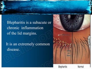
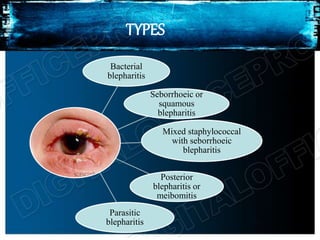
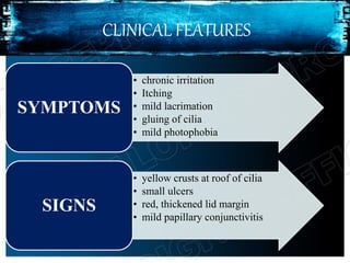
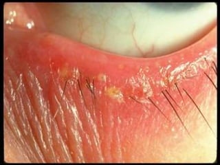
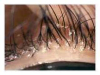
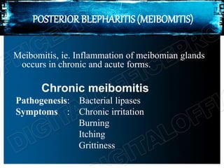
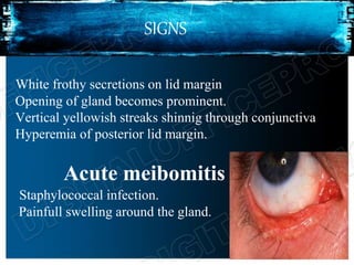
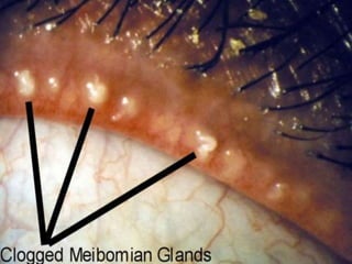
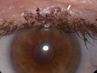
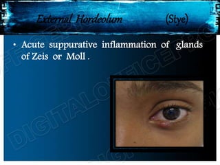
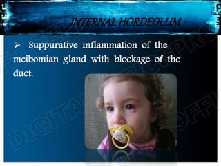
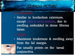
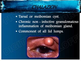
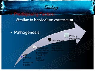
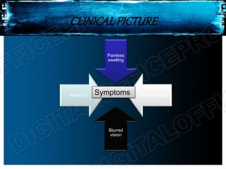
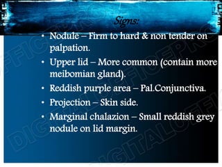
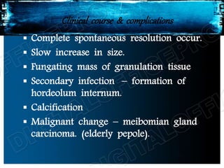
BLEPHARITIS
dr.T. KURINCHI, MS
Blepharitis is a subacute or
chronic inflammation
of the lid margins.
It is an extremely common
disease.
TYPES
Bacterial
blepharitis
Seborrhoeic or
squamous
blepharitis
Mixed staphylococcal
with seborrhoeic
blepharitis
Posterior
blepharitis or
meibomitis
Parasitic
blepharitis
BACTERIAL BLEPHARITIS
Also known as chronic anterior blepharitis or
staphylococcal blepharitis or ulcerative blepharitis
is a chronic infection of the anterior part of lid
margin.
ETIOLOGY
Causative organisms : staphylococci
streptococci
propionibacterium acnes
CLINICAL FEATURES
• chronic irritation
• Itching
• mild lacrimation
• gluing of cilia
• mild photophobia
SYMPTOMS
• yellow crusts at roof of cilia
• small ulcers
• red, thickened lid margin
• mild papillary conjunctivitis
SIGNS
COMPLICATIONS AND SEQUELAE
• Lash abnormalities like madarosis, trichiasis,
poliosis
• Tylosis, i.e. thickening and scaring of lid margins
• Eversion of punctum leading to epiphora
• Eczema of skin and ectropion
• Marginal keratitis
• Tear film instability
• Secondary inflammatory and mechanical changes.
TREATMENT
1. Lid hygiene
Warm compresses for 5-10mins
Crust removal and lid margin cleaning
2. Antibiotic eye ointment
Applied at lid margin after removal of crust.
Eye drops 3-4 times a day
Oral antibiotics like erythromycin or doxycycline
3. Topical steroids : fluoromethalon
4. Ocular lubricant ie artificial tear drops.
SEBORRHOEIC OR SQUAMOUS BLEPHARITIS
Anterior blepharitis with some spill over posteriorly.
ETIOLOGY
Seborrhea of scalp
SYMPTOMS
Whitish material at lid margin
Mild discomfort
Irritation
Falling of eyelashes
SIGNS
• Accumulation of white dandruff like scales on lid
margin
• Lashes fall out easily
• Lid margin thickned, posterior border rounded
leading to epiphora.
• Signs of bacterial blepharitis in patients with mixed
seborrhoeic and bacterial blepharitis.
TREATMENT
• Improvement of health and balanced diet
• Treatment of seborrhoea of scalp
• Removal of scales with lukewarm solution of 3%
soda bicarb or baby shampoo.
• Application of combined antibiotic and steroid eye
ointment at lid margin.
• Antibiotics
POSTERIOR BLEPHARITIS (MEIBOMITIS)
Meibomitis, ie. Inflammation of meibomian glands
occurs in chronic and acute forms.
Chronic meibomitis
Pathogenesis: Bacterial lipases
Symptoms : Chronic irritation
Burning
Itching
Grittiness
SIGNS
White frothy secretions on lid margin
Opening of gland becomes prominent.
Vertical yellowish streaks shinnig through conjunctiva
Hyperemia of posterior lid margin.
Acute meibomitis
Staphylococcal infection.
Painfull swelling around the gland.
Treatment of meibomitis
1. Lid hygiene
Warm compresses
Expression of secretions by vertical massage of lid.
2. Topical antibiotics and eye drops used 3-4 times a day.
3. Systemic tetracyclines
Doxycycline 100mg bdfor 1 week then od for 6-12wks
4. Ocular lubricants
5. Topical steroids like fluromethalon.
PARASITIC BLEPHARITIS
ETIOLOGY
Infestation of lashes by lice.
Phthiriasis palpebrum: infestation by
phthirus pubis (crab louse)
Pediculosis : infestation by pediculus
corporis (head louse)
CLINICAL FEATURES
SYMPTOMS
chronic
irritation
Itching
Burning
Mild
lacrimation
SIGNS
lid margins
red and
inflamed
lice on
lashes
nits (eggs)
seen on
base of cilia
conjunctival
congestion
TREATMENT
• Mechanical removal of lices and nits with
forceps
• Application of antibiotic ointmentsand yellow
mercuric oxide 1% to the lid margins and
lashes.
• Delousing of the patient , family members,
clothing and bedding is important to prevent
recurrences.
External Hordeolum (Stye)
• Acute suppurative inflammation of glands
of Zeis or Moll .
ETIOLOGY
Predisposing Factors :
• Common in children & young adults
• Patients with eye strain – muscle
imbalance / refractive errors
• Habitual rubbing of eyes
• Chronic blepharitis & DM
• Metabolic factors, ↑ intake of
carbohydrates & alcohol.
Causative Organism – Staph. aureus
CLINICALFEATURES
Symptoms
• Acute pain
• Swelling of lid
• Mild watering
Signs
• Stage of cellulitis :
Localised, firm, red, tender swelling
at lid margin with marked oedema.
• Stage of abscess :
Visible pus point on the lid margin
in relation to affected cilia.
TREATMENT
Hot compresses - 2-3 times a day
Evacuation of pus by pulling out the
infected cilia
Antibiotic eye drops – 3-4 times a
day & eye ointment – Bed time
Systemic anti inflammatory &
analgesis
Systemic antibiotics
INTERNALHORDEOLUM
 Suppurative inflammation of the
meibomian gland with blockage of the
duct.
ETIOLOGY:
• Prediposing Factors:
Similar to hordeolum externum.
• Causative Mechanism:
Occurs as:
 Primary staphylococcal infection of
meibomian gland
 Secondary infection in a chalazion.
CLINICALPICTURE
• Symptoms:
• Similar to hordeolum externum,
except pain is more intense, due to
swelling embedded in dense fibrous
tissue.
• Signs:
• Maximum tenderness & swelling away
from the lid margin.
• Pus usually points on the tarsal
conjunctiva.
TREATMENT
• Similar to externum
• When pus is formed – drained by vertical
incision from tarsal conjunctiva.
CHALAZION
 Tarsal or meibomian cyst.
 Chronic non - infective granulomatous
inflammation of meibomian gland.
 Commonest of all lid lumps.
Etiology
• Predisposing Factors:
Similar to hordeolum externaum
• Pathogenesis:
Mild
infection
of
meibomia
n gland
Proliferation
of epithelium
& infiltration
of wall of
ducts –
blocked.
Retention of
secretions (
sebum) in
the gland-
enlargemen
t.
Pent-up
secretions
(fatty in
nature)-
irritant &
excite non-
infective
lipogranulo
matous infl
of blocked
m.gland
CLINICALPICTURE
Painless
swelling
Mild heaviness
Blurred
vision
Watering Symptoms
Signs:
• Nodule – Firm to hard & non tender on
palpation.
• Upper lid – More common (contain more
meibomian gland).
• Reddish purple area – Pal.Conjunctiva.
• Projection – Skin side.
• Marginal chalazion – Small reddish grey
nodule on lid margin.
Clinical course & complications
 Complete spontaneous resolution occur.
 Slow increase in size.
 Fungating mass of granulation tissue
 Secondary infection – formation of
hordeolum internum.
 Calcification
 Malignant change – meibomian gland
carcinoma. (elderly pepole).
Conservative treatment – Hot
fomentation, topical antibiotic eye drops
& oral anti inflammatory drugs.
Intralesional injection of long acting
steroid (triamcinolone).
Diathermy
Oral tetracycline – Prophylaxis (if
ass.acne rosacea).
Incision & Currettage
Lid inflammation 17.08.16

Lid inflammation 17.08.16

  • 1.
  • 2.
    Blepharitis is asubacute or chronic inflammation of the lid margins. It is an extremely common disease.
  • 3.
    TYPES Bacterial blepharitis Seborrhoeic or squamous blepharitis Mixed staphylococcal withseborrhoeic blepharitis Posterior blepharitis or meibomitis Parasitic blepharitis
  • 4.
    BACTERIAL BLEPHARITIS Also knownas chronic anterior blepharitis or staphylococcal blepharitis or ulcerative blepharitis is a chronic infection of the anterior part of lid margin. ETIOLOGY Causative organisms : staphylococci streptococci propionibacterium acnes
  • 5.
    CLINICAL FEATURES • chronicirritation • Itching • mild lacrimation • gluing of cilia • mild photophobia SYMPTOMS • yellow crusts at roof of cilia • small ulcers • red, thickened lid margin • mild papillary conjunctivitis SIGNS
  • 7.
    COMPLICATIONS AND SEQUELAE •Lash abnormalities like madarosis, trichiasis, poliosis • Tylosis, i.e. thickening and scaring of lid margins • Eversion of punctum leading to epiphora • Eczema of skin and ectropion • Marginal keratitis • Tear film instability • Secondary inflammatory and mechanical changes.
  • 8.
    TREATMENT 1. Lid hygiene Warmcompresses for 5-10mins Crust removal and lid margin cleaning 2. Antibiotic eye ointment Applied at lid margin after removal of crust. Eye drops 3-4 times a day Oral antibiotics like erythromycin or doxycycline 3. Topical steroids : fluoromethalon 4. Ocular lubricant ie artificial tear drops.
  • 9.
    SEBORRHOEIC OR SQUAMOUSBLEPHARITIS Anterior blepharitis with some spill over posteriorly. ETIOLOGY Seborrhea of scalp SYMPTOMS Whitish material at lid margin Mild discomfort Irritation Falling of eyelashes
  • 10.
    SIGNS • Accumulation ofwhite dandruff like scales on lid margin • Lashes fall out easily • Lid margin thickned, posterior border rounded leading to epiphora. • Signs of bacterial blepharitis in patients with mixed seborrhoeic and bacterial blepharitis.
  • 12.
    TREATMENT • Improvement ofhealth and balanced diet • Treatment of seborrhoea of scalp • Removal of scales with lukewarm solution of 3% soda bicarb or baby shampoo. • Application of combined antibiotic and steroid eye ointment at lid margin. • Antibiotics
  • 13.
    POSTERIOR BLEPHARITIS (MEIBOMITIS) Meibomitis,ie. Inflammation of meibomian glands occurs in chronic and acute forms. Chronic meibomitis Pathogenesis: Bacterial lipases Symptoms : Chronic irritation Burning Itching Grittiness
  • 14.
    SIGNS White frothy secretionson lid margin Opening of gland becomes prominent. Vertical yellowish streaks shinnig through conjunctiva Hyperemia of posterior lid margin. Acute meibomitis Staphylococcal infection. Painfull swelling around the gland.
  • 16.
    Treatment of meibomitis 1.Lid hygiene Warm compresses Expression of secretions by vertical massage of lid. 2. Topical antibiotics and eye drops used 3-4 times a day. 3. Systemic tetracyclines Doxycycline 100mg bdfor 1 week then od for 6-12wks 4. Ocular lubricants 5. Topical steroids like fluromethalon.
  • 17.
    PARASITIC BLEPHARITIS ETIOLOGY Infestation oflashes by lice. Phthiriasis palpebrum: infestation by phthirus pubis (crab louse) Pediculosis : infestation by pediculus corporis (head louse)
  • 18.
  • 19.
    SIGNS lid margins red and inflamed liceon lashes nits (eggs) seen on base of cilia conjunctival congestion
  • 21.
    TREATMENT • Mechanical removalof lices and nits with forceps • Application of antibiotic ointmentsand yellow mercuric oxide 1% to the lid margins and lashes. • Delousing of the patient , family members, clothing and bedding is important to prevent recurrences.
  • 23.
    External Hordeolum (Stye) •Acute suppurative inflammation of glands of Zeis or Moll .
  • 24.
    ETIOLOGY Predisposing Factors : •Common in children & young adults • Patients with eye strain – muscle imbalance / refractive errors • Habitual rubbing of eyes • Chronic blepharitis & DM • Metabolic factors, ↑ intake of carbohydrates & alcohol. Causative Organism – Staph. aureus
  • 25.
    CLINICALFEATURES Symptoms • Acute pain •Swelling of lid • Mild watering
  • 26.
    Signs • Stage ofcellulitis : Localised, firm, red, tender swelling at lid margin with marked oedema. • Stage of abscess : Visible pus point on the lid margin in relation to affected cilia.
  • 27.
    TREATMENT Hot compresses -2-3 times a day Evacuation of pus by pulling out the infected cilia Antibiotic eye drops – 3-4 times a day & eye ointment – Bed time Systemic anti inflammatory & analgesis Systemic antibiotics
  • 28.
    INTERNALHORDEOLUM  Suppurative inflammationof the meibomian gland with blockage of the duct.
  • 29.
    ETIOLOGY: • Prediposing Factors: Similarto hordeolum externum. • Causative Mechanism: Occurs as:  Primary staphylococcal infection of meibomian gland  Secondary infection in a chalazion.
  • 30.
    CLINICALPICTURE • Symptoms: • Similarto hordeolum externum, except pain is more intense, due to swelling embedded in dense fibrous tissue. • Signs: • Maximum tenderness & swelling away from the lid margin. • Pus usually points on the tarsal conjunctiva.
  • 31.
    TREATMENT • Similar toexternum • When pus is formed – drained by vertical incision from tarsal conjunctiva.
  • 32.
    CHALAZION  Tarsal ormeibomian cyst.  Chronic non - infective granulomatous inflammation of meibomian gland.  Commonest of all lid lumps.
  • 33.
    Etiology • Predisposing Factors: Similarto hordeolum externaum • Pathogenesis: Mild infection of meibomia n gland Proliferation of epithelium & infiltration of wall of ducts – blocked. Retention of secretions ( sebum) in the gland- enlargemen t. Pent-up secretions (fatty in nature)- irritant & excite non- infective lipogranulo matous infl of blocked m.gland
  • 34.
  • 35.
    Signs: • Nodule –Firm to hard & non tender on palpation. • Upper lid – More common (contain more meibomian gland). • Reddish purple area – Pal.Conjunctiva. • Projection – Skin side. • Marginal chalazion – Small reddish grey nodule on lid margin.
  • 36.
    Clinical course &complications  Complete spontaneous resolution occur.  Slow increase in size.  Fungating mass of granulation tissue  Secondary infection – formation of hordeolum internum.  Calcification  Malignant change – meibomian gland carcinoma. (elderly pepole).
  • 37.
    Conservative treatment –Hot fomentation, topical antibiotic eye drops & oral anti inflammatory drugs. Intralesional injection of long acting steroid (triamcinolone). Diathermy Oral tetracycline – Prophylaxis (if ass.acne rosacea).
  • 38.