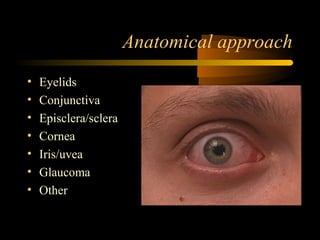
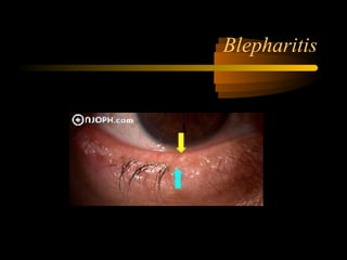
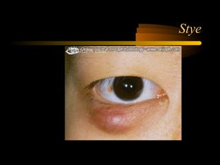
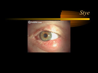
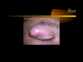
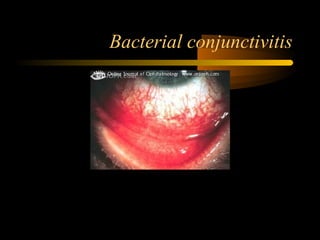
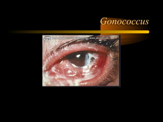
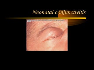
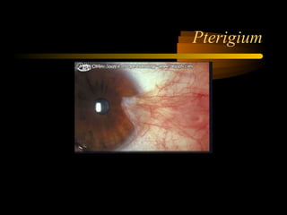
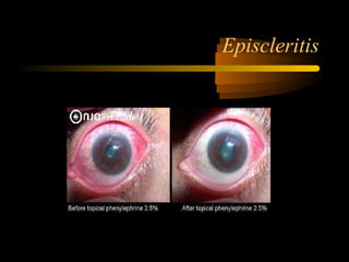
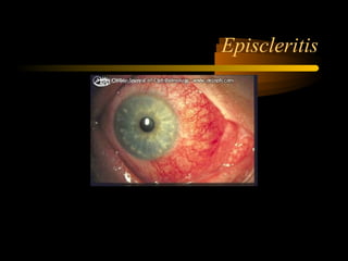
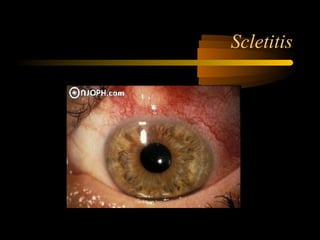
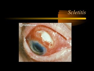
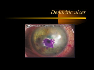
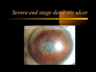
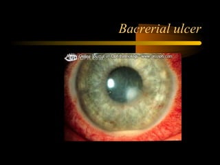
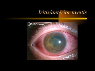
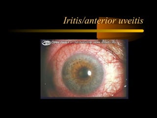
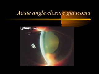
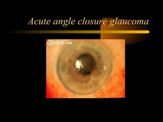
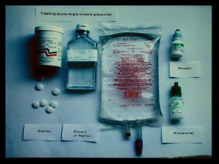
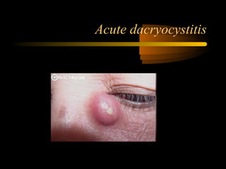
The Red Eye
Dr James Beatty
Overview
• Usually a self limiting benign disorder
• May be sight threatening
• May be the sentinel of a severe underlying
systemic disease
• Simple history and external examination
will help form a narrow differential
diagnosis
Guidelines for early referral
• Decreased vision
• Painful eye (not just discomfort)
• Unilateral red eye
• Poor response to initial therapy
Take a basic history
• Vision
• Symptoms
– Redness
– Pain
– Discharge
Anatomical approach
• Eyelids
• Conjunctiva
• Episclera/sclera
• Cornea
• Iris/uvea
• Glaucoma
• Other
The Eyelids
• Blepharitis
– Common, chronic, bilateral
– Staphylococcal or seborrhea
• Symptoms (burning, grittiness, photophobia, dry
eye, no loss of vision)
• Signs (crusty, hyperemia, irregular lid)
• Treatment (lid scrubs, O.Fucithalmic, warm
compress, tear replacement)
Blepharitis
Blepharitis
The Eyelids
• Stye or acute chalazion
– Red mass on the eyelid
– May respond to hot compress + oral and topical
antibiotics
– Move quickly to I & D if no improvement
Acute chalazion
Stye
Stye
Stye
The Conjunctiva
• Conjunctivitis
– Allergic
• Seasonal, perennial, vernal, contact
– Infective
• Viral
• Bacterial
Allergic
• Seasonal often with hayfever
• Perennial throughout the year
• Vernal often with atopy
• Bilateral and watery
• Itchy
• No decrease in VA or significant pain
• Rx remove allergen, antihistamine plus mast cell
stabalizer occasionally steroids
Allergic conjunctivitis
Vernal conjunctivitis
Viral conjunctivitis
• Usually adenovirus
• Contagious
• Occurs in epidemics
• May have flu-like symptoms
• O/E: Lymph nodes,VA fine, Follicles +- Hx
• Rx: Self limiting, relief of symptoms
Viral Conjunctivitis
Bacterial conjunctivitis
• Usually one eye
• Common in children
• O/E: Purulent discharge, eyelids stuck
together, VA is fine
• Rx: usually self limiting, antibacterial oint.
Bacterial conjunctivitis
Gonococcus
Neonatal conjunctivitis
Pterigium
• Localized area of redness
• Palperable fissure
• Active and inactive phases
• Rx: lubricants, dark glasses, surgery
• Recurrences fairly common
Pterigium
Subconjunctival haemorrhage
Episcleritis
• Idiopathic, self limiting, focal inflammation
• Usually young adults often recurrent
• O/E: unilateral area of redness, no
decreased VA, some discomfort
• Self limiting 2 weeks
• NSAID’s, lubricants
Episcleritis
Episcleritis
Scletitis
• Very painful
• Unilateral
• Sectoral, nodular, diffuse
• Necrotising or non necrotising with or without
inflammation
• VA may be decreased
• Systemic association in 50 % (RA, Sarcoid, SLE,
Zoster, Wegners)
• Needs further investigation
• Rx: Oral NSAID, Steroids, Antimetabolites
Scletitis
Scletitis
Cornea
• Very well supplied by pain receptors
• Fluorescein very helpful to identify
epithelial defects
• Trauma common
Abrassion
Arc eyes
• Arc welding without visor or light filter (intense
UV light)
• Extremely painful and photophobic
• Self limiting
• O/E:Need local for exam
• Multiple small punctate burns with Fluoroscein
• Rx: antibacterial oint, cold compress, analgesia,
NSAID drops +- cycloplegic
Foreign body
• Grinding
• Remove with local and sterile needle or
cotton bud
• Then patch and antibacterial ointment
• Check under lids
• If residual material refer ophthalmologist
Foreign body
Rust Ring
Corneal ulcers
• An ophthalmic emergency, refer
ophthalmologist ASAP
• Painful, red, decreased vision
• Staining with flouroscein
Viral ulcer
• Hepes simplex
• Recurrent, often a history of oral/nasal
herpes
• Typical branching/dendritic staining pattern
• Steroids a big no no!
• When treating a red painful eye of unknown
cause, steroids should be avoided!
• Rx: Acyclovir
Dendritic ulcer
Severe end stage dendritic ulcer
Bacterial and Fungal ulcers
• Even more sight threatening
• White or yellowish
• Stain with flourescein
• Painful with decreased vision
• Often contact lens wearers/trauma
• May need microscopy and cultures
Bacrerial ulcer
Iritis/anterior uveitis
• Inflammation of the uvea is known as
uveitis
• Anterior uveitis common, but many other
types of uveitis
• Very often recurrent and idiopathic
• May be associated with systemic disease
(collagen vascular, sarcoid, syphalis, TB)
• Often following blunt trauma
Iritis/anterior uveitis
• Symptoms: photophobia, pain, decreased
VA
• O/E: uni or bilateral, circumcorneal
injection, synechia, hypopeon
• Lots of long term complications (glaucoma,
cataracts)
• Rx: steroids and atropine
Iritis/anterior uveitis
Acute angle closure glaucoma
• Glaucoma is usually chronic, painless with
loss of vision in the late stages
• Angle closure glaucoma is acute, painful,
sight threatening.
• An emergency…refer ophthalmologist
ASAP
Acute angle closure glaucoma
• Peripheral iris blocks the trabecular meshwork
• Aqueous unable to drain
• Very high pressure (>40mmHg)
• Symptoms: severe pain, headache, nausea,
vomiting, decreased vision
• O/E: red eye, cloudy cornea, pupil non responsive,
eye is hard on palpation, eclipse sign
Acute angle closure glaucoma
• Rx: reduce the pressure with
– Diamox
– Oral glycerol
– Topical pressure lowering drops
– Miotics/pilocarpine
– Oral analgesia and aniemetics
– Peripheral iridectomies when clear cornea and
lower pressure
Other
Acute dacryocystitis
Orbital cellulitis
Graves or Thyroid eye disease
Trauma
Trauma
Trauma
Conclusion
• Short history and exam usually determines
the cause
• Think anatomically
• Refer early if in doubt, especially if there is
severe pain, corneal staining or decreased
VA
• Be careful of using steroids!
• Thank you

The Red Eye

  • 1.
    The Red Eye DrJames Beatty
  • 2.
    Overview • Usually aself limiting benign disorder • May be sight threatening • May be the sentinel of a severe underlying systemic disease • Simple history and external examination will help form a narrow differential diagnosis
  • 3.
    Guidelines for earlyreferral • Decreased vision • Painful eye (not just discomfort) • Unilateral red eye • Poor response to initial therapy
  • 4.
    Take a basichistory • Vision • Symptoms – Redness – Pain – Discharge
  • 5.
    Anatomical approach • Eyelids •Conjunctiva • Episclera/sclera • Cornea • Iris/uvea • Glaucoma • Other
  • 6.
    The Eyelids • Blepharitis –Common, chronic, bilateral – Staphylococcal or seborrhea • Symptoms (burning, grittiness, photophobia, dry eye, no loss of vision) • Signs (crusty, hyperemia, irregular lid) • Treatment (lid scrubs, O.Fucithalmic, warm compress, tear replacement)
  • 7.
  • 8.
  • 9.
    The Eyelids • Styeor acute chalazion – Red mass on the eyelid – May respond to hot compress + oral and topical antibiotics – Move quickly to I & D if no improvement
  • 10.
  • 11.
  • 12.
  • 13.
  • 14.
    The Conjunctiva • Conjunctivitis –Allergic • Seasonal, perennial, vernal, contact – Infective • Viral • Bacterial
  • 15.
    Allergic • Seasonal oftenwith hayfever • Perennial throughout the year • Vernal often with atopy • Bilateral and watery • Itchy • No decrease in VA or significant pain • Rx remove allergen, antihistamine plus mast cell stabalizer occasionally steroids
  • 16.
  • 17.
  • 18.
    Viral conjunctivitis • Usuallyadenovirus • Contagious • Occurs in epidemics • May have flu-like symptoms • O/E: Lymph nodes,VA fine, Follicles +- Hx • Rx: Self limiting, relief of symptoms
  • 19.
  • 20.
    Bacterial conjunctivitis • Usuallyone eye • Common in children • O/E: Purulent discharge, eyelids stuck together, VA is fine • Rx: usually self limiting, antibacterial oint.
  • 21.
  • 22.
  • 23.
  • 24.
    Pterigium • Localized areaof redness • Palperable fissure • Active and inactive phases • Rx: lubricants, dark glasses, surgery • Recurrences fairly common
  • 25.
  • 26.
  • 27.
    Episcleritis • Idiopathic, selflimiting, focal inflammation • Usually young adults often recurrent • O/E: unilateral area of redness, no decreased VA, some discomfort • Self limiting 2 weeks • NSAID’s, lubricants
  • 28.
  • 29.
  • 30.
    Scletitis • Very painful •Unilateral • Sectoral, nodular, diffuse • Necrotising or non necrotising with or without inflammation • VA may be decreased • Systemic association in 50 % (RA, Sarcoid, SLE, Zoster, Wegners) • Needs further investigation • Rx: Oral NSAID, Steroids, Antimetabolites
  • 31.
  • 32.
  • 33.
    Cornea • Very wellsupplied by pain receptors • Fluorescein very helpful to identify epithelial defects • Trauma common
  • 34.
  • 35.
    Arc eyes • Arcwelding without visor or light filter (intense UV light) • Extremely painful and photophobic • Self limiting • O/E:Need local for exam • Multiple small punctate burns with Fluoroscein • Rx: antibacterial oint, cold compress, analgesia, NSAID drops +- cycloplegic
  • 36.
    Foreign body • Grinding •Remove with local and sterile needle or cotton bud • Then patch and antibacterial ointment • Check under lids • If residual material refer ophthalmologist
  • 37.
  • 38.
  • 39.
    Corneal ulcers • Anophthalmic emergency, refer ophthalmologist ASAP • Painful, red, decreased vision • Staining with flouroscein
  • 40.
    Viral ulcer • Hepessimplex • Recurrent, often a history of oral/nasal herpes • Typical branching/dendritic staining pattern • Steroids a big no no! • When treating a red painful eye of unknown cause, steroids should be avoided! • Rx: Acyclovir
  • 41.
  • 42.
    Severe end stagedendritic ulcer
  • 43.
    Bacterial and Fungalulcers • Even more sight threatening • White or yellowish • Stain with flourescein • Painful with decreased vision • Often contact lens wearers/trauma • May need microscopy and cultures
  • 44.
  • 45.
    Iritis/anterior uveitis • Inflammationof the uvea is known as uveitis • Anterior uveitis common, but many other types of uveitis • Very often recurrent and idiopathic • May be associated with systemic disease (collagen vascular, sarcoid, syphalis, TB) • Often following blunt trauma
  • 46.
    Iritis/anterior uveitis • Symptoms:photophobia, pain, decreased VA • O/E: uni or bilateral, circumcorneal injection, synechia, hypopeon • Lots of long term complications (glaucoma, cataracts) • Rx: steroids and atropine
  • 47.
  • 48.
  • 49.
    Acute angle closureglaucoma • Glaucoma is usually chronic, painless with loss of vision in the late stages • Angle closure glaucoma is acute, painful, sight threatening. • An emergency…refer ophthalmologist ASAP
  • 50.
    Acute angle closureglaucoma • Peripheral iris blocks the trabecular meshwork • Aqueous unable to drain • Very high pressure (>40mmHg) • Symptoms: severe pain, headache, nausea, vomiting, decreased vision • O/E: red eye, cloudy cornea, pupil non responsive, eye is hard on palpation, eclipse sign
  • 51.
  • 52.
  • 54.
    Acute angle closureglaucoma • Rx: reduce the pressure with – Diamox – Oral glycerol – Topical pressure lowering drops – Miotics/pilocarpine – Oral analgesia and aniemetics – Peripheral iridectomies when clear cornea and lower pressure
  • 55.
  • 56.
  • 57.
  • 58.
    Graves or Thyroideye disease
  • 59.
    Graves or Thyroideye disease
  • 60.
  • 61.
  • 62.
  • 63.
    Conclusion • Short historyand exam usually determines the cause • Think anatomically • Refer early if in doubt, especially if there is severe pain, corneal staining or decreased VA • Be careful of using steroids!
  • 64.

Editor's Notes

  • #2 Try and give you is an approach Feel free to stop and ask questions Thousands of different causes
  • #3 Try not to do something that makes it worse On the basis of this a decision can be made for early referral Special investigations are seldom needed before management is instituted
  • #4 Decreased vision something in the eye or problem with the cornea The more of these features present the earlier one should refer
  • #5 Vision…….how long acute or chronic, both eyes or one eye Redness………pattern Pain…nil or wakes at night Discharge………….watery, thick and purulent, color
  • #6 All you need is a snellen chart, ophthalmoscope, LA flourescein 2fingers Lacrimal system problem lacrimal gland adenitis or Dacryocystitis Orbital problem……Orbital cellulitis or idiopathic orbital inflammatory disease or thyroid eye disease Trauma
  • #7 Worse in the morning characterized by remissions and exacerbations Usually older patients Oral tetracyclines…..doxycycline 100mg bd 1 week then 100mg daily for 6 weeks
  • #15 Allergic: bilateral, watery, no decrease in va or significant pain Rx remove allergen, antihistamine plus mast cell stabalizer occasionally steroids
  • #16 Rellestat
  • #19 NSAID such as voltared, lubricants………..steroids ophthalmological supervision
  • #21 Mucopurulent discharge, corneal ulceration…hospital topical and systemic antibiotics
  • #23 Neisseria gonorroea, witch doctors using urine to try and treat eye problems Able to attach an intact cornea Rx wash out, topical antibiotics and IV (cefotaxime 1g bd few days) treat the partner
  • #24 <5 days gonococcus Ceftriaxone 125mg imi stat irrigation chloro >5days non gonococcal erythromycin 2,5ml6hrly chloro
  • #44 Pseudomonus acanthoemba
  • #45 In contrast fungal ulcer: vegetable matter, satelite lesions, doesn’t respond well When in doubt……corneal scrape for MC and S
  • #49 Clues for systemic causes are bilateral, granulomatus, posterios