SlideShare a Scribd company logo
1 of 61
DR. V SRI HARISH
The diffuse parenchymal lung diseases (DPLDs), also
called interstitial lung diseases (ILDs), are a
heterogeneous group of rare disorders that cause
expansion of the interstitial compartment by varying
degrees of inflammation and fibrosis, resulting in
parenchymal damage.
 Among the 300-plus interstitial lung diseases, the
most important are sarcoidosis, idiopathic pulmonary
fibrosis, extrinsic allergic alveolitis, interstitial lung
disease as a feature of connectivetissuedisease,drug-
induced interstitial lung disease and pneumoconiosis.
 Diffuse parenchymal lung diseases (DPLD) are
classified into four groups:
 (1) DPLD of known causes;
 (2) idiopathic interstitial pneumonias (IIP);
 (3) granulomatous DPLD and
 (4) DPLD with well defined clinicopathologic features
 Major ILDs of known aetiology (~35% of all patients
with ILDs) :
 Pneumoconioses (e.g. asbestosis, silicosis)
 Extrinsic allergic alveolitis (hypersensitivity pneumonitis)
 Drug toxicity.
 Associated with collagen vascular diseases.
 Major ILDs of unknown aetiology (~65% of all patients with ILDs)
 Idiopathic interstitial pneumonias :
 Idiopathic pulmonary fibrosis with a histopathological pattern of usual
interstitial pneumonia (~55% of IIPs)
 Nonspecific interstitial pneumonia (~25% of IIPs)
 Respiratory bronchiolitis ILD, in smokers (~10% of IIPs)
 Desquamative interstitial pneumonia (~5% of IIPs)
 Cryptogenic organising pneumonia (~3% of IIPs)
 Lymphoid interstitial pneumonia (~1% of IIPs)
 Acute interstitial pneumonia (~1% of IIPs)
 Sarcoidosis is a multisystem granulomatous disease of
unknown etiology characterized histologically by
noncaseating granulomas that may progress to fibrosis.
 Pulmonary manifestations are present in 90% of patients.
Systemic symptoms such as fatigue, night sweats and
weight loss are common.
 Loefgren's syndrome, an acute presentation of sarcoidosis,
consists of arthritis, erythema nodosum, bilateral hilar
adenopathy and occurs in 9-34% of patients.
 Erythema nodosum is seen predominantly in women and
arthritis is more common in men.
 There are five radiologic stages of intrathoracic abnormalities:
 Stage 0: Normal chest radiograph
 Stage 1: Mediastinal and hilar lymphadenopathy
 Stage 2: Lymphadenopathy with pulmonary parenchymal lesions
 Stage 3: Pulmonary parenchymal lesions in the absence of hilar
lymphadenopathy
 Stage 4: Significant lung fibrosis with architectural distortion or bullae
 The main utility of On follow-up,
 These stages do not indicate disease chronicity or correlate with changes in
pulmonary function.65 percent of the
 HRCT findings in Sarcoidosis.
 Common findings:
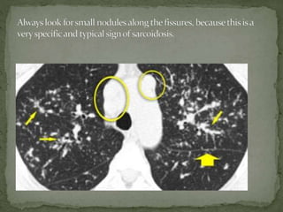
 Small nodules in a perilymphatic distribution (i.e. along
subpleural surface and fissures, along interlobular septa
and the peribronchovascular bundle).
 Upper and middle zone predominance.
 Lymphadenopathy in left hilus, right hilus and
paratracheal (1-2-3 sign). mediastinal lymphadenopathy in
the paratracheal, aortopulmonary window and subcarinal
region. 20 % show egg shell calcifications.
STAGE 1
 Uncommon findings:
 Conglomerate masses in a perihilar location.
 Larger nodules (> 1cm in diameter, in Grouped nodules
or coalescent nodlues surrounded by multiple satellite
nodules (Galaxy sign)
 Nodules so small and dense that they appear as ground
glass or even as consolidations (alveolar sarcoidosis)
 Fibrosis in Sarcoidosis.
Progressive fibrosis in sarcoidosis may lead to
peribronchovascular (perihilar) conglomerate masses of fibrous
tissue.

The typical location is posteriorly in the upper lobes, leading
to volume loss of the upper lobes with displacement of the
interlobar fissure.
 Other diseases that commonly result in this appearance are:
 Silicosis
 Tuberculosis
 Talcosis
 Alveolar sarcoidosis :
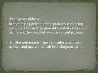
 In about 15–25 percent of the patients, coalescing
granulomas form large mass-like nodules (1–4 cm in
diameter), the so-called ‘alveolar sarcoid pattern,
 Unlike metastasis, these nodules are poorly
defined and may contain air bronchogram within.
 Differential diagnosis of Sarcoidosis.
 Lymphadenopathy:
 Primary TB: asymmetrical adenopathy , non discreate, central
necrosis.
 Histoplasmosis
 Lymphoma ( in sarcoidosis with 2 yr lymph nodes regress
then parenchymal opacticies develop. )
 Small cell lung cancer with nodal metastases
 Nodular pattern:
 Silicosis / Pneumoconiosis: predominantly centrilobular and
subpleural nodules.
 Miliary TB: random nodules.
 Fibrotic pattern:
 Usual Interstitial Pneumonia (UIP): basal and peripheral
fibrosis, honeycombing.
 Chronic Hypersensitivity Pneumonitis: mid zone fibrosis
with mosaic pattern.
 Tuberculosis (more unilateral).
some diseases with a nodular pattern.
 Hypersensitivity pneumonitis: ill defined centrilobular
nodules.
 Miliary TB: random nodules of the same size.
 Sarcoidosis: nodules with perilymphatic distribution, along
fissures, adenopathy.
 Hypersensitivity pneumonitis: centrilobular nodules,
notice sparing of the subpleural area.
 Sarcoidosis: nodules with perilymphatic distribution, along
fissures, adenopathy.
 TB: Tree-in-bud appearance in a patient with active TB.
 Langerhans cell histiocytosis: early nodular stage before the
typical cysts appear.
 Respiratory bronchiolitis: ill defined centrilobular nodules
of ground-glass opacity.
 Extrinsic allergic alveolitis is an immunologic pulmonary
disorder associated with the inhalation of one of the antigenic
organic dusts.
 Etiologic agents have been implicated, including many
thermophilic bacteria, true fungi, and various animal proteins.
 Some of the more common disease entities include
- farmer’s lung(exposure to moldy hay),
- humidifier lung (exposure to water contaminated by
thermophilic bacteria),
-birdfancier’slung (exposure to avian proteins in feathers and
excreta)
 Acute phase :
 The chest radiograph may be normal early in the acute
stage of disease.
 Within hours, fine nodular or ground glass opacities
develop, most often in the lower lobes; progressive
airspace opacifi cation may simulate pulmonary edema.
 Within hours to days, the opacities resolve and the chest
radiograph becomes normal.
 With continued or repeated exposures, the chest
radiograph will remain abnormal between acute episodes.
 Subacute phase-
 Ill-defined centrilobular nodules of ground-glass
opacity (80% of cases) or
 mosaic pattern of a combination of patchy ground-
glass opacity due to lung infiltration and
patchy lucency due to bronchiolitis with air trapping
 Chronic phase :
 Mosaic pattern with areas of ground-glass atenuation and areas
of low attenuation.
 Fibrosis and parenchymal distortion in a mid zone and
upper zone distribution with interlobular and intralobular
interstitial thickening, honeycombing, and traction
bronchiectasis.
 Appear as diffuse coarse reticular or reticulonodular opacities in
the midlung and upper lung zones;
 The diagnosis of hypersensitivity pneumonitis should be
considered when repeated episodes of rapidly changing ground
glass or airspace opacifi cation are seen in a patient with
underlying coarse interstitial lung disease.
 Hilar or mediastinal lymph node enlargement and pleural
effusion are uncommon findings in patients with
hypersensitivity pneumonitis
 Differential diagnosis of Hypersensitivity
Pneumonitis
 Subacute stage:
 RB-ILD: seen in smokers, upper lobe predilection,
usually associated with centrilobular emphysema.
 Alveolar proteinosis.
 Chronic stage:
 UIP: may show very similar HRCT findings.
UIP has a strong lower zone distribution.
In chronic HP fibrotic changes are typically seen
throughout the whole lung parenchyma from the
periphery towards the centrum.
 Centrilobular nodules and airtrapping is not seen in UIP.
 The term pneumoconiosis is used to describe the
nonneoplastic reaction of the lungs to inhaled
inorganic dust particles.
 Silicosis and CWP occur in a specific patient group
(construction workers, mining workers, workers
exposed to sandblasting, glass blowing and pottery).
 The radiographic and HRCT appearances of these
diseases, however, may not be distinguishable from
each other .
 HRCT findings in Silicosis/CWP
 Small well-defined nodules of 2 to 5mm in diameter in
both lungs.
 Upper lobe predominance
 Nodules may be calcified
 Centrilobular and subpleural distribution
 Sometimes random distribution
 Irregular conglomerate masses, known as progressive
massive fibrosis (upper , posterior , b/l )
 Masses may cavitate due to ischemic necrosis.
 Often hilar and mediastinal lymphnodes.
 Differential diagnosis of Silicosis / Pneumoconiosis.
 Sarcoidosis : can be difficult to distinguish (look for distribution
of nodules).
 Infection: miliairy TB, fungus.
 Hematogenous metastases: silicotic nodules in subpleural and
peribronchiolar location up to the level of the secundary
pulmonary lobule, may have a seemingly random distribution
and simulate metastases and miliary infections.
 Langerhans cell histiocytosis: can be difficult to distinguish from
silicosis in the early stage, when LCH is solely characterized by
the presence of small nodules. Look for nodules with cavitation.
 Collagen vascular diseases that show features of
interstitial lung disease include systemic lupus
erythematosus, rheumatoid arthritis, progressive
systemic sclerosis, polymyositis and dermatomyositis,
Sjögren syndrome and mixed connective tissue
disease.
 At histopathologic examination, these are diverse and
include usual interstitial pneumonia, nonspecific
interstitial pneumonia, COP, diffuse alveolar damage,
lymphocytic interstitial pneumonia, and apical
fibrosis.
 HRCT has proved to be very sensitive in the detection
and characterization of various histo pathologically
confirmed interstitial lung diseases.
 There is evidence that the pattern of abnormality at
HRCT reflects the relative proportions of fibrosis and
inflammation.
 A reticular pattern with traction bronchiectasis at CT
is associated with a predominantly fibrotic process,
 whereas ground-glass attenuation without a reticular
pattern or traction bronchiectasis is associated with an
inflammatory process.
 MANIFESTATIONS
 Serositis- Pleuritis Pleural effusion, thickening ,Pericarditis
Pericardial effusion
 Interstitial pneumonitis Pulmonary fibrosis (basilarpredominance)
 Necrobiotic nodules Multiple peripheral cavitatingnodules
 Caplan syndrome Multiple peripheral cavitating nodules
 Pulmonary arteritis Pulmonary arterial hypertension ,and right heart
enlargement Pulmonary hemorrhage
 The most common radiographic manifestation of
parenchymal lung involvement is an interstitial
pneumonitis and fibrosis, which histologically is a
form of UIP.
 This begins as an alveolitis (infl ammation of the
alveolar interstitium) that is seen radiographically as
fine reticular or ground glass opacities with a
lower zone predominance.
 There is gradual progression to end-stage pulmonary
fibrosis with the development of a bibasilar medium or
coarse reticular or reticulonodular pattern
(honeycombing)
 Less common parenchymal manifestations of
rheumatoid disease are lung nodules and changes
attributable to COP.
 Necrobiotic (rheumatoid) nodules in the lung can
produce peripheral well-defi ned nodular opacities on
chest radiographs.
 Similar nodules may develop in the lungs of coal
miners and silica or asbestos workers with rheumatoid
arthritis as a hypersensitivity response to inhaled dust
particles (Caplan syndrome)
 The most common radiologic abnormality is pleural
effusion(25 to 35%). Pericardial effusion is seen in
approximately 20 %.
 Pulmonary manifestations of comprise both acute and
chronic lesions.
 Acute disease includes pulmonary hemorrhage, acute
lupus pneumonitis, and pulmonary edema.
 Chronic disease, such as interstitial pneumonitis and
fibrosis, is less common than in other connective
tissue disorders.
 Ground-glass attenuation and consolidation at high-
resolution CT may reflect the presence of interstitial
pneumonitis and fibrosis, acute lupus pneumonitis.
 Radiographic evidence of interstitial fibrosis,
consisting of a reticular pattern that involves mainly
the lower lung zones, include inter lobularseptal
thickening (33%), irregular linear hyperattenuating
areas(33%), and architectural distortion (22%).
 Honey combing is very rare. So radiologic evidence of
UIP is uncommon.
 Pulmonary involvement is more common and more
severe in systemic sclerosis than in other types of
collagen vascular disease.
 The most common pulmonary manifestation is
interstitial fibrosis, which occurs in approximately
80percent of patients.
 The histological features are those of NSIP.
 The most common thin-section CT fi ndings are ground
glass opacities, reticulation and eventually traction
bronchiectasis in a lower zone, subpleural distribution.
 Pulmonary arterial hypertension with enlarged central
pulmonary arteries and RV dilatation is seen in up to 50%
of patients with scleroderma and may be seen in the
absence of interstitial fi brosis.
 Pleural effusions are signifi cantly less common in
scleroderma than in rheumatoid disease or SLE.
 A dilated air-filled esophagus may be identifi ed on the
upright chest radiograph and is a manifestation of
esophageal dysmotility from smooth muscle atrophy.
 This autoimmune disorder of middleaged women is
characterized by the sicca syndrome
keratoconjunctivitis sicca ,xerostomia], and xerorhinia
which results from lymphocytic infiltration.
 The most common manifestation is
interstitial fibrosis, which is indistinguishable from
that seen with other collagen vascular disorders.
 Involvement of tracheobronchial mucous glands leads
to thickened sputum with mucus pluggingand
recurrent bronchitis, bronchiectasis, atelectasis.
 CT demonstrates both interstitial opacities and the
presence of small airways involvement with
bronchiolectasis and a “tree-in-bud” appearance.
 Pleuritis and pleural effusion are less common.
 Patients with Sjögren syndrome are at increased risk for
developing lymphocytic interstitial pneumonitis (LIP) and
non-Hodgkin pulmonary lymphoma.
 The radiographic appearance of LIP is lower lobe coarse
reticular or reticulonodular opacities that are
indistinguishable from interstitial fi brosis.
 Thin-section CT shows ground glass opacity with scattered,
thin-walled cysts.
 Drug-induced lung disease is a major source of
iatrogenic lung injury.
 The major diagnostic problem is, that it may present
with a large variety of radiologic patterns.
 It may present as organizing pneumonia, eosinophilic
pneumonia, fibrosis, hypersensitivity pneumonitis or
even as ARDS.
 The diagnosis of drug-induced pulmonary disease is
usually one of exclusion.
 Drug-induced organizing pneumonia is commonly
caused by Bleomycin and Cyclophosphamide and
other drugs like Methotrexate, Amiodarone,
Nitrofurantoin and Penicillamine .
 Diffuse alveolar damage most common pattern
 The initial manifestation is pulmonary edema, frequently in a
geographic or nondependent distribution without much
associated pleural fl uid or interlobular septal thickening.
 After discontinuation of the offending drug, it may resolve,
stabilize or progress to fibrosis, frequently in an UIP pattern .
 Drugs that commonly cause diffuse alveolar damage include
 chemotherapeutic agents
 busulphan, bleomycin, cyclophosphamide,
 gold salts, mitomycin, and melphalan.
 Opiates
 UIP can be the result of diffuse alveolar damage or
occur as a result of chronic drug toxicity.
 The drugs most commonly implicated in this form of lung
disease are amiodarone, nitrofurantoin, and the
chemotherapeutic agents cyclophosphamide, bleomycin,
and methotrexate.
 Radiographically, patients present with bilateral,
predominantly lower lobe, coarse reticular and linear
opacities with diminished lung volumes.
 NSIP (also referred to as chronic interstitial
pneumonia when known to result from drug toxicity) is
most commonly encountered with amiodarone,
methotrexate, and BCNU therapy
 Mosaic attenuation is the description given to the
appearance at CT where there is a patchwork of regions of
differing attenuation. It is a non-specific finding, which
may be seen in any of the following:
 obstructive small airways disease: low attenuation
regions are abnormal and reflect decreased perfusion of
the poorly ventilated regions, e.g. bronchiectasis, cystic
fibrosis, constrictive bronchiolitis
 occlusive vascular disease (can be termed a mosaic
perfusion pattern in this setting ): low attenuation regions
are abnormal and reflect relative oligaemia, e.g. chronic
pulmonary embolism
 parenchymal disease: high attenuation regions are
abnormal and represent ground-glass opacity
Decreased vessel size Uniform sized vessels
Some regions too lucent Some regions too dense
No reticulation Associated reticulation,
No Nodules Nodules
Mosaic perfusion Ground glass opacity
Decreased size of vessels uniform sized vessels
(suspect mosaic perfusion) (suspect GGO)
No air trapping air trapping no air
trapping
Vascular disease obstructive disease infiltrative
disease
Chronic PE small airways disease GGO DD’s
large airways disease
Diffuse Parenchymal Lung Diseases (DPLDs) Guide

More Related Content

What's hot

Intrestitial lung disease 9 5-2016
Intrestitial  lung disease 9 5-2016Intrestitial  lung disease 9 5-2016
Intrestitial lung disease 9 5-2016pathologydept
 
Interstitial lung diseases radiology
Interstitial lung diseases radiologyInterstitial lung diseases radiology
Interstitial lung diseases radiologyShrikant Nagare
 
Interstitial Lung Diseases [ILD] Approach to Management
Interstitial Lung Diseases [ILD] Approach to ManagementInterstitial Lung Diseases [ILD] Approach to Management
Interstitial Lung Diseases [ILD] Approach to ManagementArun Vasireddy
 
Hrct in diagnosis of diffuse lung diseases
Hrct in diagnosis of  diffuse lung diseasesHrct in diagnosis of  diffuse lung diseases
Hrct in diagnosis of diffuse lung diseasesAhmed Bahnassy
 
Connective tissue disease associated ILD
Connective tissue disease associated ILDConnective tissue disease associated ILD
Connective tissue disease associated ILDRMLIMS
 
An approach to Interstitial Lung Disease / Diffuse Parenchymal Lung Disease
An approach to Interstitial Lung Disease / Diffuse Parenchymal Lung DiseaseAn approach to Interstitial Lung Disease / Diffuse Parenchymal Lung Disease
An approach to Interstitial Lung Disease / Diffuse Parenchymal Lung DiseaseThomas Kurian
 
Interstitial lung diseases- HRCT
Interstitial lung diseases- HRCTInterstitial lung diseases- HRCT
Interstitial lung diseases- HRCTNavdeep Shah
 
Idiopathic Interstitial Pneumonia
Idiopathic Interstitial PneumoniaIdiopathic Interstitial Pneumonia
Idiopathic Interstitial PneumoniaGabriel Werder
 
Interstitial lung-disease
Interstitial lung-diseaseInterstitial lung-disease
Interstitial lung-diseaseHira Saghir
 
Presentation1.pptx, radiological imaging of diffuse lung disease.
Presentation1.pptx, radiological imaging of diffuse lung disease.Presentation1.pptx, radiological imaging of diffuse lung disease.
Presentation1.pptx, radiological imaging of diffuse lung disease.Abdellah Nazeer
 
Approach To Diffuse Parenchymal Lung Diseases
Approach To  Diffuse Parenchymal Lung DiseasesApproach To  Diffuse Parenchymal Lung Diseases
Approach To Diffuse Parenchymal Lung DiseasesGamal Agmy
 
Diffuse Parenchymal Lung Disease : Radiological Approach
Diffuse Parenchymal Lung Disease : Radiological ApproachDiffuse Parenchymal Lung Disease : Radiological Approach
Diffuse Parenchymal Lung Disease : Radiological ApproachMuhammad Shoyab
 
Hrct chest in interstitial lung diseases
Hrct chest in interstitial lung diseasesHrct chest in interstitial lung diseases
Hrct chest in interstitial lung diseasesAhmed Bahnassy
 
HRCT chest Ground glass opacities
HRCT chest Ground glass opacitiesHRCT chest Ground glass opacities
HRCT chest Ground glass opacitiesMitusha Verma
 

What's hot (20)

Intrestitial lung disease 9 5-2016
Intrestitial  lung disease 9 5-2016Intrestitial  lung disease 9 5-2016
Intrestitial lung disease 9 5-2016
 
Interstitial lung diseases radiology
Interstitial lung diseases radiologyInterstitial lung diseases radiology
Interstitial lung diseases radiology
 
Interstitial Lung Diseases [ILD] Approach to Management
Interstitial Lung Diseases [ILD] Approach to ManagementInterstitial Lung Diseases [ILD] Approach to Management
Interstitial Lung Diseases [ILD] Approach to Management
 
Hrct in diagnosis of diffuse lung diseases
Hrct in diagnosis of  diffuse lung diseasesHrct in diagnosis of  diffuse lung diseases
Hrct in diagnosis of diffuse lung diseases
 
Connective tissue disease associated ILD
Connective tissue disease associated ILDConnective tissue disease associated ILD
Connective tissue disease associated ILD
 
An approach to Interstitial Lung Disease / Diffuse Parenchymal Lung Disease
An approach to Interstitial Lung Disease / Diffuse Parenchymal Lung DiseaseAn approach to Interstitial Lung Disease / Diffuse Parenchymal Lung Disease
An approach to Interstitial Lung Disease / Diffuse Parenchymal Lung Disease
 
Interstitial lung disease
Interstitial lung diseaseInterstitial lung disease
Interstitial lung disease
 
Radiographic Evaluation of Interstitial Lung Disease Laura E. Heyneman, MD Du...
Radiographic Evaluation of Interstitial Lung Disease Laura E. Heyneman, MD Du...Radiographic Evaluation of Interstitial Lung Disease Laura E. Heyneman, MD Du...
Radiographic Evaluation of Interstitial Lung Disease Laura E. Heyneman, MD Du...
 
Mac mahon venice ild pdf f
Mac mahon venice  ild pdf fMac mahon venice  ild pdf f
Mac mahon venice ild pdf f
 
Ild & old
Ild & oldIld & old
Ild & old
 
Interstitial lung diseases- HRCT
Interstitial lung diseases- HRCTInterstitial lung diseases- HRCT
Interstitial lung diseases- HRCT
 
Idiopathic Interstitial Pneumonia
Idiopathic Interstitial PneumoniaIdiopathic Interstitial Pneumonia
Idiopathic Interstitial Pneumonia
 
interstitial lung disease
interstitial lung diseaseinterstitial lung disease
interstitial lung disease
 
Interstitial lung-disease
Interstitial lung-diseaseInterstitial lung-disease
Interstitial lung-disease
 
Presentation1.pptx, radiological imaging of diffuse lung disease.
Presentation1.pptx, radiological imaging of diffuse lung disease.Presentation1.pptx, radiological imaging of diffuse lung disease.
Presentation1.pptx, radiological imaging of diffuse lung disease.
 
Approach To Diffuse Parenchymal Lung Diseases
Approach To  Diffuse Parenchymal Lung DiseasesApproach To  Diffuse Parenchymal Lung Diseases
Approach To Diffuse Parenchymal Lung Diseases
 
Diffuse Parenchymal Lung Disease : Radiological Approach
Diffuse Parenchymal Lung Disease : Radiological ApproachDiffuse Parenchymal Lung Disease : Radiological Approach
Diffuse Parenchymal Lung Disease : Radiological Approach
 
CT: Interstitial lung disease
CT: Interstitial lung diseaseCT: Interstitial lung disease
CT: Interstitial lung disease
 
Hrct chest in interstitial lung diseases
Hrct chest in interstitial lung diseasesHrct chest in interstitial lung diseases
Hrct chest in interstitial lung diseases
 
HRCT chest Ground glass opacities
HRCT chest Ground glass opacitiesHRCT chest Ground glass opacities
HRCT chest Ground glass opacities
 

Viewers also liked

INTERSTITIAL LUNG DISEASE PHARMACY PRESENTATION
INTERSTITIAL LUNG DISEASE PHARMACY PRESENTATIONINTERSTITIAL LUNG DISEASE PHARMACY PRESENTATION
INTERSTITIAL LUNG DISEASE PHARMACY PRESENTATIONfareedresidency
 
Approach to interstitial lung disease
Approach to interstitial lung diseaseApproach to interstitial lung disease
Approach to interstitial lung diseaseNahid Sherbini
 
Practical approach to interstitial lung diseases
Practical approach to interstitial lung diseases Practical approach to interstitial lung diseases
Practical approach to interstitial lung diseases Hamdi Turkey
 
Introduction to Interstitial Lung Disease(ILD) or Diffuse Parenchymal Lung ...
Introduction to Interstitial Lung Disease(ILD)  or  Diffuse Parenchymal Lung ...Introduction to Interstitial Lung Disease(ILD)  or  Diffuse Parenchymal Lung ...
Introduction to Interstitial Lung Disease(ILD) or Diffuse Parenchymal Lung ...Dr.Aslam calicut
 
Interstitial Lung Disease
Interstitial Lung DiseaseInterstitial Lung Disease
Interstitial Lung Diseasedranimesharya
 
Interstitial lung diseases 2012_pdf
Interstitial lung diseases 2012_pdfInterstitial lung diseases 2012_pdf
Interstitial lung diseases 2012_pdfDr.Manish Kumar
 
Diffuse Parenchymal Lung Diseases
Diffuse Parenchymal Lung DiseasesDiffuse Parenchymal Lung Diseases
Diffuse Parenchymal Lung DiseasesAshraf ElAdawy
 
Childhood interstitial lung disease
Childhood interstitial lung diseaseChildhood interstitial lung disease
Childhood interstitial lung diseaseKhaled Saad
 
Linear lung density x ray Dr Ahmed Esawy
Linear lung density x ray Dr Ahmed EsawyLinear lung density x ray Dr Ahmed Esawy
Linear lung density x ray Dr Ahmed EsawyAHMED ESAWY
 
Diffuse interstitial (infiltrative, restrictive) diseases. Pathology. mpiloe
Diffuse interstitial (infiltrative, restrictive) diseases. Pathology. mpiloeDiffuse interstitial (infiltrative, restrictive) diseases. Pathology. mpiloe
Diffuse interstitial (infiltrative, restrictive) diseases. Pathology. mpiloempiloenhle sibanda
 
Interstitial lung disease
Interstitial lung diseaseInterstitial lung disease
Interstitial lung diseasesu darto
 
Ipf or non ipf interstitial lung diseases
Ipf or non ipf interstitial lung diseasesIpf or non ipf interstitial lung diseases
Ipf or non ipf interstitial lung diseasesGamal Agmy
 
Chemotherapy induced lung toxicity dr. varun
Chemotherapy induced lung toxicity dr. varunChemotherapy induced lung toxicity dr. varun
Chemotherapy induced lung toxicity dr. varunVarun Goel
 
interstitial lung diseases
interstitial lung diseasesinterstitial lung diseases
interstitial lung diseasesDrBasith Lateef
 
Approach To Diffuse Parenchymal Lung Diseases
Approach To Diffuse Parenchymal Lung DiseasesApproach To Diffuse Parenchymal Lung Diseases
Approach To Diffuse Parenchymal Lung DiseasesGamal Agmy
 
Cecil Chaper 92. ILD(interstitial lung disease)
Cecil Chaper 92. ILD(interstitial lung disease)Cecil Chaper 92. ILD(interstitial lung disease)
Cecil Chaper 92. ILD(interstitial lung disease)KangCheonJi
 
Updates On Pharmacological Management Of Stable COPD 2017
Updates On Pharmacological Management Of Stable COPD 2017Updates On Pharmacological Management Of Stable COPD 2017
Updates On Pharmacological Management Of Stable COPD 2017Ashraf ElAdawy
 

Viewers also liked (20)

INTERSTITIAL LUNG DISEASE PHARMACY PRESENTATION
INTERSTITIAL LUNG DISEASE PHARMACY PRESENTATIONINTERSTITIAL LUNG DISEASE PHARMACY PRESENTATION
INTERSTITIAL LUNG DISEASE PHARMACY PRESENTATION
 
Approach to interstitial lung disease
Approach to interstitial lung diseaseApproach to interstitial lung disease
Approach to interstitial lung disease
 
Practical approach to interstitial lung diseases
Practical approach to interstitial lung diseases Practical approach to interstitial lung diseases
Practical approach to interstitial lung diseases
 
Introduction to Interstitial Lung Disease(ILD) or Diffuse Parenchymal Lung ...
Introduction to Interstitial Lung Disease(ILD)  or  Diffuse Parenchymal Lung ...Introduction to Interstitial Lung Disease(ILD)  or  Diffuse Parenchymal Lung ...
Introduction to Interstitial Lung Disease(ILD) or Diffuse Parenchymal Lung ...
 
Interstitial Lung Disease
Interstitial Lung DiseaseInterstitial Lung Disease
Interstitial Lung Disease
 
ild
ildild
ild
 
Interstitial Lung Diseases
Interstitial Lung DiseasesInterstitial Lung Diseases
Interstitial Lung Diseases
 
Interstitial lung diseases 2012_pdf
Interstitial lung diseases 2012_pdfInterstitial lung diseases 2012_pdf
Interstitial lung diseases 2012_pdf
 
Arthritis
ArthritisArthritis
Arthritis
 
Diffuse Parenchymal Lung Diseases
Diffuse Parenchymal Lung DiseasesDiffuse Parenchymal Lung Diseases
Diffuse Parenchymal Lung Diseases
 
Childhood interstitial lung disease
Childhood interstitial lung diseaseChildhood interstitial lung disease
Childhood interstitial lung disease
 
Linear lung density x ray Dr Ahmed Esawy
Linear lung density x ray Dr Ahmed EsawyLinear lung density x ray Dr Ahmed Esawy
Linear lung density x ray Dr Ahmed Esawy
 
Diffuse interstitial (infiltrative, restrictive) diseases. Pathology. mpiloe
Diffuse interstitial (infiltrative, restrictive) diseases. Pathology. mpiloeDiffuse interstitial (infiltrative, restrictive) diseases. Pathology. mpiloe
Diffuse interstitial (infiltrative, restrictive) diseases. Pathology. mpiloe
 
Interstitial lung disease
Interstitial lung diseaseInterstitial lung disease
Interstitial lung disease
 
Ipf or non ipf interstitial lung diseases
Ipf or non ipf interstitial lung diseasesIpf or non ipf interstitial lung diseases
Ipf or non ipf interstitial lung diseases
 
Chemotherapy induced lung toxicity dr. varun
Chemotherapy induced lung toxicity dr. varunChemotherapy induced lung toxicity dr. varun
Chemotherapy induced lung toxicity dr. varun
 
interstitial lung diseases
interstitial lung diseasesinterstitial lung diseases
interstitial lung diseases
 
Approach To Diffuse Parenchymal Lung Diseases
Approach To Diffuse Parenchymal Lung DiseasesApproach To Diffuse Parenchymal Lung Diseases
Approach To Diffuse Parenchymal Lung Diseases
 
Cecil Chaper 92. ILD(interstitial lung disease)
Cecil Chaper 92. ILD(interstitial lung disease)Cecil Chaper 92. ILD(interstitial lung disease)
Cecil Chaper 92. ILD(interstitial lung disease)
 
Updates On Pharmacological Management Of Stable COPD 2017
Updates On Pharmacological Management Of Stable COPD 2017Updates On Pharmacological Management Of Stable COPD 2017
Updates On Pharmacological Management Of Stable COPD 2017
 

Similar to Diffuse Parenchymal Lung Diseases (DPLDs) Guide

Typical and Atypical Presentations of Sarcoidosis
Typical and Atypical Presentations of SarcoidosisTypical and Atypical Presentations of Sarcoidosis
Typical and Atypical Presentations of SarcoidosisGamal Agmy
 
Sarcoidosis radiology pulmonary neuro abdominal .ppt
Sarcoidosis radiology pulmonary neuro abdominal .pptSarcoidosis radiology pulmonary neuro abdominal .ppt
Sarcoidosis radiology pulmonary neuro abdominal .pptranjitharadhakrishna3
 
Presentation1.pptx, radiological imaging of sarcoidosis.
Presentation1.pptx, radiological imaging of sarcoidosis.Presentation1.pptx, radiological imaging of sarcoidosis.
Presentation1.pptx, radiological imaging of sarcoidosis.Abdellah Nazeer
 
presentation1-160607012326 (1).pdf
presentation1-160607012326 (1).pdfpresentation1-160607012326 (1).pdf
presentation1-160607012326 (1).pdfssuser227d6b
 
Role of hrct in interstitial lung diseases pk upload
Role of hrct in interstitial lung diseases pk uploadRole of hrct in interstitial lung diseases pk upload
Role of hrct in interstitial lung diseases pk uploadDr pradeep Kumar
 
Pneumonia.pptxojasojioiosiokiiijijidjijioaj
Pneumonia.pptxojasojioiosiokiiijijidjijioajPneumonia.pptxojasojioiosiokiiijijidjijioaj
Pneumonia.pptxojasojioiosiokiiijijidjijioajAmarReddy75
 
Interstitial lung fibrosis diseases
Interstitial lung fibrosis diseasesInterstitial lung fibrosis diseases
Interstitial lung fibrosis diseasesMEEQAT HOSPITAL
 
Imaging of pulmonary infections
Imaging of pulmonary infectionsImaging of pulmonary infections
Imaging of pulmonary infectionsSahroz Khan
 
HRCT chest in Diffuse lung disease by Dr. Subash Pathak
HRCT chest in Diffuse lung disease by Dr. Subash PathakHRCT chest in Diffuse lung disease by Dr. Subash Pathak
HRCT chest in Diffuse lung disease by Dr. Subash PathakMilan Silwal
 
Presentation1.pptx, radiological imaging of restrictive lung diseases.
Presentation1.pptx, radiological imaging of restrictive lung diseases.Presentation1.pptx, radiological imaging of restrictive lung diseases.
Presentation1.pptx, radiological imaging of restrictive lung diseases.Abdellah Nazeer
 
pulmonary sarcoidosis
pulmonary sarcoidosispulmonary sarcoidosis
pulmonary sarcoidosisShubham661884
 
Occupational lung diseases radiology
Occupational lung diseases radiologyOccupational lung diseases radiology
Occupational lung diseases radiologySatish Naga
 

Similar to Diffuse Parenchymal Lung Diseases (DPLDs) Guide (20)

DIFFUSE LUNG DISEASES.pptx
DIFFUSE LUNG DISEASES.pptxDIFFUSE LUNG DISEASES.pptx
DIFFUSE LUNG DISEASES.pptx
 
Typical and Atypical Presentations of Sarcoidosis
Typical and Atypical Presentations of SarcoidosisTypical and Atypical Presentations of Sarcoidosis
Typical and Atypical Presentations of Sarcoidosis
 
Sarcoidosis radiology pulmonary neuro abdominal .ppt
Sarcoidosis radiology pulmonary neuro abdominal .pptSarcoidosis radiology pulmonary neuro abdominal .ppt
Sarcoidosis radiology pulmonary neuro abdominal .ppt
 
Presentation1.pptx, radiological imaging of sarcoidosis.
Presentation1.pptx, radiological imaging of sarcoidosis.Presentation1.pptx, radiological imaging of sarcoidosis.
Presentation1.pptx, radiological imaging of sarcoidosis.
 
presentation1-160607012326 (1).pdf
presentation1-160607012326 (1).pdfpresentation1-160607012326 (1).pdf
presentation1-160607012326 (1).pdf
 
Role of hrct in interstitial lung diseases pk upload
Role of hrct in interstitial lung diseases pk uploadRole of hrct in interstitial lung diseases pk upload
Role of hrct in interstitial lung diseases pk upload
 
Hrct
HrctHrct
Hrct
 
HRCT Reticular pattern
HRCT Reticular pattern HRCT Reticular pattern
HRCT Reticular pattern
 
Pneumonia.pptxojasojioiosiokiiijijidjijioaj
Pneumonia.pptxojasojioiosiokiiijijidjijioajPneumonia.pptxojasojioiosiokiiijijidjijioaj
Pneumonia.pptxojasojioiosiokiiijijidjijioaj
 
Interstitial lung fibrosis diseases
Interstitial lung fibrosis diseasesInterstitial lung fibrosis diseases
Interstitial lung fibrosis diseases
 
Casos clinico-Radiológicos
Casos clinico-RadiológicosCasos clinico-Radiológicos
Casos clinico-Radiológicos
 
Cystic Lung Diseases
Cystic Lung DiseasesCystic Lung Diseases
Cystic Lung Diseases
 
Sarcoidosis imaging
Sarcoidosis imagingSarcoidosis imaging
Sarcoidosis imaging
 
Dev
DevDev
Dev
 
Imaging of pulmonary infections
Imaging of pulmonary infectionsImaging of pulmonary infections
Imaging of pulmonary infections
 
HRCT chest in Diffuse lung disease by Dr. Subash Pathak
HRCT chest in Diffuse lung disease by Dr. Subash PathakHRCT chest in Diffuse lung disease by Dr. Subash Pathak
HRCT chest in Diffuse lung disease by Dr. Subash Pathak
 
Presentation1.pptx, radiological imaging of restrictive lung diseases.
Presentation1.pptx, radiological imaging of restrictive lung diseases.Presentation1.pptx, radiological imaging of restrictive lung diseases.
Presentation1.pptx, radiological imaging of restrictive lung diseases.
 
Cystic lung disease
Cystic lung disease   Cystic lung disease
Cystic lung disease
 
pulmonary sarcoidosis
pulmonary sarcoidosispulmonary sarcoidosis
pulmonary sarcoidosis
 
Occupational lung diseases radiology
Occupational lung diseases radiologyOccupational lung diseases radiology
Occupational lung diseases radiology
 

Recently uploaded

Call Girls Bhubaneswar Just Call 9907093804 Top Class Call Girl Service Avail...
Call Girls Bhubaneswar Just Call 9907093804 Top Class Call Girl Service Avail...Call Girls Bhubaneswar Just Call 9907093804 Top Class Call Girl Service Avail...
Call Girls Bhubaneswar Just Call 9907093804 Top Class Call Girl Service Avail...Dipal Arora
 
VIP Russian Call Girls in Varanasi Samaira 8250192130 Independent Escort Serv...
VIP Russian Call Girls in Varanasi Samaira 8250192130 Independent Escort Serv...VIP Russian Call Girls in Varanasi Samaira 8250192130 Independent Escort Serv...
VIP Russian Call Girls in Varanasi Samaira 8250192130 Independent Escort Serv...Neha Kaur
 
Premium Call Girls Cottonpet Whatsapp 7001035870 Independent Escort Service
Premium Call Girls Cottonpet Whatsapp 7001035870 Independent Escort ServicePremium Call Girls Cottonpet Whatsapp 7001035870 Independent Escort Service
Premium Call Girls Cottonpet Whatsapp 7001035870 Independent Escort Servicevidya singh
 
Call Girls Darjeeling Just Call 9907093804 Top Class Call Girl Service Available
Call Girls Darjeeling Just Call 9907093804 Top Class Call Girl Service AvailableCall Girls Darjeeling Just Call 9907093804 Top Class Call Girl Service Available
Call Girls Darjeeling Just Call 9907093804 Top Class Call Girl Service AvailableDipal Arora
 
Vip Call Girls Anna Salai Chennai 👉 8250192130 ❣️💯 Top Class Girls Available
Vip Call Girls Anna Salai Chennai 👉 8250192130 ❣️💯 Top Class Girls AvailableVip Call Girls Anna Salai Chennai 👉 8250192130 ❣️💯 Top Class Girls Available
Vip Call Girls Anna Salai Chennai 👉 8250192130 ❣️💯 Top Class Girls AvailableNehru place Escorts
 
Bangalore Call Girls Hebbal Kempapura Number 7001035870 Meetin With Bangalor...
Bangalore Call Girls Hebbal Kempapura Number 7001035870  Meetin With Bangalor...Bangalore Call Girls Hebbal Kempapura Number 7001035870  Meetin With Bangalor...
Bangalore Call Girls Hebbal Kempapura Number 7001035870 Meetin With Bangalor...narwatsonia7
 
Lucknow Call girls - 8800925952 - 24x7 service with hotel room
Lucknow Call girls - 8800925952 - 24x7 service with hotel roomLucknow Call girls - 8800925952 - 24x7 service with hotel room
Lucknow Call girls - 8800925952 - 24x7 service with hotel roomdiscovermytutordmt
 
Call Girls Aurangabad Just Call 9907093804 Top Class Call Girl Service Available
Call Girls Aurangabad Just Call 9907093804 Top Class Call Girl Service AvailableCall Girls Aurangabad Just Call 9907093804 Top Class Call Girl Service Available
Call Girls Aurangabad Just Call 9907093804 Top Class Call Girl Service AvailableDipal Arora
 
Low Rate Call Girls Kochi Anika 8250192130 Independent Escort Service Kochi
Low Rate Call Girls Kochi Anika 8250192130 Independent Escort Service KochiLow Rate Call Girls Kochi Anika 8250192130 Independent Escort Service Kochi
Low Rate Call Girls Kochi Anika 8250192130 Independent Escort Service KochiSuhani Kapoor
 
Russian Escorts Girls Nehru Place ZINATHI 🔝9711199012 ☪ 24/7 Call Girls Delhi
Russian Escorts Girls  Nehru Place ZINATHI 🔝9711199012 ☪ 24/7 Call Girls DelhiRussian Escorts Girls  Nehru Place ZINATHI 🔝9711199012 ☪ 24/7 Call Girls Delhi
Russian Escorts Girls Nehru Place ZINATHI 🔝9711199012 ☪ 24/7 Call Girls DelhiAlinaDevecerski
 
Artifacts in Nuclear Medicine with Identifying and resolving artifacts.
Artifacts in Nuclear Medicine with Identifying and resolving artifacts.Artifacts in Nuclear Medicine with Identifying and resolving artifacts.
Artifacts in Nuclear Medicine with Identifying and resolving artifacts.MiadAlsulami
 
VIP Call Girls Indore Kirti 💚😋 9256729539 🚀 Indore Escorts
VIP Call Girls Indore Kirti 💚😋  9256729539 🚀 Indore EscortsVIP Call Girls Indore Kirti 💚😋  9256729539 🚀 Indore Escorts
VIP Call Girls Indore Kirti 💚😋 9256729539 🚀 Indore Escortsaditipandeya
 
Night 7k to 12k Chennai City Center Call Girls 👉👉 7427069034⭐⭐ 100% Genuine E...
Night 7k to 12k Chennai City Center Call Girls 👉👉 7427069034⭐⭐ 100% Genuine E...Night 7k to 12k Chennai City Center Call Girls 👉👉 7427069034⭐⭐ 100% Genuine E...
Night 7k to 12k Chennai City Center Call Girls 👉👉 7427069034⭐⭐ 100% Genuine E...hotbabesbook
 
Kesar Bagh Call Girl Price 9548273370 , Lucknow Call Girls Service
Kesar Bagh Call Girl Price 9548273370 , Lucknow Call Girls ServiceKesar Bagh Call Girl Price 9548273370 , Lucknow Call Girls Service
Kesar Bagh Call Girl Price 9548273370 , Lucknow Call Girls Servicemakika9823
 
Call Girls Ludhiana Just Call 9907093804 Top Class Call Girl Service Available
Call Girls Ludhiana Just Call 9907093804 Top Class Call Girl Service AvailableCall Girls Ludhiana Just Call 9907093804 Top Class Call Girl Service Available
Call Girls Ludhiana Just Call 9907093804 Top Class Call Girl Service AvailableDipal Arora
 
💎VVIP Kolkata Call Girls Parganas🩱7001035870🩱Independent Girl ( Ac Rooms Avai...
💎VVIP Kolkata Call Girls Parganas🩱7001035870🩱Independent Girl ( Ac Rooms Avai...💎VVIP Kolkata Call Girls Parganas🩱7001035870🩱Independent Girl ( Ac Rooms Avai...
💎VVIP Kolkata Call Girls Parganas🩱7001035870🩱Independent Girl ( Ac Rooms Avai...Taniya Sharma
 
Night 7k to 12k Navi Mumbai Call Girl Photo 👉 BOOK NOW 9833363713 👈 ♀️ night ...
Night 7k to 12k Navi Mumbai Call Girl Photo 👉 BOOK NOW 9833363713 👈 ♀️ night ...Night 7k to 12k Navi Mumbai Call Girl Photo 👉 BOOK NOW 9833363713 👈 ♀️ night ...
Night 7k to 12k Navi Mumbai Call Girl Photo 👉 BOOK NOW 9833363713 👈 ♀️ night ...aartirawatdelhi
 
Bangalore Call Girls Nelamangala Number 7001035870 Meetin With Bangalore Esc...
Bangalore Call Girls Nelamangala Number 7001035870  Meetin With Bangalore Esc...Bangalore Call Girls Nelamangala Number 7001035870  Meetin With Bangalore Esc...
Bangalore Call Girls Nelamangala Number 7001035870 Meetin With Bangalore Esc...narwatsonia7
 
Call Girl Number in Panvel Mumbai📲 9833363713 💞 Full Night Enjoy
Call Girl Number in Panvel Mumbai📲 9833363713 💞 Full Night EnjoyCall Girl Number in Panvel Mumbai📲 9833363713 💞 Full Night Enjoy
Call Girl Number in Panvel Mumbai📲 9833363713 💞 Full Night Enjoybabeytanya
 
VIP Service Call Girls Sindhi Colony 📳 7877925207 For 18+ VIP Call Girl At Th...
VIP Service Call Girls Sindhi Colony 📳 7877925207 For 18+ VIP Call Girl At Th...VIP Service Call Girls Sindhi Colony 📳 7877925207 For 18+ VIP Call Girl At Th...
VIP Service Call Girls Sindhi Colony 📳 7877925207 For 18+ VIP Call Girl At Th...jageshsingh5554
 

Recently uploaded (20)

Call Girls Bhubaneswar Just Call 9907093804 Top Class Call Girl Service Avail...
Call Girls Bhubaneswar Just Call 9907093804 Top Class Call Girl Service Avail...Call Girls Bhubaneswar Just Call 9907093804 Top Class Call Girl Service Avail...
Call Girls Bhubaneswar Just Call 9907093804 Top Class Call Girl Service Avail...
 
VIP Russian Call Girls in Varanasi Samaira 8250192130 Independent Escort Serv...
VIP Russian Call Girls in Varanasi Samaira 8250192130 Independent Escort Serv...VIP Russian Call Girls in Varanasi Samaira 8250192130 Independent Escort Serv...
VIP Russian Call Girls in Varanasi Samaira 8250192130 Independent Escort Serv...
 
Premium Call Girls Cottonpet Whatsapp 7001035870 Independent Escort Service
Premium Call Girls Cottonpet Whatsapp 7001035870 Independent Escort ServicePremium Call Girls Cottonpet Whatsapp 7001035870 Independent Escort Service
Premium Call Girls Cottonpet Whatsapp 7001035870 Independent Escort Service
 
Call Girls Darjeeling Just Call 9907093804 Top Class Call Girl Service Available
Call Girls Darjeeling Just Call 9907093804 Top Class Call Girl Service AvailableCall Girls Darjeeling Just Call 9907093804 Top Class Call Girl Service Available
Call Girls Darjeeling Just Call 9907093804 Top Class Call Girl Service Available
 
Vip Call Girls Anna Salai Chennai 👉 8250192130 ❣️💯 Top Class Girls Available
Vip Call Girls Anna Salai Chennai 👉 8250192130 ❣️💯 Top Class Girls AvailableVip Call Girls Anna Salai Chennai 👉 8250192130 ❣️💯 Top Class Girls Available
Vip Call Girls Anna Salai Chennai 👉 8250192130 ❣️💯 Top Class Girls Available
 
Bangalore Call Girls Hebbal Kempapura Number 7001035870 Meetin With Bangalor...
Bangalore Call Girls Hebbal Kempapura Number 7001035870  Meetin With Bangalor...Bangalore Call Girls Hebbal Kempapura Number 7001035870  Meetin With Bangalor...
Bangalore Call Girls Hebbal Kempapura Number 7001035870 Meetin With Bangalor...
 
Lucknow Call girls - 8800925952 - 24x7 service with hotel room
Lucknow Call girls - 8800925952 - 24x7 service with hotel roomLucknow Call girls - 8800925952 - 24x7 service with hotel room
Lucknow Call girls - 8800925952 - 24x7 service with hotel room
 
Call Girls Aurangabad Just Call 9907093804 Top Class Call Girl Service Available
Call Girls Aurangabad Just Call 9907093804 Top Class Call Girl Service AvailableCall Girls Aurangabad Just Call 9907093804 Top Class Call Girl Service Available
Call Girls Aurangabad Just Call 9907093804 Top Class Call Girl Service Available
 
Low Rate Call Girls Kochi Anika 8250192130 Independent Escort Service Kochi
Low Rate Call Girls Kochi Anika 8250192130 Independent Escort Service KochiLow Rate Call Girls Kochi Anika 8250192130 Independent Escort Service Kochi
Low Rate Call Girls Kochi Anika 8250192130 Independent Escort Service Kochi
 
Russian Escorts Girls Nehru Place ZINATHI 🔝9711199012 ☪ 24/7 Call Girls Delhi
Russian Escorts Girls  Nehru Place ZINATHI 🔝9711199012 ☪ 24/7 Call Girls DelhiRussian Escorts Girls  Nehru Place ZINATHI 🔝9711199012 ☪ 24/7 Call Girls Delhi
Russian Escorts Girls Nehru Place ZINATHI 🔝9711199012 ☪ 24/7 Call Girls Delhi
 
Artifacts in Nuclear Medicine with Identifying and resolving artifacts.
Artifacts in Nuclear Medicine with Identifying and resolving artifacts.Artifacts in Nuclear Medicine with Identifying and resolving artifacts.
Artifacts in Nuclear Medicine with Identifying and resolving artifacts.
 
VIP Call Girls Indore Kirti 💚😋 9256729539 🚀 Indore Escorts
VIP Call Girls Indore Kirti 💚😋  9256729539 🚀 Indore EscortsVIP Call Girls Indore Kirti 💚😋  9256729539 🚀 Indore Escorts
VIP Call Girls Indore Kirti 💚😋 9256729539 🚀 Indore Escorts
 
Night 7k to 12k Chennai City Center Call Girls 👉👉 7427069034⭐⭐ 100% Genuine E...
Night 7k to 12k Chennai City Center Call Girls 👉👉 7427069034⭐⭐ 100% Genuine E...Night 7k to 12k Chennai City Center Call Girls 👉👉 7427069034⭐⭐ 100% Genuine E...
Night 7k to 12k Chennai City Center Call Girls 👉👉 7427069034⭐⭐ 100% Genuine E...
 
Kesar Bagh Call Girl Price 9548273370 , Lucknow Call Girls Service
Kesar Bagh Call Girl Price 9548273370 , Lucknow Call Girls ServiceKesar Bagh Call Girl Price 9548273370 , Lucknow Call Girls Service
Kesar Bagh Call Girl Price 9548273370 , Lucknow Call Girls Service
 
Call Girls Ludhiana Just Call 9907093804 Top Class Call Girl Service Available
Call Girls Ludhiana Just Call 9907093804 Top Class Call Girl Service AvailableCall Girls Ludhiana Just Call 9907093804 Top Class Call Girl Service Available
Call Girls Ludhiana Just Call 9907093804 Top Class Call Girl Service Available
 
💎VVIP Kolkata Call Girls Parganas🩱7001035870🩱Independent Girl ( Ac Rooms Avai...
💎VVIP Kolkata Call Girls Parganas🩱7001035870🩱Independent Girl ( Ac Rooms Avai...💎VVIP Kolkata Call Girls Parganas🩱7001035870🩱Independent Girl ( Ac Rooms Avai...
💎VVIP Kolkata Call Girls Parganas🩱7001035870🩱Independent Girl ( Ac Rooms Avai...
 
Night 7k to 12k Navi Mumbai Call Girl Photo 👉 BOOK NOW 9833363713 👈 ♀️ night ...
Night 7k to 12k Navi Mumbai Call Girl Photo 👉 BOOK NOW 9833363713 👈 ♀️ night ...Night 7k to 12k Navi Mumbai Call Girl Photo 👉 BOOK NOW 9833363713 👈 ♀️ night ...
Night 7k to 12k Navi Mumbai Call Girl Photo 👉 BOOK NOW 9833363713 👈 ♀️ night ...
 
Bangalore Call Girls Nelamangala Number 7001035870 Meetin With Bangalore Esc...
Bangalore Call Girls Nelamangala Number 7001035870  Meetin With Bangalore Esc...Bangalore Call Girls Nelamangala Number 7001035870  Meetin With Bangalore Esc...
Bangalore Call Girls Nelamangala Number 7001035870 Meetin With Bangalore Esc...
 
Call Girl Number in Panvel Mumbai📲 9833363713 💞 Full Night Enjoy
Call Girl Number in Panvel Mumbai📲 9833363713 💞 Full Night EnjoyCall Girl Number in Panvel Mumbai📲 9833363713 💞 Full Night Enjoy
Call Girl Number in Panvel Mumbai📲 9833363713 💞 Full Night Enjoy
 
VIP Service Call Girls Sindhi Colony 📳 7877925207 For 18+ VIP Call Girl At Th...
VIP Service Call Girls Sindhi Colony 📳 7877925207 For 18+ VIP Call Girl At Th...VIP Service Call Girls Sindhi Colony 📳 7877925207 For 18+ VIP Call Girl At Th...
VIP Service Call Girls Sindhi Colony 📳 7877925207 For 18+ VIP Call Girl At Th...
 

Diffuse Parenchymal Lung Diseases (DPLDs) Guide

  • 1. DR. V SRI HARISH
  • 2. The diffuse parenchymal lung diseases (DPLDs), also called interstitial lung diseases (ILDs), are a heterogeneous group of rare disorders that cause expansion of the interstitial compartment by varying degrees of inflammation and fibrosis, resulting in parenchymal damage.  Among the 300-plus interstitial lung diseases, the most important are sarcoidosis, idiopathic pulmonary fibrosis, extrinsic allergic alveolitis, interstitial lung disease as a feature of connectivetissuedisease,drug- induced interstitial lung disease and pneumoconiosis.
  • 3.  Diffuse parenchymal lung diseases (DPLD) are classified into four groups:  (1) DPLD of known causes;  (2) idiopathic interstitial pneumonias (IIP);  (3) granulomatous DPLD and  (4) DPLD with well defined clinicopathologic features
  • 4.
  • 5.  Major ILDs of known aetiology (~35% of all patients with ILDs) :  Pneumoconioses (e.g. asbestosis, silicosis)  Extrinsic allergic alveolitis (hypersensitivity pneumonitis)  Drug toxicity.  Associated with collagen vascular diseases.
  • 6.  Major ILDs of unknown aetiology (~65% of all patients with ILDs)  Idiopathic interstitial pneumonias :  Idiopathic pulmonary fibrosis with a histopathological pattern of usual interstitial pneumonia (~55% of IIPs)  Nonspecific interstitial pneumonia (~25% of IIPs)  Respiratory bronchiolitis ILD, in smokers (~10% of IIPs)  Desquamative interstitial pneumonia (~5% of IIPs)  Cryptogenic organising pneumonia (~3% of IIPs)  Lymphoid interstitial pneumonia (~1% of IIPs)  Acute interstitial pneumonia (~1% of IIPs)
  • 7.  Sarcoidosis is a multisystem granulomatous disease of unknown etiology characterized histologically by noncaseating granulomas that may progress to fibrosis.  Pulmonary manifestations are present in 90% of patients. Systemic symptoms such as fatigue, night sweats and weight loss are common.  Loefgren's syndrome, an acute presentation of sarcoidosis, consists of arthritis, erythema nodosum, bilateral hilar adenopathy and occurs in 9-34% of patients.  Erythema nodosum is seen predominantly in women and arthritis is more common in men.
  • 8.  There are five radiologic stages of intrathoracic abnormalities:  Stage 0: Normal chest radiograph  Stage 1: Mediastinal and hilar lymphadenopathy  Stage 2: Lymphadenopathy with pulmonary parenchymal lesions  Stage 3: Pulmonary parenchymal lesions in the absence of hilar lymphadenopathy  Stage 4: Significant lung fibrosis with architectural distortion or bullae  The main utility of On follow-up,  These stages do not indicate disease chronicity or correlate with changes in pulmonary function.65 percent of the
  • 9.  HRCT findings in Sarcoidosis.  Common findings:  Small nodules in a perilymphatic distribution (i.e. along subpleural surface and fissures, along interlobular septa and the peribronchovascular bundle).  Upper and middle zone predominance.  Lymphadenopathy in left hilus, right hilus and paratracheal (1-2-3 sign). mediastinal lymphadenopathy in the paratracheal, aortopulmonary window and subcarinal region. 20 % show egg shell calcifications.
  • 11.
  • 12.  Uncommon findings:  Conglomerate masses in a perihilar location.  Larger nodules (> 1cm in diameter, in Grouped nodules or coalescent nodlues surrounded by multiple satellite nodules (Galaxy sign)  Nodules so small and dense that they appear as ground glass or even as consolidations (alveolar sarcoidosis)
  • 13.
  • 14.  Fibrosis in Sarcoidosis. Progressive fibrosis in sarcoidosis may lead to peribronchovascular (perihilar) conglomerate masses of fibrous tissue.  The typical location is posteriorly in the upper lobes, leading to volume loss of the upper lobes with displacement of the interlobar fissure.  Other diseases that commonly result in this appearance are:  Silicosis  Tuberculosis  Talcosis
  • 15.
  • 16.  Alveolar sarcoidosis :  In about 15–25 percent of the patients, coalescing granulomas form large mass-like nodules (1–4 cm in diameter), the so-called ‘alveolar sarcoid pattern,  Unlike metastasis, these nodules are poorly defined and may contain air bronchogram within.
  • 17.
  • 18.  Differential diagnosis of Sarcoidosis.  Lymphadenopathy:  Primary TB: asymmetrical adenopathy , non discreate, central necrosis.  Histoplasmosis  Lymphoma ( in sarcoidosis with 2 yr lymph nodes regress then parenchymal opacticies develop. )  Small cell lung cancer with nodal metastases  Nodular pattern:  Silicosis / Pneumoconiosis: predominantly centrilobular and subpleural nodules.  Miliary TB: random nodules.  Fibrotic pattern:  Usual Interstitial Pneumonia (UIP): basal and peripheral fibrosis, honeycombing.  Chronic Hypersensitivity Pneumonitis: mid zone fibrosis with mosaic pattern.  Tuberculosis (more unilateral).
  • 19. some diseases with a nodular pattern.  Hypersensitivity pneumonitis: ill defined centrilobular nodules.  Miliary TB: random nodules of the same size.  Sarcoidosis: nodules with perilymphatic distribution, along fissures, adenopathy.  Hypersensitivity pneumonitis: centrilobular nodules, notice sparing of the subpleural area.  Sarcoidosis: nodules with perilymphatic distribution, along fissures, adenopathy.  TB: Tree-in-bud appearance in a patient with active TB.  Langerhans cell histiocytosis: early nodular stage before the typical cysts appear.  Respiratory bronchiolitis: ill defined centrilobular nodules of ground-glass opacity.
  • 20.  Extrinsic allergic alveolitis is an immunologic pulmonary disorder associated with the inhalation of one of the antigenic organic dusts.  Etiologic agents have been implicated, including many thermophilic bacteria, true fungi, and various animal proteins.  Some of the more common disease entities include - farmer’s lung(exposure to moldy hay), - humidifier lung (exposure to water contaminated by thermophilic bacteria), -birdfancier’slung (exposure to avian proteins in feathers and excreta)
  • 21.  Acute phase :  The chest radiograph may be normal early in the acute stage of disease.  Within hours, fine nodular or ground glass opacities develop, most often in the lower lobes; progressive airspace opacifi cation may simulate pulmonary edema.  Within hours to days, the opacities resolve and the chest radiograph becomes normal.  With continued or repeated exposures, the chest radiograph will remain abnormal between acute episodes.
  • 22.  Subacute phase-  Ill-defined centrilobular nodules of ground-glass opacity (80% of cases) or  mosaic pattern of a combination of patchy ground- glass opacity due to lung infiltration and patchy lucency due to bronchiolitis with air trapping
  • 23.
  • 24.  Chronic phase :  Mosaic pattern with areas of ground-glass atenuation and areas of low attenuation.  Fibrosis and parenchymal distortion in a mid zone and upper zone distribution with interlobular and intralobular interstitial thickening, honeycombing, and traction bronchiectasis.  Appear as diffuse coarse reticular or reticulonodular opacities in the midlung and upper lung zones;  The diagnosis of hypersensitivity pneumonitis should be considered when repeated episodes of rapidly changing ground glass or airspace opacifi cation are seen in a patient with underlying coarse interstitial lung disease.  Hilar or mediastinal lymph node enlargement and pleural effusion are uncommon findings in patients with hypersensitivity pneumonitis
  • 25.
  • 26.  Differential diagnosis of Hypersensitivity Pneumonitis  Subacute stage:  RB-ILD: seen in smokers, upper lobe predilection, usually associated with centrilobular emphysema.  Alveolar proteinosis.  Chronic stage:  UIP: may show very similar HRCT findings. UIP has a strong lower zone distribution. In chronic HP fibrotic changes are typically seen throughout the whole lung parenchyma from the periphery towards the centrum.  Centrilobular nodules and airtrapping is not seen in UIP.
  • 27.  The term pneumoconiosis is used to describe the nonneoplastic reaction of the lungs to inhaled inorganic dust particles.  Silicosis and CWP occur in a specific patient group (construction workers, mining workers, workers exposed to sandblasting, glass blowing and pottery).  The radiographic and HRCT appearances of these diseases, however, may not be distinguishable from each other .
  • 28.  HRCT findings in Silicosis/CWP  Small well-defined nodules of 2 to 5mm in diameter in both lungs.  Upper lobe predominance  Nodules may be calcified  Centrilobular and subpleural distribution  Sometimes random distribution  Irregular conglomerate masses, known as progressive massive fibrosis (upper , posterior , b/l )  Masses may cavitate due to ischemic necrosis.  Often hilar and mediastinal lymphnodes.
  • 29.
  • 30.
  • 31.  Differential diagnosis of Silicosis / Pneumoconiosis.  Sarcoidosis : can be difficult to distinguish (look for distribution of nodules).  Infection: miliairy TB, fungus.  Hematogenous metastases: silicotic nodules in subpleural and peribronchiolar location up to the level of the secundary pulmonary lobule, may have a seemingly random distribution and simulate metastases and miliary infections.  Langerhans cell histiocytosis: can be difficult to distinguish from silicosis in the early stage, when LCH is solely characterized by the presence of small nodules. Look for nodules with cavitation.
  • 32.  Collagen vascular diseases that show features of interstitial lung disease include systemic lupus erythematosus, rheumatoid arthritis, progressive systemic sclerosis, polymyositis and dermatomyositis, Sjögren syndrome and mixed connective tissue disease.  At histopathologic examination, these are diverse and include usual interstitial pneumonia, nonspecific interstitial pneumonia, COP, diffuse alveolar damage, lymphocytic interstitial pneumonia, and apical fibrosis.
  • 33.  HRCT has proved to be very sensitive in the detection and characterization of various histo pathologically confirmed interstitial lung diseases.  There is evidence that the pattern of abnormality at HRCT reflects the relative proportions of fibrosis and inflammation.  A reticular pattern with traction bronchiectasis at CT is associated with a predominantly fibrotic process,  whereas ground-glass attenuation without a reticular pattern or traction bronchiectasis is associated with an inflammatory process.
  • 34.  MANIFESTATIONS  Serositis- Pleuritis Pleural effusion, thickening ,Pericarditis Pericardial effusion  Interstitial pneumonitis Pulmonary fibrosis (basilarpredominance)  Necrobiotic nodules Multiple peripheral cavitatingnodules  Caplan syndrome Multiple peripheral cavitating nodules  Pulmonary arteritis Pulmonary arterial hypertension ,and right heart enlargement Pulmonary hemorrhage
  • 35.  The most common radiographic manifestation of parenchymal lung involvement is an interstitial pneumonitis and fibrosis, which histologically is a form of UIP.  This begins as an alveolitis (infl ammation of the alveolar interstitium) that is seen radiographically as fine reticular or ground glass opacities with a lower zone predominance.  There is gradual progression to end-stage pulmonary fibrosis with the development of a bibasilar medium or coarse reticular or reticulonodular pattern (honeycombing)
  • 36.  Less common parenchymal manifestations of rheumatoid disease are lung nodules and changes attributable to COP.  Necrobiotic (rheumatoid) nodules in the lung can produce peripheral well-defi ned nodular opacities on chest radiographs.  Similar nodules may develop in the lungs of coal miners and silica or asbestos workers with rheumatoid arthritis as a hypersensitivity response to inhaled dust particles (Caplan syndrome)
  • 37.
  • 38.
  • 39.  The most common radiologic abnormality is pleural effusion(25 to 35%). Pericardial effusion is seen in approximately 20 %.  Pulmonary manifestations of comprise both acute and chronic lesions.  Acute disease includes pulmonary hemorrhage, acute lupus pneumonitis, and pulmonary edema.  Chronic disease, such as interstitial pneumonitis and fibrosis, is less common than in other connective tissue disorders.
  • 40.  Ground-glass attenuation and consolidation at high- resolution CT may reflect the presence of interstitial pneumonitis and fibrosis, acute lupus pneumonitis.  Radiographic evidence of interstitial fibrosis, consisting of a reticular pattern that involves mainly the lower lung zones, include inter lobularseptal thickening (33%), irregular linear hyperattenuating areas(33%), and architectural distortion (22%).  Honey combing is very rare. So radiologic evidence of UIP is uncommon.
  • 41.
  • 42.  Pulmonary involvement is more common and more severe in systemic sclerosis than in other types of collagen vascular disease.  The most common pulmonary manifestation is interstitial fibrosis, which occurs in approximately 80percent of patients.  The histological features are those of NSIP.
  • 43.  The most common thin-section CT fi ndings are ground glass opacities, reticulation and eventually traction bronchiectasis in a lower zone, subpleural distribution.  Pulmonary arterial hypertension with enlarged central pulmonary arteries and RV dilatation is seen in up to 50% of patients with scleroderma and may be seen in the absence of interstitial fi brosis.  Pleural effusions are signifi cantly less common in scleroderma than in rheumatoid disease or SLE.  A dilated air-filled esophagus may be identifi ed on the upright chest radiograph and is a manifestation of esophageal dysmotility from smooth muscle atrophy.
  • 44.
  • 45.
  • 46.  This autoimmune disorder of middleaged women is characterized by the sicca syndrome keratoconjunctivitis sicca ,xerostomia], and xerorhinia which results from lymphocytic infiltration.  The most common manifestation is interstitial fibrosis, which is indistinguishable from that seen with other collagen vascular disorders.  Involvement of tracheobronchial mucous glands leads to thickened sputum with mucus pluggingand recurrent bronchitis, bronchiectasis, atelectasis.
  • 47.  CT demonstrates both interstitial opacities and the presence of small airways involvement with bronchiolectasis and a “tree-in-bud” appearance.  Pleuritis and pleural effusion are less common.  Patients with Sjögren syndrome are at increased risk for developing lymphocytic interstitial pneumonitis (LIP) and non-Hodgkin pulmonary lymphoma.  The radiographic appearance of LIP is lower lobe coarse reticular or reticulonodular opacities that are indistinguishable from interstitial fi brosis.  Thin-section CT shows ground glass opacity with scattered, thin-walled cysts.
  • 48.
  • 49.
  • 50.  Drug-induced lung disease is a major source of iatrogenic lung injury.  The major diagnostic problem is, that it may present with a large variety of radiologic patterns.  It may present as organizing pneumonia, eosinophilic pneumonia, fibrosis, hypersensitivity pneumonitis or even as ARDS.  The diagnosis of drug-induced pulmonary disease is usually one of exclusion.
  • 51.  Drug-induced organizing pneumonia is commonly caused by Bleomycin and Cyclophosphamide and other drugs like Methotrexate, Amiodarone, Nitrofurantoin and Penicillamine .
  • 52.  Diffuse alveolar damage most common pattern  The initial manifestation is pulmonary edema, frequently in a geographic or nondependent distribution without much associated pleural fl uid or interlobular septal thickening.  After discontinuation of the offending drug, it may resolve, stabilize or progress to fibrosis, frequently in an UIP pattern .  Drugs that commonly cause diffuse alveolar damage include  chemotherapeutic agents  busulphan, bleomycin, cyclophosphamide,  gold salts, mitomycin, and melphalan.  Opiates
  • 53.
  • 54.  UIP can be the result of diffuse alveolar damage or occur as a result of chronic drug toxicity.  The drugs most commonly implicated in this form of lung disease are amiodarone, nitrofurantoin, and the chemotherapeutic agents cyclophosphamide, bleomycin, and methotrexate.  Radiographically, patients present with bilateral, predominantly lower lobe, coarse reticular and linear opacities with diminished lung volumes.  NSIP (also referred to as chronic interstitial pneumonia when known to result from drug toxicity) is most commonly encountered with amiodarone, methotrexate, and BCNU therapy
  • 55.
  • 56.  Mosaic attenuation is the description given to the appearance at CT where there is a patchwork of regions of differing attenuation. It is a non-specific finding, which may be seen in any of the following:  obstructive small airways disease: low attenuation regions are abnormal and reflect decreased perfusion of the poorly ventilated regions, e.g. bronchiectasis, cystic fibrosis, constrictive bronchiolitis  occlusive vascular disease (can be termed a mosaic perfusion pattern in this setting ): low attenuation regions are abnormal and reflect relative oligaemia, e.g. chronic pulmonary embolism  parenchymal disease: high attenuation regions are abnormal and represent ground-glass opacity
  • 57. Decreased vessel size Uniform sized vessels Some regions too lucent Some regions too dense No reticulation Associated reticulation, No Nodules Nodules Mosaic perfusion Ground glass opacity
  • 58. Decreased size of vessels uniform sized vessels (suspect mosaic perfusion) (suspect GGO) No air trapping air trapping no air trapping Vascular disease obstructive disease infiltrative disease Chronic PE small airways disease GGO DD’s large airways disease