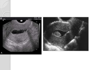
Fibroids and Adenomyosis
Dr.Aftab Qadir
1.Uterine leiomyoma
 Benign tumours of myometrium
 Most common solid benign uterine
neoplasm
 ~25% of women of reproductive age
 Responsive to hormones
Often asymptomatic
 Menorrhagia
 Pain
 Infertility
 Palpable mass
Radiographic features
Conventional radiography
 Popcorn calcification or peripheral rim of
calcification
 Displacement of bowel gas by a pelvic mass
Ultrasound
 Usually hypoechoic, but can be isoechoic, or
even hyperechoic
 Calcification
 Cystic areas of necrosis or degeneration
CT
 Usually of soft tissue density
 May exhibit coarse peripheral or
central calcification
 May distort the usually smooth uterine
contour
 Enhancement pattern is variable
Pelvic MRI
Low to intermediate signal intensity
on T1 and T2 weighted images
compared with the normal
myometrium
 High central signal intensity on T2
from hemorrhage
Complications
 Malignant degeneration
into leiomyosarcomas
 May torse, leading to acute pelvic pain
 Pregnancy may cause fibroid growth
Differential diagnosis
 Uterine leiomyosarcoma
 Uterine lipoleiomyoma
 Ovarian masses
 Focal myometrial contraction during
pregnancy
 Focal adenomyosis
2.Adenomyosis
 Ectopic endometrial tissue in the
myometrium
 Spectrum of endometriosis
 Women of reproductive age
 Higher frequency history of surgical
uterine procedures
Symptomatic:
 Menorrhagia and dysmenorrhea
 May present with chronic pelvic pain
 In 20% of cases is associated with co-
existent endometriosis
Types
 Diffuse adenomyosis: most common
 Focal adenomyosis
 Cystic adenomyosis: rare
Ultrasound
Sonographic features are variable.
 Normal appearing uterus
 Focal or diffuse myometrial bulkiness,
typically of the posterior wall
 Thickening of the transition zone
 Subendometrial echogenic linear striations
 Subendometrial echogenic nodules
 Small myometrial cysts / sub endometrial
cysts
 Heterogeneous myometrial echotexture
Hysterosalpingogram (HSG)
 May show diverticula extending into
the myometrium
CT
 May suggest its presence when
uterine enlargement is present.
 Distinguishing between adenomyosis
and uterine fibroids is difficult
Pelvic MRI
 Modality of choice to diagnose
and characterise adenomyosis
 T2 weighted images are most useful
 Thickening of the junctional zone of
the uterus to more than 12 mm
 T2
◦ Appears as an ill-defined focal/diffuse
region of thickening, often with small high
T2 signal regions representing small
regions of cystic change
 T1
◦ Foci of high T1 signal are often seen
Differences on sonography
Few cases
Thank You

Fibroids&adenomyosis

  • 1.
  • 2.
    1.Uterine leiomyoma  Benigntumours of myometrium  Most common solid benign uterine neoplasm  ~25% of women of reproductive age  Responsive to hormones
  • 4.
    Often asymptomatic  Menorrhagia Pain  Infertility  Palpable mass
  • 5.
    Radiographic features Conventional radiography Popcorn calcification or peripheral rim of calcification  Displacement of bowel gas by a pelvic mass Ultrasound  Usually hypoechoic, but can be isoechoic, or even hyperechoic  Calcification  Cystic areas of necrosis or degeneration
  • 23.
    CT  Usually ofsoft tissue density  May exhibit coarse peripheral or central calcification  May distort the usually smooth uterine contour  Enhancement pattern is variable
  • 27.
    Pelvic MRI Low tointermediate signal intensity on T1 and T2 weighted images compared with the normal myometrium  High central signal intensity on T2 from hemorrhage
  • 31.
    Complications  Malignant degeneration intoleiomyosarcomas  May torse, leading to acute pelvic pain  Pregnancy may cause fibroid growth
  • 32.
    Differential diagnosis  Uterineleiomyosarcoma  Uterine lipoleiomyoma  Ovarian masses  Focal myometrial contraction during pregnancy  Focal adenomyosis
  • 33.
    2.Adenomyosis  Ectopic endometrialtissue in the myometrium  Spectrum of endometriosis  Women of reproductive age  Higher frequency history of surgical uterine procedures
  • 35.
    Symptomatic:  Menorrhagia anddysmenorrhea  May present with chronic pelvic pain  In 20% of cases is associated with co- existent endometriosis
  • 36.
    Types  Diffuse adenomyosis:most common  Focal adenomyosis  Cystic adenomyosis: rare
  • 37.
    Ultrasound Sonographic features arevariable.  Normal appearing uterus  Focal or diffuse myometrial bulkiness, typically of the posterior wall  Thickening of the transition zone  Subendometrial echogenic linear striations  Subendometrial echogenic nodules  Small myometrial cysts / sub endometrial cysts  Heterogeneous myometrial echotexture
  • 43.
    Hysterosalpingogram (HSG)  Mayshow diverticula extending into the myometrium CT  May suggest its presence when uterine enlargement is present.  Distinguishing between adenomyosis and uterine fibroids is difficult
  • 47.
    Pelvic MRI  Modalityof choice to diagnose and characterise adenomyosis  T2 weighted images are most useful  Thickening of the junctional zone of the uterus to more than 12 mm
  • 48.
     T2 ◦ Appearsas an ill-defined focal/diffuse region of thickening, often with small high T2 signal regions representing small regions of cystic change  T1 ◦ Foci of high T1 signal are often seen
  • 53.
  • 55.
  • 61.

Editor's Notes

  • #3 Fibroids are responsive to hormones (e.g stimulated by oestrogens). Being rare in prepubertal females, they commonly accelerate in growth during pregnancy and involutes with menopause
  • #5 Subserosal fibroids may be pedunculated and predominantly extra-uterine, simulating an adnexal mass Any fibroid may undergo atrophy, internal haemorrhage, fibrosis, and calcification. They can also undergo several types of degeneration hyaline degeneration: focal or generalized hyalinization: this is the most common type of degeneration (can occur in ~60% of cases)  cystic degeneration myxoid degeneration generally considered uncommon although reported as high a 50% by some authors  red degeneration due to haemorrhagic infarction, which can occur particularly during pregnancy, and may present with acute abdominal pain
  • #8 Conventional radiography Soft tissue mass arising from the pelvis but separate from the urinary bladder Amorphous, flocculent calcifications in the pelvis May resemble “popcorn” or may calcify the outer rim of fibroid  ultrasound is used to diagnose the presence and monitor the growth of fibroids uncomplicated leiomyomas are usually hypoechoic
  • #9 CT scout view
  • #10 1. hip screw 2. calcified fibroid 3. unidentified curvilinear artifact 4. calcification in iliac artery wall 5 ? small geode 6. ECG dot 7. right lamina- ? partial spina bifida occulta 8. surgical staples
  • #11 Distorted uterus with filling defect
  • #12 HSG. Cavity enlarged by fibroids.
  • #13 HSG. Cavity and right fallopian tube being distorted by large mural fibroid. Note small calcified fibroid on the left (arrows).
  • #14 HSG. Filling defect due to submucousal fibroid.
  • #15 The fibroid shows a dense peripheral calcific rim, within the intramural mass that occupies the fundus and body of the uterus.
  • #17 Sonography of the uterus shows that the mass is possibly within the endomterial cavity. Power Doppler image (top-right) shows that there is a rim of vascularity around the mass. The mass measures more than 4 cm. in size. These ultrasound findings/ images suggest submucous fibroid of the uterus. The main differential diagnosis in this case is endometrial polyp. However, the larger size (polyps are usually less than 2 cm in size) and lack of a single vascular pedicle
  • #18  Large uterine fibroid and a tiny one
  • #19 Intramural leiomyomata are frequently visualized. Examples of fibroids which compromise the contours of the endometrial cavity are shown (A–D). Refraction artifacts resulting from tissue density interfaces and the texture of the fibroids often aid in their identification.
  • #20  TAS. Fibroids. Typical recurrent shadowing (arrowheads)
  • #21 Fibroid abutting but not displacing the cavity (arrowheads). The larger fibroid shows typical recurrent shadowing
  • #22 Subserosal fibroid
  • #23 Saline-Infused Sonography sonohysterogram
  • #25 endometrial polyps and adhesions on hysterosalpingosonography
  • #26 Mass containing mixed densities, low attenuation if necrotic and higher attenuation if calcified or hemorrhagic 
  • #27 CT through the pelvis demonstrates a large mass continuous with the uterus, which has central fluid density.  There is no free peritoneal fluid and the bladder is compressed and empty. 
  • #28 CT Calcified Fibroid
  • #29 CT. Bulky uterus with low-density areas due to fibroids. One small fleck of calcification.
  • #30 the most accurate modality for detecting, localizing and characterising fibroids MRI is of significant value in the symptomatic patient when surgery and uterine salvage therapy is considered. It is also of great value in differentiating a pedunculated fibroid from an adnexal mass MRI Low/intermediate signal intensity of T1 and T2 weighted images High central signal intensity on T2 from hemorrhage May have hyperintense rim With contrast, most are hypointense, about 25% isointense and 10% hyperintense to myometrium
  • #31 as they are usually hypervascular, flow voids are often observed around them marked high signal intensity with gradual enhancement (albeit mild) suggests myxoid degeneration
  • #32 Sagittal T2 Multiple subserosal, intramural and submucosal fibroid, with one of the submucosal fibroid projecting in endometrial cavity.
  • #34 T2 sagittal and T1 fat sat post contrast The uterus is largely replaced by multiple innumerable varying size and well demarcated low T2 SI lesions that show enhancement on T1 post Gad sequences, that is somewhat more heterogeneous in the large lesions. These lesion represents uterine leiomyomas (fibroids), which are intramural in the majority of the lesions, except for one small subserosal and another submucosal lesion. Since the lesions are enhancing, this reflect expected favorable response to UAE if conducted.
  • #35 rarely invasion of adjacent venous channels leading to intravenous leiomyomatosis
  • #37 Risk Factors for Developing Adenomyosis Multipariety Elevated estrogen levels History of aggressive curettage 
  • #39 Benign invasion of the myometrium by the endometrium also results in adjacent smooth muscle hyperplasia
  • #40 Adenomyosis is usually relatively generalized, affecting large portions of the uterus (typically the posterior wall), but sparing the cervix. Despite often marked enlargement of the uterus, the overall contour is usually preserved 
  • #41 zone can sometimes be visualised as a hypoechoic halo surrounding the endometrial layer of ≥12 mm thickness (heterogenous myometrial echotexture)  hyperechoic: islands of endometrial glands hypoechoic: associated muscle hypertrophy a "Venetian blind" appearance may be seen due to subendometrial echogenic linear striations and acoustic shadowing where endometrial tissues cause a hyperplastic reaction
  • #42 This transabdominal ultrasound image shows a large (bulky) uterus with a diffusely infiltrative, in homogenous appearance of the myometrium with dirty, streaky shadowing posteriorly. The uterus shows a globular shape with the endometrium almost obscured. These features are  typically seen in adenomyosis. However, both fibroids/ leiomyoma of the uterus can simultaneously be present with diffuse adenomyosis as in this case
  • #43 Asymmetrical uterine wall thickening anteriorly with focal heterogeneity and associated increased blood flow, as well as the "venetian blind" pattern of acoustic shadowing.
  • #44 Transvaginal scan showing asymmetric thickness of the myometrium with tiny subendometrial cysts representing ectopic endometrial glands (Adenomyosis)
  • #45 Sonographic Characteristics of Adenomyosis  Focal or diffuse enlargement of the uterus Heterogeneous myometrium Myometrial cysts Striations of acoustic shadowing (venetian blinds) Normal or poorly defined endometrium-myometrium border
  • #46 Bulky retroverted uterus 168cc volume, mildy probe tender. Diffuse myometrial hyperemia on colour doppler imaging. Subtle uterine "Venetian blind" artefact (arrows).
  • #47 CT Is unable to diagnose adenomyosis although the presence of calcifications strongly fibroid
  • #48 The opacified uterine cavity presents irregular contour with small outpouchings of contrast.
  • #49 HSG. Enlarged uterus with multiple diverticular-like projections of contrast into the myometrium typical of adenomyosis.
  • #50 CT. Bulky mildly heterogeneous uterus with posterior displacement of the cavity due to adenomyosis rather than fibroids. Differential is of a fibroid or other uterine mass. 
  • #51 Normal junctional zone measures no more than 5 mm On MRI, adenomyosis demonstrates either diffuse or focal thickening of the junctional zone. On T2 images, there are low signal areas corresponding to muscle hyperplasia and increased sign areas, which represent ectopic endometrial tissue. When hemorrhage occurs, T1 images show foci of increased signal. A junctional zone thickness > 12 mm is diagnostic of adenomyosis on MRI. Generally, the uterus is enlarged as well. There is also a focal form (adenomyoma) which presents as an ovoid, T2 dark mass often situated in the junctional zone.
  • #52 contrast enhanced MR evaluation is usually not indicated for evaluation of adenomyosis, however, if performed it shows enhancement of the ectopic endometrial glands
  • #55 US and MR images demonstrate adenomyosis
  • #56 Diffuse adenomyosis on sagittal T2 - weighted spin-echo (TSE 3500/100) image showing diffuse irregular low-signal thickening of the junctional zone. b = bladder.
  • #58 Enlargement of uterus and heterogeneous myometrium in patient with adenomyosis Tiny subendometrial cysts (arrow) representing dilated glands of ectopic endometrium, highly specific for diffuse adenomyosis. Marked enlargement of the junctional zone (arrows) SagittalT2- weighted image shows an ill defined myometrial lesion of low signal intensity in the anterior mypmetrium. Innumerable hyperintense foci (arrows) are embedded in the lesion.
  • #60 Uterine Fibroids. Conventional radiograph on the left show displacement of the bowel gas out of the pelvis because of a large soft tissue mass (white arrows) arising from the pelvis and extending into the lower abdomen. Contrast-enhanced coronal reformatted CT scan of the abdomen on right demonstrates a large pelvic mass comprised of degenerating fibroids (black arrow), hemorrhage (white arrow) and calcification (dotted white arrow).
  • #61 Uterine leiomyoma with IUCD
  • #62 A well defined submucosal filling defect is seen along right lateral wall of fundus. HSG reveals a submucosal leiomyoma
  • #63 Two large hypointense intramural leiomyomas left to the amnion cavity. The more caudally located lesion is surrounded by a large rim of haemorrhage appearing bright on T1- and T2-weighted images. It contains a more recent bleeding component with T2-dephasing—Red degeneration. No relation with the placenta. No extrauterine or intraperitoneal fluid collections.
  • #64 Sag T2 Diffuse junctional zone thickening is in keeping with adenomyosis Sag T1 FS the peripheral high T1WI rim of the ovarian mass is seen The left ovary shows complex cystic mass that has low T1WI, high T2WI central content. The key finding for the diagnosis is the high T1WI, dark T2WI rim which is not suppressed by fat saturation. These are in keeping with blood and in the presence of adenomyosis this is highly suggestive of an endometrioma.