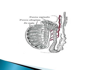
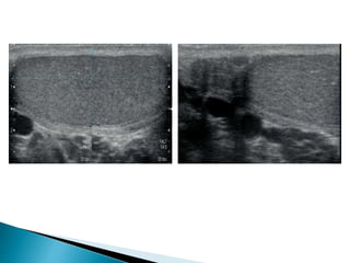
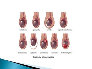
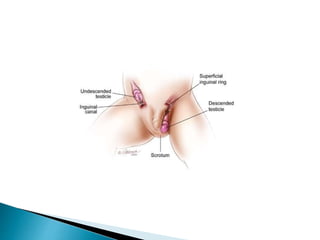
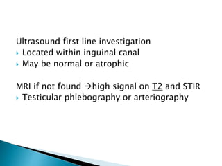
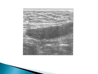
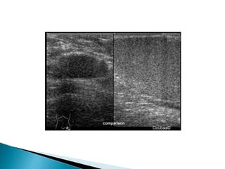
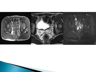
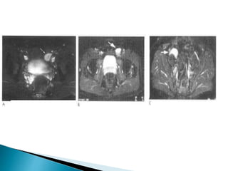
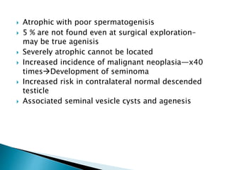
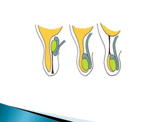
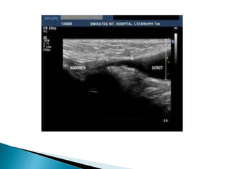
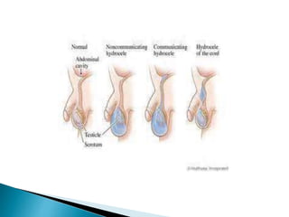
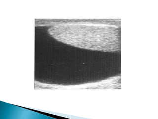
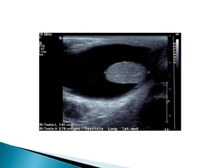
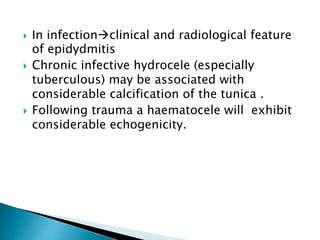
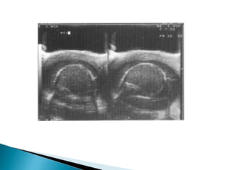
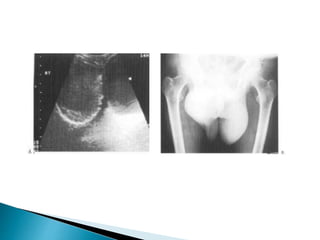
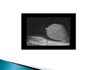
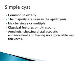
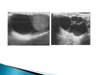
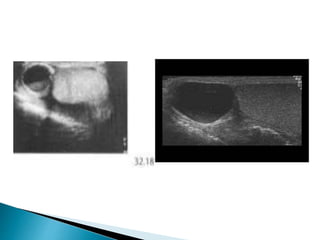
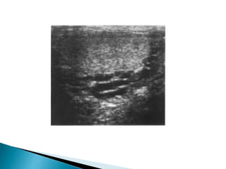
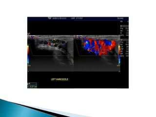
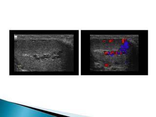
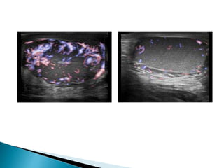
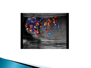
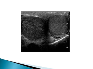
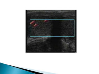
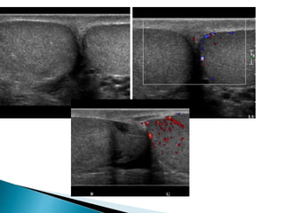
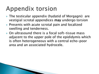
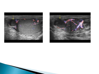
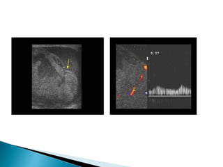
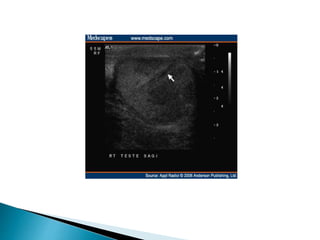
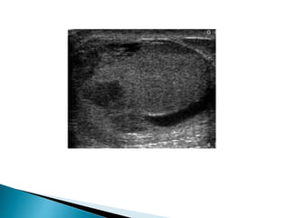
 Congenital disorders
 Extra testicular scrotal disorders
 Testicular trauma
 Ambiguous genitalia
 Undescended testes
 Patent process vaginalis
 Radiologist's role is determination of which
internal pelvic structures are present
 Associated congenital abnormalities of the
urinary tract.
 Chromosomal, endocrine and clinical
assessment are required
 Ultrasound and MRI are the preferred
modalities
 Majority of prepubertal males
 Most commonly lies in the inguinal canal
(canalicular).
 May lie higher up along the normal line of
descent (abdominal testicle)
 Most abdominal testicles lie just proximal to
the inguinal ring
 In a site away from the normal line of
descent (ectopic).
Ultrasound first line investigation
 Located within inguinal canal
 May be normal or atrophic
MRI if not found high signal on T2 and STIR
 Testicular phlebography or arteriography
 Atrophic with poor spermatogenisis
 5 % are not found even at surgical exploration–
may be true agenisis
 Severely atrophic cannot be located
 Increased incidence of malignant neoplasia—x40
timesDevelopment of seminoma
 Increased risk in contralateral normal descended
testicle
 Associated seminal vesicle cysts and agenesis
 Failure of closure of the processes vaginalis
after testicular descent
 Results in a persistent communication with
the abdomen
 Incomplete closure of the processes may lead
to a developmental cyst,
 Hydrocele
 Cysts
 Varicocele
 Epidydmitis
 Orcitis
 Extratesticular tumors
 Formation of fluid between the two layers
(visceral and parietal) of the tunica vaginalis.
 Mostly idiopathic
 May develop as a result of infection
(epididymo-orchitis), trauma, malignant
testicular tumour or infarction (including
torsion).
 Sonographically it is seen as an anechoic area
partly surrounding the testicle
 In infectionclinical and radiological feature
of epidydmitis
 Chronic infective hydrocele (especially
tuberculous) may be associated with
considerable calcification of the tunica .
 Following trauma a haematocele will exhibit
considerable echogenicity.
 Common in elderly
 The majority are seen in the epididymis.
 May be single or multiple.
 Classical features on ultrasound
 Anechoic, showing distal acoustic
enhancement and having no appreciable wall
thickness.
 Dilatation of the network of veins draining the
testicle
 Usually asymptomatic.
 Association with subfertility, reported in 21-39% of
males
 Most frequent between 15 and 25 years of age
 Almost always left-sided when symptomatic
 Present with scrotal aching and/or soft scrotal
mass.
 Classically these symptoms worsen during the day
while the patient is upright.
 Vast majority are primary
 Due to developmental abnormalities of the
valves and/or the veins themselves.
 More likely on the left, where at least 95%
are encountered.
 Minority occur secondary to a lesion
compressing or occluding the testicular
vein.
 On ultrasound varicoceles are seen as a
echo-free serpiginous structures measuring
more than 2 mm maximum diameter .
 Visible flow may he seen within larger
varicoceles.
 Their prominence is increased in the
upright position and with the Valsalva
manoeuvre.
 Inflammation of the epididymis
 Mostly seen in young adult males
 The clinical presentation varies in severity
and acuteness from mild pain, tenderness
and scrotal swelling
 Severe pyrexal illness with marked scrotal
pain and swelling.
 On ultrasound the epididymis shows swelling,
 Diffuse or patchy reduction in echogenicity,
 Doppler ultrasound demonstrates
hypervascularity.
 A heterogeneous pattern of predominant
increase in echogenicity is more frequently
associated with chronic epididymitis.
 Often an associated hydrocele
 Edematous thickening of the overlying skin
 There may be coexisting orchitis
 Inflammation of the testicle
 2/3rd are unilateral.
 In the acute phase ultrasound show testicular
swelling with patchy or diffuse reduction in
echogenicity.
 Doppler ultrasound show increased
vascularity
 Severe orchitis may be associated with
ischaemia and infarction with reduced or
absent vascularity.
 Following resolution the testicle may return
to normal.
 Severe orchitis heterogeneous areas may
develop with a potential for intra testicular
abscess formation
 May result in atrophy with reduction in size
and echogenicity  with little or no
spermatogenesis.
 Twisting of spermatic cord with ischemia of
testes
 Most frequent in the first year of life or in
adolescence, when the testicle is rapidly enlarges
 The loose attachment of the testicle and
spermatic cord to the scrotum in infants and
neonates predisposes to torsion of the entire
cord above the level of the scrotum (extravaginal
torsion).
 Rotation of the cord within the tunica vaginalis
(intravaninal torsion) is the commonest situation
in the older age group.
 Torsion may be complete or incomplete and
spontaneous torsion and detorsion may occur
 The degree of torsion determines the severity of
the ischemia
 In the acute stage ultrasound may be normal or
demonstrate a swollen testicle with patchy or
diffuse hypoechogenicity .
 The epididymis may also become swollen and
echo-poor.
 There may be a reactive hydrocele and the
overlying scrotal skin may be thickened and
oedematous.
 Doppler ultrasound with sensitivity of 85% in the
diagnosis of torsion reduced vascularity
(absence or poor colour flow, reduced peak
systolic velocities) compared with the unaffected
side.
 Given the importance of operating within a few
hours of the onset of symptoms
 Neither the performance nor interpretation of an
ultrasound examination should delay surgical
treatment.
 If there is doubt the urologist should operate on
clinical grounds
Is there torsion?
 The testicular appendix (hydatid of Morgagni) are
vestigial scrotal appendices may undergo torsion
 Presents with acute scrotal pain and localized
swelling and tenderness.
 On ultrasound there is a focal soft-tissue mass
adjacent to the upper pole of the epididymis which
is often heterogeneous with a central echo-poor
area and an associated hydrocele.
 Blunt scrotal trauma commonly results in
hemorrhage around the testicle (haematocele)
and intratesticular hematoma,
 May be associated with a tear of the tunica
albuginea.
 More significant trauma may be associated
with demonstrable fragmentation of the
testicle.
 Ultrasound is the imaging modality.
 The commonest finding is a complex
haemorrhagic hydrocele (haematocele)
 The underlying testicle may show areas of
contusion or haematoma, visible as echo-
poor areas, often with a relatively linear
configuration.
 The testicle may be deformed by subcapsular
haemorrhage.
 There may be rupture of the capsule (tunica
albuginca ) with disruption of the underlying
testicle and associated haematoma .
 The testicular tear may be linear or complex,
 May be fragmentation of the testicle.
Scrotal disorders
Scrotal disorders
Scrotal disorders
Scrotal disorders
Scrotal disorders
Scrotal disorders
Scrotal disorders
Scrotal disorders
Scrotal disorders
Scrotal disorders
Scrotal disorders

Scrotal disorders

  • 2.
     Congenital disorders Extra testicular scrotal disorders  Testicular trauma
  • 6.
     Ambiguous genitalia Undescended testes  Patent process vaginalis
  • 7.
     Radiologist's roleis determination of which internal pelvic structures are present  Associated congenital abnormalities of the urinary tract.  Chromosomal, endocrine and clinical assessment are required  Ultrasound and MRI are the preferred modalities
  • 9.
     Majority ofprepubertal males  Most commonly lies in the inguinal canal (canalicular).  May lie higher up along the normal line of descent (abdominal testicle)  Most abdominal testicles lie just proximal to the inguinal ring  In a site away from the normal line of descent (ectopic).
  • 12.
    Ultrasound first lineinvestigation  Located within inguinal canal  May be normal or atrophic MRI if not found high signal on T2 and STIR  Testicular phlebography or arteriography
  • 17.
     Atrophic withpoor spermatogenisis  5 % are not found even at surgical exploration– may be true agenisis  Severely atrophic cannot be located  Increased incidence of malignant neoplasia—x40 timesDevelopment of seminoma  Increased risk in contralateral normal descended testicle  Associated seminal vesicle cysts and agenesis
  • 18.
     Failure ofclosure of the processes vaginalis after testicular descent  Results in a persistent communication with the abdomen  Incomplete closure of the processes may lead to a developmental cyst,
  • 21.
     Hydrocele  Cysts Varicocele  Epidydmitis  Orcitis  Extratesticular tumors
  • 22.
     Formation offluid between the two layers (visceral and parietal) of the tunica vaginalis.  Mostly idiopathic  May develop as a result of infection (epididymo-orchitis), trauma, malignant testicular tumour or infarction (including torsion).  Sonographically it is seen as an anechoic area partly surrounding the testicle
  • 26.
     In infectionclinicaland radiological feature of epidydmitis  Chronic infective hydrocele (especially tuberculous) may be associated with considerable calcification of the tunica .  Following trauma a haematocele will exhibit considerable echogenicity.
  • 31.
     Common inelderly  The majority are seen in the epididymis.  May be single or multiple.  Classical features on ultrasound  Anechoic, showing distal acoustic enhancement and having no appreciable wall thickness.
  • 34.
     Dilatation ofthe network of veins draining the testicle  Usually asymptomatic.  Association with subfertility, reported in 21-39% of males  Most frequent between 15 and 25 years of age  Almost always left-sided when symptomatic  Present with scrotal aching and/or soft scrotal mass.  Classically these symptoms worsen during the day while the patient is upright.
  • 36.
     Vast majorityare primary  Due to developmental abnormalities of the valves and/or the veins themselves.  More likely on the left, where at least 95% are encountered.  Minority occur secondary to a lesion compressing or occluding the testicular vein.
  • 37.
     On ultrasoundvaricoceles are seen as a echo-free serpiginous structures measuring more than 2 mm maximum diameter .  Visible flow may he seen within larger varicoceles.  Their prominence is increased in the upright position and with the Valsalva manoeuvre.
  • 41.
     Inflammation ofthe epididymis  Mostly seen in young adult males  The clinical presentation varies in severity and acuteness from mild pain, tenderness and scrotal swelling  Severe pyrexal illness with marked scrotal pain and swelling.
  • 42.
     On ultrasoundthe epididymis shows swelling,  Diffuse or patchy reduction in echogenicity,  Doppler ultrasound demonstrates hypervascularity.  A heterogeneous pattern of predominant increase in echogenicity is more frequently associated with chronic epididymitis.  Often an associated hydrocele  Edematous thickening of the overlying skin  There may be coexisting orchitis
  • 44.
     Inflammation ofthe testicle  2/3rd are unilateral.  In the acute phase ultrasound show testicular swelling with patchy or diffuse reduction in echogenicity.  Doppler ultrasound show increased vascularity  Severe orchitis may be associated with ischaemia and infarction with reduced or absent vascularity.
  • 45.
     Following resolutionthe testicle may return to normal.  Severe orchitis heterogeneous areas may develop with a potential for intra testicular abscess formation  May result in atrophy with reduction in size and echogenicity  with little or no spermatogenesis.
  • 48.
     Twisting ofspermatic cord with ischemia of testes  Most frequent in the first year of life or in adolescence, when the testicle is rapidly enlarges  The loose attachment of the testicle and spermatic cord to the scrotum in infants and neonates predisposes to torsion of the entire cord above the level of the scrotum (extravaginal torsion).  Rotation of the cord within the tunica vaginalis (intravaninal torsion) is the commonest situation in the older age group.
  • 53.
     Torsion maybe complete or incomplete and spontaneous torsion and detorsion may occur  The degree of torsion determines the severity of the ischemia  In the acute stage ultrasound may be normal or demonstrate a swollen testicle with patchy or diffuse hypoechogenicity .  The epididymis may also become swollen and echo-poor.  There may be a reactive hydrocele and the overlying scrotal skin may be thickened and oedematous.
  • 54.
     Doppler ultrasoundwith sensitivity of 85% in the diagnosis of torsion reduced vascularity (absence or poor colour flow, reduced peak systolic velocities) compared with the unaffected side.  Given the importance of operating within a few hours of the onset of symptoms  Neither the performance nor interpretation of an ultrasound examination should delay surgical treatment.  If there is doubt the urologist should operate on clinical grounds
  • 55.
  • 56.
     The testicularappendix (hydatid of Morgagni) are vestigial scrotal appendices may undergo torsion  Presents with acute scrotal pain and localized swelling and tenderness.  On ultrasound there is a focal soft-tissue mass adjacent to the upper pole of the epididymis which is often heterogeneous with a central echo-poor area and an associated hydrocele.
  • 58.
     Blunt scrotaltrauma commonly results in hemorrhage around the testicle (haematocele) and intratesticular hematoma,  May be associated with a tear of the tunica albuginea.  More significant trauma may be associated with demonstrable fragmentation of the testicle.
  • 59.
     Ultrasound isthe imaging modality.  The commonest finding is a complex haemorrhagic hydrocele (haematocele)  The underlying testicle may show areas of contusion or haematoma, visible as echo- poor areas, often with a relatively linear configuration.
  • 60.
     The testiclemay be deformed by subcapsular haemorrhage.  There may be rupture of the capsule (tunica albuginca ) with disruption of the underlying testicle and associated haematoma .  The testicular tear may be linear or complex,  May be fragmentation of the testicle.

Editor's Notes

  • #4 Anatomy Scrotum
  • #5 Normal Sagittal Image Testis. Normal Sagittal image head of epididymis
  • #8 Wide variety of phenotypes. This is a complex situation with a wide variety of phenotypes.
  • #9 Neonate with bifid scrotum, micropenis, and hypospadius.
  • #10 the testes may normally be retractile into the groin Cryptorchidism because of the cremasteric muscle reflex retractile. If, however, the testicle can never be located within the scrotum, it can be considered undescended. they may i.e. further cranially within the pelvis or retroperitoneum.
  • #12 Ectopic testicles are uncommon
  • #13 Ultrasound can be regarded as the first-line investigation to locate an undescended testicle, being quick and able to locate the testicle at its commonest sites (within the inguinal canal or just proximal to it ). The testicle may look relatively normal, although the longer it has been undescended the more likely it is to be small. If the testicle cannot be identified on ultrasound, a more extensive search may be performed with MRI. This is a better modality than CT as it avoids radiation and the testicle shows a conspicuous high signal on T2 weighted and STIR sequences. Testicular phlebography or arteriography bas been employed in the search for undescended testes. If both ultrasound and MRI are negative it is unlikely that these angio graphic procedures will detect the missing organ, as it is probably absent or extremely atrophic.
  • #16 The testicle is typically low signal on T1 and high signal on T2. The normal testicle exhibits intense diffusion restriction, and these sequences can aid confident identification.
  • #17 T1 and T2 weighted axial and coronal sequences, especially with a small field of view (FOV) are advisable as well as diffusion weighted imaging. The testicle is typically low signal on T1 and high signal on T2. The normal testicle exhibits intense diffusion restriction The hyperintensity is consistent with restricted diffusion The normal left testicle is identified lying within the left side of the scrotal sac.  The undescended smaller right testicle is intra-peritoneal in location, lying medial to the right external iliac vessels.
  • #18  Transverse STiR images from MRI examinations of patients with undescended testicles (arrow) in the proximal end of the inguinal canal (A) suprapubic pouch (B) and pelvis (C).
  • #19 Failure of descent by the age of 2-3 years is associated with abnormal development of the testicle and this is particularly severe if it continues beyond puberty. it is distinctly possible that a number are so severely atrophic that they cannot be located. Testes that remain undescended (especially abdominal testes) in boys above the age of 5 years also suffer from an increased incidence of malignant neoplasia, up to 40 times normal, usually with the development of seminoma.
  • #20 Cyst-usually in the upper scrotum or inguinal region.
  • #21  the normal embryologic development of the processus vaginalis, which arises as an out pouching of the parietal peritoneum at the beginning of the 3rd month of gestation. After the testis descends into the scrotum (between the 7th and 9th months of gestation), the processus vaginalis is obliterated.
  • #22 transmit disease processes (ascites) or become the site of a hernia.
  • #24 (in comparison to cysts, which do not surround the testicle).
  • #26 Ultrasound demonstration of a hydrocele seen as an echofree area partly surrounding a normal testicle
  • #28 When infection is a cause there are usually obvious clinical and radiological features of epididymitis. especially on the rare occasions when infection is sufficiently severe for frank pus (pyocele) to develop. but a variety of other agents may be responsible, although now tuberculosis is rare. The hydrocele may demonstrate internal echoes , commonest infective organisms are usually bowel-related Gram-negative bacilli
  • #29 Ultrasound showing an infected, partly septated echogenic hydrocele
  • #30 Ultrasound (A) showing dense peripheral calcification around the exterior of a chronic inflammatory hydrocele. This is also visible on the plain film (B).
  • #31  Encysted hydrocele. (a) Diagram shows a fluid collection that does not communicate with the peritoneum or the scrotum. (b) Longitudinal US image in a 12-year-old boy shows a complex ovoid encysted lesion proximal to the testis (T), a finding indicative of an encysted hydrocele with protein and cholesterol contents.
  • #32 Large hydrocele with peritesticular fluid in the tunica vaginalis (A small appendix testis can be seen)
  • #33 Simple cysts are extremely common particularly in the upper pole of epidydmis and in the scrotum. They may be seen at any age from adolescence onwards but are most common in the elderly. Haemorrhage or infection may alter the appearance of a pre- existing cyst to show some degree of echogenicity, occasionally with a visible fluid level infection or trauma (including vasectomy) may provoke the creation of a cystic lesion with internal echoes, partly due to the presence of spermatazoa spermatocele. These again are more common in the upper pole of the epididymis.
  • #34 Ultrasound showing (A) a classical echo-free well-defined thin-walled solitary epididymal cyst and (B) a cluster of simple cysts.
  • #35 Ultrasound of infected epididymal cyst showing debris and fluid level. Spermatocele
  • #36 Occasionally they present relatively acutely as a manifestation of a renal carcinoma. Treatment may be offered for discomfort or as part of the management of subfertility. confusion with cysts is unlikely, even when the varicocele is small, but confirmation of their nature can he obtained with slow-flow Doppler Ultrasound. The classical cause is a left-sided renal cell carcinoma extending along the renal vein as far as the termination of the testicular vein, but benign (for example, hydronephrosis) and other malignant conditions (including abdominal lymphadenopathy) can provoke a varicocele on either side. There is a higher risk of an underlying cause, particularly a tumour if the varicocele is of recent onset with an acute presentation, the patient is older than 40 years.
  • #40 Ultrasound of varicocele seen as echo-free serpiginous structures.
  • #42 Intratesticular varicocele with a tortuous dilatated intra testicular vein with reflux during valsalva
  • #43 Testicular phlebography and percutaneous embolisation of varicocele. The testicular vein in this patient has a wide termination unprotected by a valve and at least one small accessory connection to the renal vein (A). The catheter is manipulated into the distal testicular vein (B) to commence deploying the coils (C).
  • #44 usually due to ascending infection with Gram-negative bacilli is common. with a second peak in late middle age and the elderly, when it may be associated with cystitis and/or prostatitis in association with benign prostatic disease and bladder outflow obstruction. Although usually asymmetrical, epididymitis is not uncommonly bilateral.
  • #45 often nodular and particularly affecting the lower or upper pole. although sometimes the epididymis shows considerable heterogeneity and even rarely a diffuse increase in echogenicity. Rarely the condition progresses so that there is demonstrable abscess formation within the epididymis. There is of variable size, which may be septated and contain echogenic fluid. Severe cases may be associated with and (in up to 20% of cases).
  • #46 Epididyimitis with a thickened hypervascularized epididymis. The testis is normal.
  • #47 may be seen in systemic viral illness (classically mumps) or in association with bacterial epididymitis. Up to 25% of postpubertal males with mumps will suffer some degree of orchitis, usually 7-10 days after the parotitis. Other viruses implicated include echoviruses, group B arboviruses and the lymphocytic choriomeningitis virus. Again there is a substantial risk of subsequent atrophy leading to a small echo-poor testicle The underlying organisms are usually the same as for epididymids, although rarely chronic epididymitis, especially with abscess formation, may be due to tuberculosis. Calcific scars may be seen following any form of orchids.
  • #49 Ultrasound of aggressive epididymitis showing a heterogeneous mass with areas of reduced and increased echogenicity adjacent to the lower pole of the testicle. Ultrasound of severe orchitis. The bulk of the testicle shows diffuse reduction in echogenicity. The heterogeneous area in the lower pole represents a developing abscess.
  • #52 extra-vaginal (supra-vaginal) torsion occurs at the level of the external inguinal ring seen in neonates intra-vaginal more common variety due to bell clapper deformity  typically occurs in adolescents and young adults Different patterns of torsion have been described. This is most often due to poor testicular attachment to the posterior scrotal wall by an abnormally narrow mesentery, which predisposes to rotation of the cord.
  • #54 Transverse scan of both testicles showing normal left testicle and right testicular torsion. Note the hypoechogenicity of the right testicle
  • #55 Transverse plane through both testes.  The power Doppler image of the scrotum demonstrates right testicular perfusion.  The swollen left testicle is not perfused
  • #57 If torsion is incomplete, the testicle remains viable longer: 80% up to 12 h and 40% up to 24 h. Even in this situation, however, viability falls to 10% after 24 h and the rapidity with which irreversible changes occur within the testicle. Surgical treatment of complete torsion within 5hour is associated with a testicular salvage rate of 80%, which falls to 20 17- or less after 12 h. False positives (for example ischaemia associated with severe epididymo-orchids) and false negatives (due to difficulty in obtaining an adequate colour flow, intermittent nature of torsion, etc.) are encountered and there is still considerable controversy as to the role of this investigation in the management of these patients.
  • #59 Incomplete torsion of testis with high resistance to-and-fro flow Spectral Doppler shows high resistance flow in left testis with diastolic reversal, while the right testis shows normal low resistance flow.
  • #60 Ultrasound of an infarcting testicle. There are extensive areas of reduced echogenicity within the substance of the testicle. The adjacent epididymis is also markedly diseased and swollen.
  • #61 (such as the epididymal appendix) This may slough off and become calcified, giving rise to mobile highly echogenic shadowing foci between the layers of the tunica vaginalis (scrotoliths or scrotal pearls).
  • #62 Torsion of the appendix epididymis in an adult with a swollen appendix and increased vascularity of the epididymis
  • #63  The role of imaging depends on the surgical approach. Where there is substantial trauma, surgical exploration, drainage of hematoma and repair of testicular tears may enhance the subsequent viability of the testicle
  • #65 Scrotal hematoma may he complicated by abscess formation. severe testicular damage may be followed by subsequent atrophy.
  • #66 Traumatic changes in the testis with a contusion and hematoma without vascularity
  • #68 This sonogram of a different patient following trauma reveals in homogeneous testicular echotexture and a fracture line (arrow).
  • #69 Ultrasonography of a three-days old testicular rupture: the tunica cannot longer be seen, the pro lapsing testicular tissue appears inhomogeneous.
  • #72 Scrotal sonography - reveals three testes Magnetic resonance imaging (MRI) examination was done, which showed normal T 1 and T 2 images of right testis and lower testis of left hemiscrotum.The upper testis of left hemiscrotum showed increase signal intensity on T1 and decreased signal intensity on T2 images, suggestive of torsion and hemorrhagic necrosis .Minimal hydrocele was noted in both hemiscrota.
  • #73  Longitudinal US image of the spermatic cord in a 1-year-old boy shows an inguinoscrotal hernia that contains bowel (arrowheads). A patent internal inguinal ring also is seen (arrows).
  • #74 Pyocele with an irregular septated cystic mass
  • #75 Epididymo-orchitis with a hypervascularized swollen testis and epididymis
  • #76 Cystic dilatation of the rete tesis with intratesticular cysts left testis