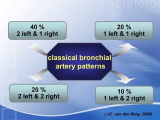
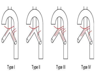
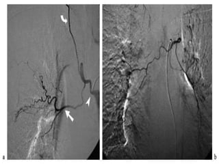
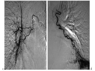
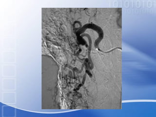
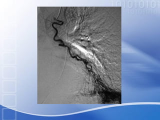
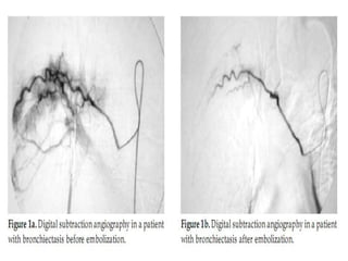
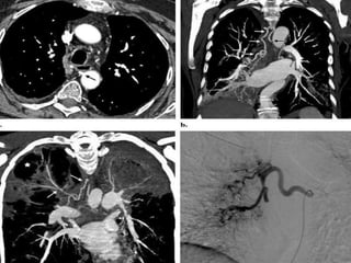
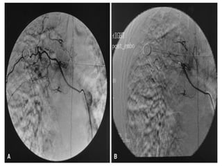
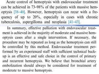
Embolotherapy
In Patients With Massive
Hemoptysis
By
Mahmoud E. Abou El-Magd
Assistant lecturer of pulmonary and critical care medicine
Hemoptysis
• Hemoptysis is classified as massive or
nonmassive based on the rate of bleeding.
• The most commonly used definition of
massive hemoptysis is 600 mL in 24
hours .
(Ibrahim , 2008).
• Life-threatening (massive) hemoptysis was
defined as expectoration of at least 200 mL of
blood per hour in a patient with normal or nearly
normal lung function.
• Production of at least 50 mL of blood per hour in
a patient with a chronic respiratory failure, or
more than two episodes of moderate hemoptysis
within a 24 h period .
• Moderate hemoptysis was defined as more than
or equal to three episodes of 100 mL of bleeding
per day within 1 week.
(Agmy, 2013).
• The evaluation of hemoptysis involves a careful
history, physical examination, and a chest
radiograph. Initial studies also include a
complete blood count.
• In the vast majority (90%) of cases the source of
the bleeding is the bronchial circulation.
• Massive hemoptysis may be due to active TB,
prior TB, bronchiectasis, mycetoma, Tight mitral
stenosis, and lung cancer.
(Corder, 2003 )
Pathophysiology
Hemoptysis
Hypoxic pul . V.C
Direct erosion
Elevated local Bl . Pr
angiogenetic growth factors
(Yoon et al., 2002)
• Conservative management of massive
hemoptysis has a 50–100% mortality rate
.
• The diagnostic accuracy of FOB is 0%–
30% in patients with normal chest
radiograph and 10%–43% in all patients
with hemoptysis.
(Yoon et al., 2002)
• Two decades ago,surgery was regarded
as the treatment of choice for hemoptysis.
but mortality rates were up to 40%
following emergency surgery .
• The mortality rate in patients who are
incapable of tolerating surgical procedure
was 80%.
(Yoon et al., 2002)
Indications
for
Recurrent
hemoptys
-is
Pre-
operati-
ve
Bronchial angiography
Massive
hemoptysis
Undiagnosed
hemoptysis
(Sirajuddin and Lucien, 2008)
Pulmonary
vascular Anatomy
Bronchial Arteries
supply
trachea
Pulmonary airways
Visceral pleura
esophagus
Vasa vasorum of aorta,
Pul. A & V
(van den Berg, 2006)
classical bronchial
artery patterns
40 %
2 left & 1 right
20 %
1 left & 1 right
20 %
2 left & 2 right
10 %
1 left & 2 right
( JC. van den Berg, 2006)
Pathologic Features Of
The Bronchial Artery
Non-bronchial
Systemic
Artery
Anatomy
brachiocephalic arterythe aortic arch
internal mammary
artery
Pericardiacophrenic
subclavian artery
thyrocervical trunk
(Yoon et al., 2002)
Embolization
tools
A - Particulate Agents
The potential to occlude a target vessel at a
desired point (proximal or distal) by selecting
a particle size that corresponds to that
diameter.
1- Polyvinyl Alcohol Particles
( JC. van den Berg, 2006)
1-Polyvinyl Alcohol Particles
• 50 and 2000 µm, the typical size ranges
used clinically are 300 to 500 µm or 500
to 700 µm.
• Irregular shape .
(J. Golzarian et al., 2006 )
Polyvinyl alcohol
Focal angio-
necrosis
Thrombus
formation
Plt
aggregation
Slow
flow
Adherence
Mechanism of action
(J. Golzarian et al., 2006 )
A - Particulate Agents
The potential to occlude a target vessel at a
desired point (proximal or distal) by selecting
a particle size that corresponds to that
diameter.
1- Polyvinyl Alcohol Particles
2- Spherical Embolic Agents
( JC. van den Berg, 2006)
2-Spherical Embolic Agents
• Trisacryl Gelatin Microspheres
40–120, 100–300, 500–700, and 700–900 mm,
In its original form, these spheres are clear.
• Polyvinyl Alcohol microspheres (Contour
SE particles)
100–300,300-500, 500–700, 700–900, and 900–1,200
m m.
(Avritscher and Wallace, 2012 )
A - Particulate Agents
The potential to occlude a target vessel at a
desired point (proximal or distal) by selecting
a particle size that corresponds to that
diameter.
1- Polyvinyl Alcohol Particles
2- Spherical Embolic Agents
3- Gelfoam
( JC. van den Berg, 2006)
3-Gelfoam
• Gelatin sponge (Gelfoam) is a white,
water-insoluble, prepared from purified
pig skin gelatin.
• Temporary.
• 4w to 4m
(Golzarian et al., 2006 )
B - Liquid Agents
1- Ethanol or Absolute Alcohol
not radiopaque and highly diffusible ,Severe
complications such as cardiac arrest and pulmonary
embolism have been reported.
2- Cyanoacrylate
(Burrows and Mason, 2004)
C- Coils and Metallic Embolization
• The metallic coils range in size from 0.018 in.
(microcoils) to the standard 0.035–0.038 in.
sizes. made from either stainless steel or
platinum , all coils are permanent Devices.
• Coils should not be used in combination with
particulate embolization for the treatment of
tumors .
• Gugliemi detachable coil (GDC) system
(Golzarian et al., 2006 )
D – Balloons
E – Microcatheters
come in a variety of sizes from the larger bore
(outer diameter 3 F) to standard size (2.7 F) to
very small bore (2 F).
(Golzarian et al., 2006 )
Chylothorax 55
Embolotherapy
Technique
PREPARATION
• consent .
• sedatives or narcotic analgesics .
• monitoring .
• contrast .
• Fasting 4h before examination, except diabetic
patient.
• Stop
(Aspocid, Clexane) 1 day before examination.
(Marivane) 3 days before examination.
• Control of hypertension.
(Bader et al., 2004 )
Contra-indications
• absolute contraindication
spinal artery !!
• relative contraindication
- Contrast allergy.
-Uncontrolled (Hypertension)
-Coagulopathy Problems.
-Renal dysfunction.
-Uncontrolled (arrhythmias).
-Pregnancy !
(Chun and Belli, 2009)
Complications
1. (transient) chest pain, in 24% up to
91% of cases .
2. dysphagia, 0.7% to 18.2%.
3. spinal cord ischemia, 1.4%–6.5%
4. “post-embolization syndrome”.
5. Non-target embolization (colon,
coronary and cerebral circulation).
(McPherson, 2010 )
6. Rare complications as Aortic and
bronchial necrosis .
7. Other complications related to
angiography in general as
-Bleeding, False aneurysm ,Reactions to the
dye and Kidney damage .
(Wong et al., 2002)
Recurrence
Recurrence of hemoptysis may
occur due to:
• Recanalization
• Incomplete embolization.
• Revascularization
• anomalous bronchial arteries .
• The underlying disease.
(Chun and Belli , 2009)
Results
• Active and old TB had the best results .
• Patients with malignancy were the most
difficult to manage … Why ?
• Poor outcomes have been observed in
patients with aspergilloma .
(Mauro et al ., 2006 )
• We recommend use of bronchial
arteriography as a routine investigatory
tool for patient with hemoptysis .
• Future researches regarding other uses of
radiological bronchial and pulmonary
arteries intevention .
Home takeaways
• Bronchial artery embolization is highly effective in the
treatment of acute hemoptysis. Short-term non
recurrence rates (with follow-up up to 1 month) range
from 77% to 99% .
• success rates of 100% can be achieved using repeat
embolization and control of underlying disease either
pharmacologically or surgically .
• The most effective nonsurgical treatment for massive
hemoptysis is bronchial artery embolization (BAE) .
(Chun and Belli , 2009)
Bronchial artery embolization

Bronchial artery embolization

  • 2.
    Embolotherapy In Patients WithMassive Hemoptysis By Mahmoud E. Abou El-Magd Assistant lecturer of pulmonary and critical care medicine
  • 3.
  • 4.
    • Hemoptysis isclassified as massive or nonmassive based on the rate of bleeding. • The most commonly used definition of massive hemoptysis is 600 mL in 24 hours . (Ibrahim , 2008).
  • 5.
    • Life-threatening (massive)hemoptysis was defined as expectoration of at least 200 mL of blood per hour in a patient with normal or nearly normal lung function. • Production of at least 50 mL of blood per hour in a patient with a chronic respiratory failure, or more than two episodes of moderate hemoptysis within a 24 h period . • Moderate hemoptysis was defined as more than or equal to three episodes of 100 mL of bleeding per day within 1 week. (Agmy, 2013).
  • 6.
    • The evaluationof hemoptysis involves a careful history, physical examination, and a chest radiograph. Initial studies also include a complete blood count. • In the vast majority (90%) of cases the source of the bleeding is the bronchial circulation. • Massive hemoptysis may be due to active TB, prior TB, bronchiectasis, mycetoma, Tight mitral stenosis, and lung cancer. (Corder, 2003 )
  • 7.
  • 8.
    Hemoptysis Hypoxic pul .V.C Direct erosion Elevated local Bl . Pr angiogenetic growth factors (Yoon et al., 2002)
  • 9.
    • Conservative managementof massive hemoptysis has a 50–100% mortality rate . • The diagnostic accuracy of FOB is 0%– 30% in patients with normal chest radiograph and 10%–43% in all patients with hemoptysis. (Yoon et al., 2002)
  • 10.
    • Two decadesago,surgery was regarded as the treatment of choice for hemoptysis. but mortality rates were up to 40% following emergency surgery . • The mortality rate in patients who are incapable of tolerating surgical procedure was 80%. (Yoon et al., 2002)
  • 11.
  • 12.
  • 13.
    Bronchial Arteries supply trachea Pulmonary airways Visceralpleura esophagus Vasa vasorum of aorta, Pul. A & V (van den Berg, 2006)
  • 14.
    classical bronchial artery patterns 40% 2 left & 1 right 20 % 1 left & 1 right 20 % 2 left & 2 right 10 % 1 left & 2 right ( JC. van den Berg, 2006)
  • 18.
  • 29.
    Non-bronchial Systemic Artery Anatomy brachiocephalic arterythe aorticarch internal mammary artery Pericardiacophrenic subclavian artery thyrocervical trunk (Yoon et al., 2002)
  • 33.
  • 34.
    A - ParticulateAgents The potential to occlude a target vessel at a desired point (proximal or distal) by selecting a particle size that corresponds to that diameter. 1- Polyvinyl Alcohol Particles ( JC. van den Berg, 2006)
  • 35.
    1-Polyvinyl Alcohol Particles •50 and 2000 µm, the typical size ranges used clinically are 300 to 500 µm or 500 to 700 µm. • Irregular shape . (J. Golzarian et al., 2006 )
  • 36.
  • 37.
    A - ParticulateAgents The potential to occlude a target vessel at a desired point (proximal or distal) by selecting a particle size that corresponds to that diameter. 1- Polyvinyl Alcohol Particles 2- Spherical Embolic Agents ( JC. van den Berg, 2006)
  • 38.
    2-Spherical Embolic Agents •Trisacryl Gelatin Microspheres 40–120, 100–300, 500–700, and 700–900 mm, In its original form, these spheres are clear. • Polyvinyl Alcohol microspheres (Contour SE particles) 100–300,300-500, 500–700, 700–900, and 900–1,200 m m. (Avritscher and Wallace, 2012 )
  • 42.
    A - ParticulateAgents The potential to occlude a target vessel at a desired point (proximal or distal) by selecting a particle size that corresponds to that diameter. 1- Polyvinyl Alcohol Particles 2- Spherical Embolic Agents 3- Gelfoam ( JC. van den Berg, 2006)
  • 43.
    3-Gelfoam • Gelatin sponge(Gelfoam) is a white, water-insoluble, prepared from purified pig skin gelatin. • Temporary. • 4w to 4m (Golzarian et al., 2006 )
  • 45.
    B - LiquidAgents 1- Ethanol or Absolute Alcohol not radiopaque and highly diffusible ,Severe complications such as cardiac arrest and pulmonary embolism have been reported. 2- Cyanoacrylate (Burrows and Mason, 2004)
  • 46.
    C- Coils andMetallic Embolization • The metallic coils range in size from 0.018 in. (microcoils) to the standard 0.035–0.038 in. sizes. made from either stainless steel or platinum , all coils are permanent Devices. • Coils should not be used in combination with particulate embolization for the treatment of tumors . • Gugliemi detachable coil (GDC) system (Golzarian et al., 2006 )
  • 52.
    D – Balloons E– Microcatheters come in a variety of sizes from the larger bore (outer diameter 3 F) to standard size (2.7 F) to very small bore (2 F). (Golzarian et al., 2006 )
  • 55.
  • 56.
  • 57.
    PREPARATION • consent . •sedatives or narcotic analgesics . • monitoring . • contrast . • Fasting 4h before examination, except diabetic patient. • Stop (Aspocid, Clexane) 1 day before examination. (Marivane) 3 days before examination. • Control of hypertension. (Bader et al., 2004 )
  • 58.
  • 59.
    • absolute contraindication spinalartery !! • relative contraindication - Contrast allergy. -Uncontrolled (Hypertension) -Coagulopathy Problems. -Renal dysfunction. -Uncontrolled (arrhythmias). -Pregnancy ! (Chun and Belli, 2009)
  • 61.
  • 62.
    1. (transient) chestpain, in 24% up to 91% of cases . 2. dysphagia, 0.7% to 18.2%. 3. spinal cord ischemia, 1.4%–6.5% 4. “post-embolization syndrome”. 5. Non-target embolization (colon, coronary and cerebral circulation). (McPherson, 2010 )
  • 63.
    6. Rare complicationsas Aortic and bronchial necrosis . 7. Other complications related to angiography in general as -Bleeding, False aneurysm ,Reactions to the dye and Kidney damage . (Wong et al., 2002)
  • 64.
  • 65.
    Recurrence of hemoptysismay occur due to: • Recanalization • Incomplete embolization. • Revascularization • anomalous bronchial arteries . • The underlying disease. (Chun and Belli , 2009)
  • 66.
  • 67.
    • Active andold TB had the best results . • Patients with malignancy were the most difficult to manage … Why ? • Poor outcomes have been observed in patients with aspergilloma . (Mauro et al ., 2006 )
  • 68.
    • We recommenduse of bronchial arteriography as a routine investigatory tool for patient with hemoptysis . • Future researches regarding other uses of radiological bronchial and pulmonary arteries intevention .
  • 81.
  • 82.
    • Bronchial arteryembolization is highly effective in the treatment of acute hemoptysis. Short-term non recurrence rates (with follow-up up to 1 month) range from 77% to 99% . • success rates of 100% can be achieved using repeat embolization and control of underlying disease either pharmacologically or surgically . • The most effective nonsurgical treatment for massive hemoptysis is bronchial artery embolization (BAE) . (Chun and Belli , 2009)