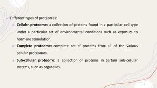
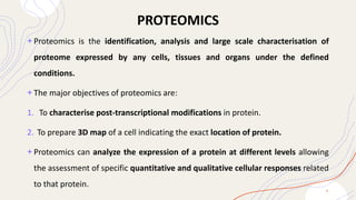
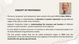
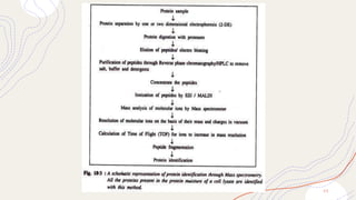
1
Proteomics
PROTEOMICS
2
+ Proteome is a set of proteins produced in an organism, system, or biological
context or entire set of proteins that is, or can be, expressed by a genome, cell,
tissue, or organism at a certain expressed time in a given set of condition.
Proteomics is the study of all the proteins produced by a cell.
+ The proteome is not constant; it differs from cell to cell and changes over time.
+ To some degree, the proteome reflects the underlying transcriptome.
+ Different types of proteomes:
1. Cellular proteome: a collection of proteins found in a particular cell type
under a particular set of environmental conditions such as exposure to
hormone stimulation.
2. Complete proteome: complete set of proteins from all of the various
cellular proteomes.
3. Sub-cellular proteome: a collection of proteins in certain sub-cellular
systems, such as organelles.
3
PROTEOMICS
+ Proteomics is the identification, analysis and large scale characterisation of
proteome expressed by any cells, tissues and organs under the defined
conditions.
+ The major objectives of proteomics are:
1. To characterise post-transcriptional modifications in protein.
2. To prepare 3D map of a cell indicating the exact location of protein.
+ Proteomics can analyze the expression of a protein at different levels allowing
the assessment of specific quantitative and qualitative cellular responses related
to that protein.
4
+ The terms ‘proteome’ and ‘proteomics’ were coined in the early 1990 by Marc Wilkins.
+ Proteomics helps in understanding of alteration in protein expression during different
stages of life cycle or under stress condition.
+ Likewise, Proteomics helps in understanding the structure and function of different
proteins as well as protein - protein interactions of an organism.
+ A minor defect in protein structure, it is function or alternation in expression pattern can
be easily detected using proteomics studies.
+ The first protein studies that can be called proteomics began in 1975 with the
introduction of the two dimensional gel and mapping of the proteins from the bacterium
Escherichia coli, guinea pig and mouse.
5
CONCEPT OF PROTEOMICS
TYPES OF PROTEOMICS
STRUCTURAL
PROTEOMICS FUNCTIONAL
PROTEOMICS
EXPRESSION
PROTEOMICS
6
1 2 3
TYPES OF PROTEOMICS
1) Structural Proteomics:
+ Structural proteomics deals with the study of structure and nature of protein
complexes present in a particular cell organelle.
+ It is mapping out the 3-D structure and nature of protein complexes present
specifically in a particular cell organelle.
+ The ultimate aim of structural proteomics is to build a body of structural
information that will help predict the probable structure and potential function
for almost any protein from knowledge of its coding sequence.
+ Structural proteomics can also help assembling information about protein-protein
interactions and about architecture of cells to explain how the expression of
certain proteins contributes in cell’s unique characteristics.
7
2. Functional Proteomics:
• Functional proteomics refers to the use of proteomics techniques to analyze the
characteristics of molecular protein-networks involved in a living cell.
• One of the recent successes of functional proteomics is the identification and analysis of
molecular protein networks involved in the nuclear pore complex (NPC) in yeast.
• This success helps understand the translocation of molecules from nucleus to the
cytoplasm and vice versa.
• It focuses on the large-scale characterization of proteins and their functions
within a biological system and involves the systematic analysis of protein
expression, localization, interactions, post-translational modifications, and their
roles in cellular processes.
8
3. Expression Proteomics:
• Expression proteomics concerned with to the quantitative study of protein expression
between samples differing by some variable.
• The pattern of expression of the complete proteome or of its part (sub-proteome)
between samples can be compared with the help of expression proteomics.
• The expression proteomics is quite useful in identifying disease specific proteins. For
example, over expression or under-expression of proteins in cancerous cells and normal
cells taken from a cancer patient and a normal individual, respectively, can be analyzed
using various techniques, such as two dimensional gel electrophoresis, mass
spectrometry, microarray, etc.
• This can help understand the development of cancer and facilitate development of drugs
to treat cancer.
9
Top-down proteomics and bottom-up
proteomics
Top-down proteomics bottom-up proteomics
Proteins in a sample of interest are first
separated before being individually
characterized.
All the proteins in the sample are first
digested into a complex mixture of
peptides and these peptides are then
analyzed to identify which proteins were
present in the sample.
1 0
1 1
PROTEIN
PROFILING
STEPS IN PROTEIN PROFILING
1 2
PROTEIN PROFILING
+ Protein profiling refers to the comprehensive analysis of proteins present in a
biological sample, typically aiming to characterize their identities, quantities,
modifications, interactions, and other properties.
+ It involves the systematic identification and quantification of proteins to gain
insights into their roles in biological processes, disease states, or other relevant
conditions.
+ Protein profiling can be conducted using various techniques and methodologies,
including mass spectrometry-based approaches, protein microarrays, and
immunoassays.
1 3
HISTORY
+ One of the earliest methods for protein analysis has been Edman
degradation (introduced in 1967) where a single peptide is subjected to
multiple steps of chemical degradation to resolve its sequence.
+ These early methods have mostly been supplanted by technologies that
offer higher throughput.
+ Introduction of Electrophoresis (1930s-1940s): Electrophoresis,
particularly techniques like gel electrophoresis, became widely used for
separating proteins based on their charge and size.
1 4
STEPS IN PROTEIN PROFILING
1. Sample Collection and Preparation:
• The first step in protein profiling is the collection of the biological sample of
interest, which could be cells, tissues, or bodily fluids.
• Sample preparation involves lysing the cells or tissues to release proteins and
removing contaminants that might interfere with subsequent analysis.
• Various extraction methods, such as cell lysis, tissue homogenization, and protein
precipitation, may be used depending on the sample type.
1 5
2. Protein Separation:
• Proteins in the sample are separated based on their physicochemical properties,
such as size, charge, hydrophobicity, or affinity to specific molecules. Common
protein separation techniques include:
• Gel Electrophoresis: Proteins are separated based on size and charge using
techniques such as SDS-PAGE or 2D gel electrophoresis.
• Chromatography: Proteins are separated based on their interactions with a
stationary phase in techniques like ion exchange chromatography, size
exclusion chromatography, or affinity chromatography.
• Capillary Electrophoresis: Proteins are separated in a capillary tube based on
charge and size differences.
1 6
3. Protein Detection and Visualization:
• Following separation, proteins are typically visualized and detected using various
staining or labeling methods:
• Coomassie Brilliant Blue or silver staining for gel-based techniques like SDS-
PAGE or 2D gel electrophoresis.
• Fluorescent dyes or specific protein stains for gel-free techniques.
• Western blotting for specific protein detection using antibodies.
4. Protein Digestion (for Mass Spectrometry-based Approaches):
• For mass spectrometry-based protein identification, proteins are enzymatically
digested into peptides using proteolytic enzymes such as trypsin. This step
converts proteins into a more suitable form for mass spectrometric analysis.
1 7
5. Mass Spectrometry Analysis:
• The digested peptides are analyzed using mass spectrometry to determine their mass-to-
charge ratio (m/z) and fragmentation patterns. Common mass spectrometry-based
techniques for protein identification include:
• Liquid Chromatography-Mass Spectrometry (LC-MS/MS)
• Matrix-Assisted Laser Desorption/Ionization Mass Spectrometry (MALDI-MS)
• Electrospray Ionization Mass Spectrometry (ESI-MS).
MALDI MS- Matrix-Assisted Laser Desorption/Ionization Mass Spectrometry:
+ In mass spectrometry MALDI is an ionization technique that uses a laser energy-
absorbing matrix to create ions from large molecules with minimal fragmentation.
+ It has been applied to the analysis of biomolecules (biopolymers such
as DNA, proteins, peptides and carbohydrates) and various organic molecules
(such as polymers, dendrimers and other macromolecules), which tend to be
fragile and fragment when ionized by more conventional ionization methods.
1 8
+ It is similar in character to electrospray ionization (ESI) in that
both techniques are relatively soft (low fragmentation) ways
of obtaining ions of large molecules in the gas phase, though
MALDI typically produces far fewer multi-charged ions.
+ MALDI methodology is a three-step process. First, the
sample is mixed with a suitable matrix material and
applied to a metal plate.
+ Second, a pulsed laser irradiates the sample,
triggering ablation and desorption of the sample and
matrix material.
+ Finally, the analyte molecules are ionized by
being protonated or deprotonated in the hot plume of
ablated gases, and then they can be accelerated into
whichever mass spectrometer is used to analyse them.
1 9
6. Protein Identification and Database Searching:
• Mass spectrometry data are processed and analyzed using bioinformatics tools to
identify the proteins present in the sample. Database search algorithms compare
experimental spectra with theoretical spectra generated from protein sequence
databases to match peptide sequences and identify proteins.
2 0
7. Protein Quantification:
• Quantitative proteomics techniques are used to measure the abundance of
identified proteins across different samples or conditions. This can be achieved using
isotopic labeling methods (e.g., SILAC, TMT, iTRAQ) or label-free quantification
approaches based on peak intensities or spectral counting.
8. Data Analysis and Interpretation:
• The identified and quantified proteins are subjected to data analysis to extract
meaningful information, such as differential expression patterns, functional
annotations, and protein-protein interactions. Statistical analysis and bioinformatics
tools are employed to interpret the proteomics data in the context of biological
processes and pathways.
2 1
9. Validation and Follow-up Studies:
• Identified proteins and findings from proteomics experiments are often validated
using independent techniques, such as western blotting, immunoprecipitation, or
functional assays. Follow-up studies may involve additional experiments to
confirm the functional significance of identified proteins or to explore specific
biological questions further.
2 2
2 3
REFERENCE
+https://www.bioradiations.com/wpcontent/uploads/2014/12/BioR
adiations-123_07-0095.pdf
+https://www.technologynetworks.com/proteomics/articles/proteo
mics-principles-techniques-and-applications-343804
+Sanchez-Pla, A.,Reverter, F.,Ruiz de Villa, M. C.,& Camabella,
M. (2012). Transcriptomics: mRNA and alternative splicing.
Journal of Neuroimmunology.
2 4

Proteomics: types, protein profiling steps etc.

  • 1.
  • 2.
    PROTEOMICS 2 + Proteome isa set of proteins produced in an organism, system, or biological context or entire set of proteins that is, or can be, expressed by a genome, cell, tissue, or organism at a certain expressed time in a given set of condition. Proteomics is the study of all the proteins produced by a cell. + The proteome is not constant; it differs from cell to cell and changes over time. + To some degree, the proteome reflects the underlying transcriptome.
  • 3.
    + Different typesof proteomes: 1. Cellular proteome: a collection of proteins found in a particular cell type under a particular set of environmental conditions such as exposure to hormone stimulation. 2. Complete proteome: complete set of proteins from all of the various cellular proteomes. 3. Sub-cellular proteome: a collection of proteins in certain sub-cellular systems, such as organelles. 3
  • 4.
    PROTEOMICS + Proteomics isthe identification, analysis and large scale characterisation of proteome expressed by any cells, tissues and organs under the defined conditions. + The major objectives of proteomics are: 1. To characterise post-transcriptional modifications in protein. 2. To prepare 3D map of a cell indicating the exact location of protein. + Proteomics can analyze the expression of a protein at different levels allowing the assessment of specific quantitative and qualitative cellular responses related to that protein. 4
  • 5.
    + The terms‘proteome’ and ‘proteomics’ were coined in the early 1990 by Marc Wilkins. + Proteomics helps in understanding of alteration in protein expression during different stages of life cycle or under stress condition. + Likewise, Proteomics helps in understanding the structure and function of different proteins as well as protein - protein interactions of an organism. + A minor defect in protein structure, it is function or alternation in expression pattern can be easily detected using proteomics studies. + The first protein studies that can be called proteomics began in 1975 with the introduction of the two dimensional gel and mapping of the proteins from the bacterium Escherichia coli, guinea pig and mouse. 5 CONCEPT OF PROTEOMICS
  • 6.
    TYPES OF PROTEOMICS STRUCTURAL PROTEOMICSFUNCTIONAL PROTEOMICS EXPRESSION PROTEOMICS 6 1 2 3
  • 7.
    TYPES OF PROTEOMICS 1)Structural Proteomics: + Structural proteomics deals with the study of structure and nature of protein complexes present in a particular cell organelle. + It is mapping out the 3-D structure and nature of protein complexes present specifically in a particular cell organelle. + The ultimate aim of structural proteomics is to build a body of structural information that will help predict the probable structure and potential function for almost any protein from knowledge of its coding sequence. + Structural proteomics can also help assembling information about protein-protein interactions and about architecture of cells to explain how the expression of certain proteins contributes in cell’s unique characteristics. 7
  • 8.
    2. Functional Proteomics: •Functional proteomics refers to the use of proteomics techniques to analyze the characteristics of molecular protein-networks involved in a living cell. • One of the recent successes of functional proteomics is the identification and analysis of molecular protein networks involved in the nuclear pore complex (NPC) in yeast. • This success helps understand the translocation of molecules from nucleus to the cytoplasm and vice versa. • It focuses on the large-scale characterization of proteins and their functions within a biological system and involves the systematic analysis of protein expression, localization, interactions, post-translational modifications, and their roles in cellular processes. 8
  • 9.
    3. Expression Proteomics: •Expression proteomics concerned with to the quantitative study of protein expression between samples differing by some variable. • The pattern of expression of the complete proteome or of its part (sub-proteome) between samples can be compared with the help of expression proteomics. • The expression proteomics is quite useful in identifying disease specific proteins. For example, over expression or under-expression of proteins in cancerous cells and normal cells taken from a cancer patient and a normal individual, respectively, can be analyzed using various techniques, such as two dimensional gel electrophoresis, mass spectrometry, microarray, etc. • This can help understand the development of cancer and facilitate development of drugs to treat cancer. 9
  • 10.
    Top-down proteomics andbottom-up proteomics Top-down proteomics bottom-up proteomics Proteins in a sample of interest are first separated before being individually characterized. All the proteins in the sample are first digested into a complex mixture of peptides and these peptides are then analyzed to identify which proteins were present in the sample. 1 0
  • 11.
  • 12.
  • 13.
    PROTEIN PROFILING + Proteinprofiling refers to the comprehensive analysis of proteins present in a biological sample, typically aiming to characterize their identities, quantities, modifications, interactions, and other properties. + It involves the systematic identification and quantification of proteins to gain insights into their roles in biological processes, disease states, or other relevant conditions. + Protein profiling can be conducted using various techniques and methodologies, including mass spectrometry-based approaches, protein microarrays, and immunoassays. 1 3
  • 14.
    HISTORY + One ofthe earliest methods for protein analysis has been Edman degradation (introduced in 1967) where a single peptide is subjected to multiple steps of chemical degradation to resolve its sequence. + These early methods have mostly been supplanted by technologies that offer higher throughput. + Introduction of Electrophoresis (1930s-1940s): Electrophoresis, particularly techniques like gel electrophoresis, became widely used for separating proteins based on their charge and size. 1 4
  • 15.
    STEPS IN PROTEINPROFILING 1. Sample Collection and Preparation: • The first step in protein profiling is the collection of the biological sample of interest, which could be cells, tissues, or bodily fluids. • Sample preparation involves lysing the cells or tissues to release proteins and removing contaminants that might interfere with subsequent analysis. • Various extraction methods, such as cell lysis, tissue homogenization, and protein precipitation, may be used depending on the sample type. 1 5
  • 16.
    2. Protein Separation: •Proteins in the sample are separated based on their physicochemical properties, such as size, charge, hydrophobicity, or affinity to specific molecules. Common protein separation techniques include: • Gel Electrophoresis: Proteins are separated based on size and charge using techniques such as SDS-PAGE or 2D gel electrophoresis. • Chromatography: Proteins are separated based on their interactions with a stationary phase in techniques like ion exchange chromatography, size exclusion chromatography, or affinity chromatography. • Capillary Electrophoresis: Proteins are separated in a capillary tube based on charge and size differences. 1 6
  • 17.
    3. Protein Detectionand Visualization: • Following separation, proteins are typically visualized and detected using various staining or labeling methods: • Coomassie Brilliant Blue or silver staining for gel-based techniques like SDS- PAGE or 2D gel electrophoresis. • Fluorescent dyes or specific protein stains for gel-free techniques. • Western blotting for specific protein detection using antibodies. 4. Protein Digestion (for Mass Spectrometry-based Approaches): • For mass spectrometry-based protein identification, proteins are enzymatically digested into peptides using proteolytic enzymes such as trypsin. This step converts proteins into a more suitable form for mass spectrometric analysis. 1 7
  • 18.
    5. Mass SpectrometryAnalysis: • The digested peptides are analyzed using mass spectrometry to determine their mass-to- charge ratio (m/z) and fragmentation patterns. Common mass spectrometry-based techniques for protein identification include: • Liquid Chromatography-Mass Spectrometry (LC-MS/MS) • Matrix-Assisted Laser Desorption/Ionization Mass Spectrometry (MALDI-MS) • Electrospray Ionization Mass Spectrometry (ESI-MS). MALDI MS- Matrix-Assisted Laser Desorption/Ionization Mass Spectrometry: + In mass spectrometry MALDI is an ionization technique that uses a laser energy- absorbing matrix to create ions from large molecules with minimal fragmentation. + It has been applied to the analysis of biomolecules (biopolymers such as DNA, proteins, peptides and carbohydrates) and various organic molecules (such as polymers, dendrimers and other macromolecules), which tend to be fragile and fragment when ionized by more conventional ionization methods. 1 8
  • 19.
    + It issimilar in character to electrospray ionization (ESI) in that both techniques are relatively soft (low fragmentation) ways of obtaining ions of large molecules in the gas phase, though MALDI typically produces far fewer multi-charged ions. + MALDI methodology is a three-step process. First, the sample is mixed with a suitable matrix material and applied to a metal plate. + Second, a pulsed laser irradiates the sample, triggering ablation and desorption of the sample and matrix material. + Finally, the analyte molecules are ionized by being protonated or deprotonated in the hot plume of ablated gases, and then they can be accelerated into whichever mass spectrometer is used to analyse them. 1 9
  • 20.
    6. Protein Identificationand Database Searching: • Mass spectrometry data are processed and analyzed using bioinformatics tools to identify the proteins present in the sample. Database search algorithms compare experimental spectra with theoretical spectra generated from protein sequence databases to match peptide sequences and identify proteins. 2 0
  • 21.
    7. Protein Quantification: •Quantitative proteomics techniques are used to measure the abundance of identified proteins across different samples or conditions. This can be achieved using isotopic labeling methods (e.g., SILAC, TMT, iTRAQ) or label-free quantification approaches based on peak intensities or spectral counting. 8. Data Analysis and Interpretation: • The identified and quantified proteins are subjected to data analysis to extract meaningful information, such as differential expression patterns, functional annotations, and protein-protein interactions. Statistical analysis and bioinformatics tools are employed to interpret the proteomics data in the context of biological processes and pathways. 2 1
  • 22.
    9. Validation andFollow-up Studies: • Identified proteins and findings from proteomics experiments are often validated using independent techniques, such as western blotting, immunoprecipitation, or functional assays. Follow-up studies may involve additional experiments to confirm the functional significance of identified proteins or to explore specific biological questions further. 2 2
  • 23.
  • 24.