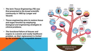
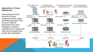
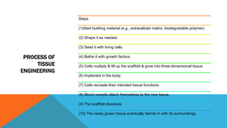
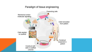
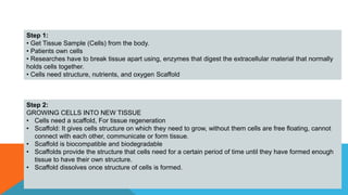
Tissue engineering involves using scaffolds, cells, and biomolecules to create functional 3D tissues. It aims to develop biological substitutes to restore tissue function and repair damaged tissues, avoiding problems with organ transplants, mechanical devices, and surgery. A major goal is designing scaffolds that recreate the in vivo microenvironment through biophysical and biochemical signaling. Stem cells are a promising cell source for their ability to integrate into tissues and secrete growth factors. Signaling molecules can also be used to modulate cell behavior. Magnetic targeting of stem cells may help with the challenge of cell retention in tissue engineering applications like cardiac repair.
Tissue engineering is the use of a combination of cells, engineering and materials methods, and suitable biochemical and physicochemical factors to improve or replace biological functions.
The term has also been applied to efforts to perform specific biochemical functions using cells within an artificially-created support system (e.g. an artificial pancreas, or a bio artificial liver).
A commonly applied definition of tissue engineering, as stated by Langer and Vacanti is “An interdisciplinary field that applies the principles of engineering and life sciences toward the development of biological substitutes that restore, maintain, or improve [Biological tissue] function or a whole organ”
This document provides an overview of tissue engineering as it relates to periodontal regeneration. It discusses the historical perspective of tissue engineering and need for strategies to regenerate lost periodontal tissues. Current strategies employed include using scaffolds, growth factors, and stem cells. Scaffolds provide a framework for cellular migration and integration. Sources of cells for tissue engineering include mesenchymal stem cells and various dental stem cells. Growth factors that have potential for periodontal regeneration include PDGF, IGF, TGF-β, and FGF. Future research directions include whole tooth regeneration using dental progenitor cells and scaffolds.
Biomaterials for tissue engineering slideshareBukar Abdullahi
An overview of Tissue Engineering with some basics in Biomaterials and Synthetic Polymers. Further references should be considered as I presented this a specific target audience.
This document summarizes tissue engineering and regeneration. It discusses sources of cells for tissue engineering including stem cells, somatic stem cells like mesenchymal stem cells, and induced pluripotent stem cells. It also discusses scaffolds for tissue engineering, both natural scaffolds derived from tissues and artificial scaffolds made from polymers or ceramics. The challenges of engineering tissues in vitro include delivering nutrients to 3D constructs, maintaining cell viability, and ensuring cell function is maintained. Safety concerns include tumor formation and transmitting infections. The future of the field involves using stem and iPS cells with improved scaffolds, and tailoring therapies to patient subgroups through molecular profiling.
The document summarizes the past, present, and future of regenerative tissue engineering. It discusses how the field began in the 1950s-60s by combining cell biology with new materials to generate living tissue components. Major advances included the use of stem cells and development of biocompatible scaffolds. The future of the field involves improved biomaterials that mimic natural extracellular matrix, bioprinting of complex tissues, and using various stem cell sources for cell therapy and organ regeneration to treat aging populations. The market for tissue engineering is estimated to grow substantially in coming years.
TISSUE DEVELOPMENT WITH TISSUE ENGINEERING APPROACHFelix Obi
Tissue Engineering is the development and practice of combining scaffolds, cells, and suitable biochemical factors (regulatory factors or Signals) into functional tissues. The goal of tissue engineering is to assemble functional constructs that restore, maintain, or improve damaged tissues or whole organs.
Cells are the building blocks of tissue, and tissues are the basic unit of function in the body. Generally, groups of cells make and secrete their own support structures, called extracellular matrix. This matrix, or scaffold, does more than just support the cells; it also acts as a relay station for various signaling molecules. Thus, cells receive messages from many sources that become available from the local environment. Each signal can start a chain of responses that determine what happens to the cell. By understanding how individual cells respond to signals, interact with their environment, and organize into tissues and organisms, Tissue Engineers are now able to manipulate these processes to amend damaged tissues or even create new ones.
Tissue engineering involves combining living cells with biomaterials to generate new living tissue. It aims to regenerate damaged or diseased tissues and organs. The process involves taking cells from a patient and growing them on a biodegradable scaffold. Once the new tissue forms, it is implanted to replace the damaged tissue. This allows tissue to be grown with the patient's own cells, avoiding rejection. Successful applications include growing skin to treat burns and cartilage to repair joints. Tissue engineering could solve the shortage of donor organs and offer permanent solutions for many medical conditions.
This document provides an overview of tissue engineering. It discusses the process of tissue engineering which involves using a scaffold material, seeding it with living cells, using growth factors, and implanting the new tissue. It also describes different types of stem cells, materials used for scaffolds, and methods to synthesize tissue engineered scaffolds. Applications of tissue engineering include bioartificial organs and tissues like skin, bone, and blood vessels. Both advantages and disadvantages of the field are mentioned.
Despite advances in organ transplantation, thousands still die each year waiting for donor organs. Tissue engineering aims to construct artificial organs and tissues in vitro by combining cells, biomaterials, and growth factors to replace diseased organs. Some key challenges include developing scaffolds that mimic the extracellular matrix, integrating multiple cell types, and applying mechanical and chemical signals to direct tissue development. While tissue engineering has shown promise for tissues like bone and skin, fully regenerating complex organs that do not naturally regenerate has yet to be achieved. Further research is still needed to meet clinical and patient expectations for safety, effectiveness and cost.
Stem Cells and Tissue Engineering: past, present and futureAna Rita Ramos
This document discusses the past, present, and future of tissue engineering and biomedicine. It covers the basics of tissue engineering, including the use of cells, scaffolds, and signals. It also discusses different cell sources for tissue engineering like embryonic stem cells, adult stem cells, and directed differentiation. Applications of stem cells in tissue engineering are also reviewed, such as engineering skin, bone, and cardiovascular tissues. The future of the field is seen to involve further understanding stem cell biology and differentiation, as well as moving from small lab experiments to large-scale production of cells and tissues.
Introduction
Definition
History
Principle
Cell sources
What cells can be used?
Scaffolds
Biomaterials
Bioreactor
How tissue engineering is done?
How does tissue engineering differ from cloning?
Tissue engineering of specific structures
Application of tissue engineering
Limitations
Conclusion
References
This document discusses 3D cell culture techniques. It defines 3D cell culture as permitting biological cells to grow and interact in all three dimensions, mimicking their natural environment. This is an improvement over 2D cultures. The document outlines various 3D culture methods including scaffold-based techniques using polymeric scaffolds, biological scaffolds, or micropatterned surfaces. Non-scaffold methods include hanging drop plates, spheroid plates, and microfluidics. Bioreactors and gels are also discussed. The document notes applications in regenerative medicine, drug testing, and modeling tumors or tissues. Considerations in choosing a 3D culture method include the specific application and ability to recreate in vivo barriers or cancers.
This document discusses 3D cell culture techniques. It defines 3D cell culture as permitting biological cells to grow and interact in all three dimensions, mimicking their natural environment. This is an improvement over 2D cultures where cells grow unnaturally. The document describes various 3D culture methods including scaffold-based techniques using polymeric scaffolds or biological scaffolds, and non-scaffold methods like hanging drop plates, spheroid plates, microfluidics, and gels. It also discusses bioreactors and lists applications of 3D cultures.
The document contains the answers to multiple choice and short answer questions regarding tissue engineering. It discusses three key components required for successful tissue engineering: implanted and cultured cells, biomaterial scaffolds, and biological signaling molecules. It also outlines growth factors involved in cell adhesion like PDGF, EGF, TGF-α, TGF-β, and IGF. Additionally, it states that current technology is not mature enough to develop adhesive tissues in the laboratory due to a need for more advanced techniques and an in-depth understanding of material ingredients and properties.
Tissue engineering involves using cells, biomaterials, and growth factors to regenerate damaged tissues and organs. There are several strategies for tissue engineering, including injecting stem cells, using scaffolds to guide cell growth, and inducing cell differentiation. Ideal scaffolds are biocompatible, porous, and gradually degrade as new tissue forms. Common scaffold materials include natural polymers, ceramics, and synthetic polymers. Tissue-engineered dental tissues are being developed by harvesting patient cells and growing them on scaffolds or as cell sheets to regenerate the periodontal ligament.
Tissue engineering aims to regenerate tissues by combining cells, scaffolds, and signaling molecules. There are two main strategies - in vitro construction of tissues in the lab prior to implantation, and in vivo regeneration of tissues at the implantation site. Successful tissue engineering requires the right cells, scaffolding for cell attachment and growth, and signaling to guide tissue development. Stem cells are promising cell sources due to their ability to differentiate into many cell types.
Three Dimensional Printing Scheme PresentationRita Barakat
3D bioprinting aims to simulate physiological environments to promote cell and tissue growth. Scaffolds allow cell attachment, migration, and diffusion of nutrients, and emulate the extracellular matrix. Common scaffold materials include hydrogels like agarose, gelatin, and collagen. 3D printing techniques like inkjet printing and extrusion methods are used to build scaffolds in a layer-by-layer process and incorporate cells and hydrogels. The goal is to develop techniques to print more complex, multicellular tissues and provide nutrients to maintain cell viability.
Revolution of 3 d organ model in pharmacological researchsyeddastagir9
3D organ models have gained interest as alternatives to animal testing in pharmacological research. This seminar discusses the revolution of 3D organ models with a focus on 3D bioprinting approaches. It describes various 3D bioprinting methods like biomimicry and autonomous self-assembly used to create tissue structures. Examples of 3D bioprinted structures for organs like liver, kidney, heart and neural tissue are provided. The seminar highlights current research using 3D bioprinting for applications like vascularization, drug development, and high-throughput screening.
Genes and Tissue Culture Assignment Presentation (Group 3)Lim Ke Wen
The culture of cells in two dimensions does not reproduce the histological characteristics of a tissue for informative or useful study. Growing cells as three-dimensional (3D) models more analogous to their existence in vivo may be more clinically relevant. Discuss the potential of using three dimensional cell cultures for anti-cancer drug screening.
3D-Bioprinting coming of age-from cells to organsDaniel Thomas
Over the past decade, annual spending on pharmaceutical development to treat many endocrinological systems has increased exponentially.
Currently, preclinical studies to test the safety and efficiency of new drugs, use laboratory animals and traditional 2D cell culture models. Neither of these methods are completely accurate reflections of how a drug will react in a human patient.
A solution has emerged in the form of 3D-Bioprinting technology, developed for the scalable, accurate and repeatable deposition of biologically active materials. With advances in this biomanufacturing technology, durable biological tissues for use in testing new pharmaceutical products are now being harnessed and refined.
Tissue engineering involves using cells, biomaterials, and suitable factors to engineer biological tissues and potentially whole organs. It combines principles of engineering and life sciences towards developing tissue substitutes that can restore, maintain, or improve tissue function. Key aspects include extracting cells like stem cells, seeding them onto biodegradable scaffolds that provide a template for new tissue growth, and using bioreactors to condition the developing tissues. Potential applications include growing skin, cartilage, bone, and other tissues for regenerative medicine purposes.
The Indian Dental Academy is the Leader in continuing dental education , training dentists in all aspects of dentistry and
offering a wide range of dental certified courses in different formats.
Bioprinting was defined as the use of material transfer processes for patterning and assembling biologically relevant materials- molecules, cells, tissues, and biodegradable biomaterials with a prescribed organization to accomplish one or more biological function. This is a developmental biology- inspired approach to tissue engineering and is based on the assumption that tissues and organs are self- organizing systems, and that cells and especially micro tissues can undergo biological self- assembly and self- organization without any external influence in the form of instructive, supporting and directing rigid templates or solid scaffolds.
Bioprinting or the biomedical application of rapid prototyping, also defined as layer- by- layer additive biomanufacturing, is an emerging transforming biomimetic technology that has potential for surpassing traditional solid scaffold- based tissue engineering. It is a rapid prototyping technology based on three dimensional, automated, computer-aided deposition of ‘‘bioink particles’’ (multicellular spheroids) into a ‘‘biopaper’’ (biocompatible gel; e.g. collagen) by a bioprinter
When I was asked to give a companion lecture in support of ‘The Philosophy of Science’ (https://shorturl.at/4pUXz) I decided not to walk through the detail of the many methodologies in order of use. Instead, I chose to employ a long standing, and ongoing, scientific development as an exemplar. And so, I chose the ever evolving story of Thermodynamics as a scientific investigation at its best.
Conducted over a period of >200 years, Thermodynamics R&D, and application, benefitted from the highest levels of professionalism, collaboration, and technical thoroughness. New layers of application, methodology, and practice were made possible by the progressive advance of technology. In turn, this has seen measurement and modelling accuracy continually improved at a micro and macro level.
Perhaps most importantly, Thermodynamics rapidly became a primary tool in the advance of applied science/engineering/technology, spanning micro-tech, to aerospace and cosmology. I can think of no better a story to illustrate the breadth of scientific methodologies and applications at their best.
PPT on Direct Seeded Rice presented at the three-day 'Training and Validation Workshop on Modules of Climate Smart Agriculture (CSA) Technologies in South Asia' workshop on April 22, 2024.
EWOCS-I: The catalog of X-ray sources in Westerlund 1 from the Extended Weste...Sérgio Sacani
Context. With a mass exceeding several 104 M⊙ and a rich and dense population of massive stars, supermassive young star clusters
represent the most massive star-forming environment that is dominated by the feedback from massive stars and gravitational interactions
among stars.
Aims. In this paper we present the Extended Westerlund 1 and 2 Open Clusters Survey (EWOCS) project, which aims to investigate
the influence of the starburst environment on the formation of stars and planets, and on the evolution of both low and high mass stars.
The primary targets of this project are Westerlund 1 and 2, the closest supermassive star clusters to the Sun.
Methods. The project is based primarily on recent observations conducted with the Chandra and JWST observatories. Specifically,
the Chandra survey of Westerlund 1 consists of 36 new ACIS-I observations, nearly co-pointed, for a total exposure time of 1 Msec.
Additionally, we included 8 archival Chandra/ACIS-S observations. This paper presents the resulting catalog of X-ray sources within
and around Westerlund 1. Sources were detected by combining various existing methods, and photon extraction and source validation
were carried out using the ACIS-Extract software.
Results. The EWOCS X-ray catalog comprises 5963 validated sources out of the 9420 initially provided to ACIS-Extract, reaching a
photon flux threshold of approximately 2 × 10⁻⁸ photons cm−2
s
−1
. The X-ray sources exhibit a highly concentrated spatial distribution,
with 1075 sources located within the central 1 arcmin. We have successfully detected X-ray emissions from 126 out of the 166 known
massive stars of the cluster, and we have collected over 71 000 photons from the magnetar CXO J164710.20-455217.
ESR spectroscopy in liquid food and beverages.pptxPRIYANKA PATEL
With increasing population, people need to rely on packaged food stuffs. Packaging of food materials requires the preservation of food. There are various methods for the treatment of food to preserve them and irradiation treatment of food is one of them. It is the most common and the most harmless method for the food preservation as it does not alter the necessary micronutrients of food materials. Although irradiated food doesn’t cause any harm to the human health but still the quality assessment of food is required to provide consumers with necessary information about the food. ESR spectroscopy is the most sophisticated way to investigate the quality of the food and the free radicals induced during the processing of the food. ESR spin trapping technique is useful for the detection of highly unstable radicals in the food. The antioxidant capability of liquid food and beverages in mainly performed by spin trapping technique.
Mending Clothing to Support Sustainable Fashion_CIMaR 2024.pdfSelcen Ozturkcan
Ozturkcan, S., Berndt, A., & Angelakis, A. (2024). Mending clothing to support sustainable fashion. Presented at the 31st Annual Conference by the Consortium for International Marketing Research (CIMaR), 10-13 Jun 2024, University of Gävle, Sweden.
The binding of cosmological structures by massless topological defectsSérgio Sacani
Assuming spherical symmetry and weak field, it is shown that if one solves the Poisson equation or the Einstein field
equations sourced by a topological defect, i.e. a singularity of a very specific form, the result is a localized gravitational
field capable of driving flat rotation (i.e. Keplerian circular orbits at a constant speed for all radii) of test masses on a thin
spherical shell without any underlying mass. Moreover, a large-scale structure which exploits this solution by assembling
concentrically a number of such topological defects can establish a flat stellar or galactic rotation curve, and can also deflect
light in the same manner as an equipotential (isothermal) sphere. Thus, the need for dark matter or modified gravity theory is
mitigated, at least in part.
Authoring a personal GPT for your research and practice: How we created the Q...Leonel Morgado
Thematic analysis in qualitative research is a time-consuming and systematic task, typically done using teams. Team members must ground their activities on common understandings of the major concepts underlying the thematic analysis, and define criteria for its development. However, conceptual misunderstandings, equivocations, and lack of adherence to criteria are challenges to the quality and speed of this process. Given the distributed and uncertain nature of this process, we wondered if the tasks in thematic analysis could be supported by readily available artificial intelligence chatbots. Our early efforts point to potential benefits: not just saving time in the coding process but better adherence to criteria and grounding, by increasing triangulation between humans and artificial intelligence. This tutorial will provide a description and demonstration of the process we followed, as two academic researchers, to develop a custom ChatGPT to assist with qualitative coding in the thematic data analysis process of immersive learning accounts in a survey of the academic literature: QUAL-E Immersive Learning Thematic Analysis Helper. In the hands-on time, participants will try out QUAL-E and develop their ideas for their own qualitative coding ChatGPT. Participants that have the paid ChatGPT Plus subscription can create a draft of their assistants. The organizers will provide course materials and slide deck that participants will be able to utilize to continue development of their custom GPT. The paid subscription to ChatGPT Plus is not required to participate in this workshop, just for trying out personal GPTs during it.
(June 12, 2024) Webinar: Development of PET theranostics targeting the molecu...Scintica Instrumentation
Targeting Hsp90 and its pathogen Orthologs with Tethered Inhibitors as a Diagnostic and Therapeutic Strategy for cancer and infectious diseases with Dr. Timothy Haystead.
3. Tissue Engineering is...
“an interdisciplinary field that applies the principles of engineering and life sciences
towards the development of biological substitutes that restore, maintain, or improve
tissue function or a whole organ”
4. • The term Tissue Engineering (TE) was
first presented to the broad scientific
community in 1993 by Langer and
Vacanti.
• Tissue engineering aims to restore tissue
and organ function by employing
biological and engineering strategies to
clinical problems.
• The functional failure of tissues and
organs is a severe and costly healthcare
problem, as their replacement is limited
by the availability of compatible donors .
5. Why Tissue Engineering is Important
• Supply of donor organs cannot keep up with demand.
• Other available therapies such as surgical reconstruction, drug therapy, synthetic
prostheses, and medical devices aren't always successful.
• It will eliminate any risk of organ rejection because the new organ would be made
from the person's own tissue.
• It repairs tissues, organs, and bones successfully
• Victims of organ/tissue defects will not have to suffer
6. Principle of Tissue Engineering:
The general principles of tissue engineering involve combining living cells with a
natural/synthetic support or scaffold that is also biodegradable to build a three dimensional living
construct that is functionally, structurally and mechanically equal to or better than the tissue that
is to be replaced.
7. Regenerative medicine is a broad field that includes tissue engineering but also incorporates
research on self-healing – where the body uses its own systems, sometimes with help foreign
biological material to recreate cells and rebuild tissues and organs.
The terms “tissue engineering” and “regenerative medicine” have become largely
interchangeable, as the field hopes to focus on cures instead of treatments for complex, often
chronic diseases.
This field continues to evolve. In addition to medical applications, non-therapeutic applications
include using tissues as biosensors to detect biological or chemical threat agents, and tissue
chips that can be used to test the toxicity of an experimental medication.
Examples include cell therapies (the injection of stem cells or progenitor cells);
immunomodulation therapy (regeneration by biologically active molecules administered alone or
as secretions by infused cells); and tissue engineering (transplantation of laboratory grown
organs and tissues).
8. The Triad of Tissue Engineering
Tissue engineering applications typically
involve the combination of three pillars: cells,
signals, and scaffolds, which represent the
“triad of tissue engineering”.
9. Approaches in Tissue
Engineering
Incorporating the three
elements of tissue
engineering needs a good
scaffolding technique. Over
the years, different
approaches have been
developed, resulting in
scaffolds that can support the
cells and encourage tissue
growth after implantation.
10. PROCESS OF
TISSUE
ENGINEERING
Steps
(1)Start building material (e.g., extracellular matrix, biodegradable polymer).
(2) Shape it as needed.
(3) Seed it with living cells.
(4) Bathe it with growth factors.
(5) Cells multiply & fill up the scaffold & grow into three-dimensional tissue.
(6) Implanted in the body.
(7) Cells recreate their intended tissue functions.
(8) Blood vessels attach themselves to the new tissue.
(9) The scaffold dissolves.
(10) The newly grown tissue eventually blends in with its surroundings.
11.
12. Step 1:
• Get Tissue Sample (Cells) from the body.
• Patients own cells
• Researches have to break tissue apart using, enzymes that digest the extracellular material that normally
holds cells together.
• Cells need structure, nutrients, and oxygen Scaffold
Step 2:
GROWING CELLS INTO NEW TISSUE
• Cells need a scaffold, For tissue regeneration
• Scaffold: It gives cells structure on which they need to grow, without them cells are free floating, cannot
connect with each other, communicate or form tissue.
• Scaffold is biocompatible and biodegradable
• Scaffolds provide the structure that cells need for a certain period of time until they have formed enough
tissue to have their own structure.
• Scaffold dissolves once structure of cells is formed.
13. Step 3:
IMPLANTING NEW TISSUE
• Bioengineered Tissue Implants Regenerate Damaged Knee Cartilage Science Daily.
• Cartilage was removed from 23 patients with an average age of 36 years.
• After growing the cells in culture for 14 days, the researchers seeded them onto scaffolds
made of esterified hyaluronic acid, grew them for another 14 days on the scaffolds, and then
implanted them into the injured knees of the study patients.
• Cartilage regeneration was seen in ten of 23 patients, including in some patients with
preexisting early osteoarthritis of the knee secondary to traumatic injury.
• Maturation of the implanted, tissue-engineered cartilage was evident as early as 11 months
after implantation.
14. The potential of stem cells
capable of self-renewal- can divide and renew themselves for long periods
unspecialized cells that can differentiate into other types of cells
15.
16. SCAFFOLDS:
• Cells are often implanted or 'seeded' into an
artificial structure capable of supporting
three-dimensional tissue formation. These
structures, typically called scaffolds usually
serve at least one of the following purposes:
• Allow cell attachment and migration.
• Deliver and retain cells and biochemical
factors.
• Enable diffusion of vital cell nutrients and
expressed products.
• Exert certain mechanical and biological
influences to modify the behavior of the cell
phase.
17. Requirements for scaffolds
• High porosity and an adequate pore size
• Biodegradability rate at which degradation occurs has to coincide as much as
possible with the rate of tissue formation
• Inject ability
18. DIFFERENT TYPES OD SCAFFOLD FABRICATION
TECHNIQUES
• The conventional tissue engineering scaffold production techniques include thermally
induced phase separation (TIPS), fiber bonding, electrospinning, solvent casting and
particulate leaching, membrane lamination, freeze-drying, and gas foaming .
• The recent development in scaffold fabrication mainly comprises integration with computer-
aided design (CAD) software such as stereolithography, bioplotting, solvent-based free
forming, combination modelling technique, fused deposition modelling, 3D printing, and
selective laser sintering (SLS) .
• The techniques retain the ability to maintain pore structures, cell–cell interaction, reduction
in mechanical instability, and control over mitigation of the cellular matrix.
19. Scaffold Fabrication:
• Scaffold fabrication is a critical step in tissue engineering as it provides the structural
support necessary for cell attachment, proliferation, and differentiation.
• There are several methods used to fabricate scaffolds:
1. 3D Printing: Also known as additive manufacturing, 3D printing allows for the precise
layer-by-layer deposition of materials to create scaffolds with complex geometries and
controlled porosity. This technique enables customization of scaffold properties such as
pore size, interconnectivity, and mechanical strength.
20. 2. Electrospinning: Electrospinning involves the use of an electric field to draw polymer solutions or melts into
ultrafine fibers. These fibers are collected to form nanofibrous scaffolds with a high surface area-to-volume ratio,
which can closely mimic the native extracellular matrix (ECM) architecture.
3. Decellularization: Decellularization is a technique where cellular components are removed from natural tissues,
leaving behind an acellular scaffold composed of the ECM. These scaffolds retain the native tissue's biochemical
composition and mechanical properties, making them suitable for tissue engineering applications.
21. Cell Seeding Techniques:
• Once scaffolds are fabricated, cells need to be introduced onto or into them to initiate tissue formation.
• Several cell seeding techniques are employed:
1. Static Seeding: In static seeding, cells are typically pipetted or seeded directly onto the scaffold surface
and allowed to adhere through gravity or gentle agitation. While simple and straightforward, static seeding
may result in uneven cell distribution.
2. Dynamic Seeding: Dynamic seeding involves the use of bioreactor systems where scaffolds are placed in
a chamber with controlled flow conditions. This allows for better distribution of cells throughout the scaffold,
enhancing cell attachment and proliferation.
22. 3. Perfusion Seeding: Perfusion seeding takes dynamic seeding a step further by continuously perfusing cell
suspension through the scaffold using a pump system. This method ensures uniform cell distribution and nutrient
delivery, promoting cell viability and tissue formation.
23. Tissue Culture Methods:
• Tissue culture techniques are employed to maintain cells or tissues in vitro under controlled
conditions.
• This is essential for studying cellular behavior, tissue development, and for producing
functional tissue constructs. Key tissue culture methods include:
1. 2D Cell Culture: In 2D cell culture, cells are typically grown as monolayers on flat surfaces
such as tissue culture plates. While simple and widely used, 2D culture lacks the three-
dimensional context present in vivo, which may affect cell behavior and function.
2. 3D Cell Culture: 3D cell culture involves culturing cells in three-dimensional environments
that mimic the in vivo tissue architecture more closely. This can be achieved using
hydrogels, scaffolds, or spheroid culture methods, providing cells with a more
physiologically relevant environment.
3. Bioreactor Culture: Bioreactors are dynamic culture systems that provide controlled
conditions such as temperature, pH, oxygen levels, and mechanical stimulation. Bioreactor
culture allows for the scaling up of tissue constructs and the simulation of physiological
conditions, making it suitable for tissue engineering applications.
24. Advantages and Disadvantages of different types of scaffolds fabrication techniques for tissue engineering
application.
27. APPLICATIONS OF TISSUE ENGINEERING
• The main goal of tissue engineering is to regenerate and replace human tissues and organs through a
combination of biological, clinical, and engineering approaches.
Replacing/Regenerating Target Organs
• Skin:
• Tissue-engineered skin aims to restore barrier function to patients for whom this has been severely
compromised, e.g., burn patients.
• Skin is a widely explored engineered tissue, and several commercial skin products are available, e.g., Epicel®
by Genzyme is a product based on autologous cells grown to cover a wound; Apligraf® by Organogenesis is a
dual layer skin equivalent with keratinocytes and fibroblasts on collagen gel; Dermagraft® by Advanced Tissue
Science uses a similar approach with dermal fibroblast on resorbable polymer, among many others .
• These products treat burns, ulcers, deep wounds, and other injuries. Collagen, fibrin, hyaluronic acid, and
poly(lactic glycolic acid) are mainly used in skin substitute matrices. Keratinocytes, melanocytes, and
fibroblasts compose the majority of cells in skin tissue, thus expanding and transplanting these cells within
biocompatible matrices is key to successful skin regeneration .
28. Liver:
• Liver transplantation is end-stage treatment for many liver diseases. Several factors including drug
use, alcohol abuse, and viruses like Hepatitis can cause acute liver failure. It is important to
completely replace the damaged liver or support the patients that wait for donor organs or suffer
from chronic liver diseases with tissue-engineered livers .
• Intense efforts exist to develop a bridging device that can support a patient’s liver function until a
donor is available, e.g., dialysis, charcoal hemoperfusion, immobilized enzymes or exchange
transfusion. Several extracorporeal systems use patients’ own cells in a hollow-fiber, spoutedbed or
flat-bed device, which reduce the chance of immune rejection.
• Several bioartificial livers (BAL) have been developed and designed to flow patient’s plasma through
a bioreactor that houses/maintains hepatocytes sandwiched between artificial plates or capillaries.
29. Pancreas:
• Diabetes is the fifth highest cause of death . One type of diabetes is type 1 diabetes mellitus
(T1DM), an autoimmune disease which destroys the insulin-secreting cells of the pancreas. T1DM is
relevant to tissue engineering, since it can be treated by replacing the destroyed pancreatic islet
cells.
• Techniques for pancreatic tissue engineering aim to release insulin from transplanted islets into the
blood to restore normal blood glucose levels.
• Three main approaches are used: a tubular membrane that encapsulates islets and connects to
blood vessels; hollow fibers containing islets embedded in a polymer matrix; and encapsulation of
islets in microcapsules .
• The membranes used in the perfusion devices and coatings in microcapsules are developed from
biocompatible polymers that allow insulin to diffuse into the bloodstream, while protecting the cells
from destruction by immune cells. An insufficient source of islet cells presents a challenge, but
recent advances with stem cells may overcome this.
30. Heart
• Many patients are left with damaged or malfunctioning cardiac tissue that lead to arrhythmias and diminished
cardiac output. Tissue engineering is actively pursing treatments for myocardial infarction, congenital heart
defects, and stenotic valves through regenerating cardiac tissues.
• Heart valves are developed by transplanting autologous cells onto a scaffold, growing and maturing the cell-
seeded scaffold, and finally transplanting the valves into the patient.
• Decellularized heart valves consist of extracellular matrix which is repopulated with host cells, but they can
potentially produce severe immune response .
• Alternatively, biomaterial-based heart valves are designed from various natural materials, e.g., collagen,
fibrin, and synthetic polymers, e.g., PLGA, poly(hydroxy butyrate) . Biomaterial-based heart valves have
advantageous characteristics, including malleability and improved mechanical strength.
• Cells for cardiovascular applications are usually obtained from donor tissues, e.g, peripheral arteries with
mixed populations of myofibroblasts and endothelial cells, as well as established cell lines of myofibroblasts
and endothelial cells. There are several successful applications of tissue-engineered heart valves for both in
vitro and in vivo models. Additionally, efforts have been made to regenerate myocardium by scaffold-based
approaches with natural and synthetic materials
31. Blood vessels:
• Poly(tetrafluoro ethylene) PTFE and Dacron® grafts have traditionally been used as vascular grafts,
particularly for large diameter vessels. However, these grafts are largely unsuccessful for small diameter
blood vessels, due to thrombogenicity and compliance mismatch.
• Tissue engineering strategies therefore provide great opportunities for development of blood vessels.
Decellularized arteries with well-preserved extra cellular proteins have been repopulated with cells both
in vitro and in vivo to generate blood vessels .
• Polymer-based scaffolds are often used as a template to guide the regeneration of blood vessels.
• Also, recently developed microfluidic and microfabrication techniques allow the complex architecture of
a blood vessel with defined microstructures to be realized .
• In an alternative approach, sheets of smooth muscle cells and extracellular matrix are generated under
flow conditions in the absence of scaffold, and are subsequently rolled over a support to mature into a
vessel structure .
• An emerging approach focuses on combining endothelial cells with mesenchymal stem cells or smooth
muscle cells to help stabilize the blood vessel and help aid in the vessel pruning process .
• It is important to note that formation of new blood vessels is critical for engineering any tissue to supply
nutrients and oxygen to cells.
32. Nervous system:
• Generation of nerve tissues is a major focus in tissue engineering . Injuries in the central nervous
system (CNS) are often accompanied by permanent functional impairment, unlike peripheral
nervous system (PNS) damage where the axons are able to re-extend and re-innervate, leading to
functional recovery .
• This is because the environment of the damaged site in CNS blocks the regeneration of neurons.
• Peripheral nerve grafts that include synthetic or biological substrates have been developed to act as
a bridge to guide the nerve regeneration process. To restore the structures lost in the
disorganization of axons during injury it is critical to build a bridge that spans the lesion gap with all
the morphological, chemical, and biological cues that mimic normal tissue.
33. Bone:
• There are numerous clinical applications that can benefit from bone regeneration therapies
including spinal fusion to alleviate back pain, temporomandibular joint reconstruction to alleviate
jaw pain, and restoration of contour and shape within reconstructed craniofacial bone.
• Significant progress in the development of bone regeneration therapies has been achieved, yet
large critical-sized defects which do not heal remain a significant clinical challenge. Although
several inorganic-, polymeric-, and hybrid-based scaffolds have been examined to engineer bone,
it has been difficult to develop a material that displays optimal mechanical properties and
degradation kinetics for bone repair .
• It is clear that the scaffolds for bone regeneration should have an interconnected macroporosity to
allow three-dimensional bone growth throughout the scaffold.
• Implantation of empty macroporous scaffolds that are devoid of cells typically do not improve the
healing response though combining biodegradable scaffolds with cells is a commonly explored
strategy
34. Other Applications of Tissue Engineering
• There are several non-traditional, yet useful and important applications of tissue engineering strategies.
In particular, the field of drug screening is in need of advanced in vitro methods for the assessment of
the activity and toxicity of drugs before clinical studies are initiated .
• Since most drugs are metabolized in the liver, microscale hepatocyte systems may allow high throughput
screening for liver toxicity . Furthermore, humans or animals “on a chip” are in development.
• These chips are essentially several reactors with different cell types connected in series which can
stimulate the pathway of a chemical substance through several parts of the body, such as the liver, brain,
and fat.
• In the future, tissue engineering-based drug testing and toxicity assays can provide an alternate strategy
to reduce the use of in vivo animal testing. Moreover, an artificially engineered tissue-engineered system
can provide enhanced control of tissue microenvironments, both in physiological and pathological
conditions (e.g., cancer tumor), contributing to the understanding of complex tissues and organs
35. • Tissue engineering is a young field that
utilizes cells, biomaterials, physical
signals (e.g., mechanical stimulation),
biochemical signals (e.g., growth factors
and cytokines), and their combinations,
to engineer tissues.
• The most common application of tissue
engineering is to create tissues that can
be used to repair or replace tissues in
the body suffering partial or complete
loss of function.
• However, tissue engineering has started
to find new applications such as the
development of extracorporeal life
support units (e.g., bioartificial liver and
kidney), in vitro disease models, tissues
for drug screening, smart diagnosis,
and personalized medicine.
36. Year Achievements Reference
1907 Dr. Harrison Ross first observed
living developing nerve fiber
Harrison Rose et al., 1907
1948 First artificial Kidney was
synthesized
Gauvin et al., 2011
1988 3D positioning of cells Klebe RJ , 1988
1993 Term "Tissue engineering" was
defined
Klebe RJ ,1988
1994 Biofabrication Fritz Monika,1994
1997 Commercially available skin Mac Neil ,2007
2002 Commercially available bone Parikh, 2002
2003 Patent for bio-printer Mironov et al., 2003
2008 Decellularized organ Ott et al., 2008
2010 10-year-old child saved Kalathur et al.,2010
2015 Development of soft tissue Pati et al ., 2015
2019 Development of new
stereolithographic process for multi-
vascular networks
Grigoryan et al.,2019
Timeline for the milestone in Tissue engineering.
37. Cao, Vacanti et al., (1997)
The Vacanti mouse ear was created using a biodegradable
polymer scaffold shaped like a human ear. The scaffold was
seeded with chondrocytes, which are cartilage cells, and implanted
under the skin on the back of a laboratory mouse. Over time, the
chondrocytes proliferated and synthesized new cartilage tissue,
resulting in the growth of an ear-shaped structure.
38. Different tissue-engineered organs.
(a) Scaffold prepared from synthetic biodegradable
polyglycolic acid (PLA) in the shape of a 3-year-old
auricle.
(b) Scaffolds implanted subcutaneously on the back of an
immunodeficient mouse.
(c) First trachea organ transplant using human's bone
marrow stem cells.
(d) Constructed artificial bladder seeded with human
bladder cells and dipped in a growth solution.
(e) Bioengineered kidney that mimics the function of a
normal kidney the control of the urinary system and
blood filtration.
(f) Tissue-engineered heart valve using human marrow
stromal cells.
39. Challenges in Tissue Engineering
• In fact, autologous, allogeneic, and xenogeneic cells are all potential sources, and each of these can be
subdivided into stem cells (adult or embryonic) or differentiated cells. Since they all have their own
advantages and disadvantages (immune reaction, differentiation, etc.), the choice of the right source for
the cells and their culture is a challenge by itself.
• The choice of scaffold biomaterials is not an easier task either. The scaffolds must actually respond to
both the structural and functional requirements of the body. It must be biocompatible and should be able
to communicate with the ECM while at the same time providing the needed mechanical support. While
natural materials have better biocompatibility and biodegradability, synthetic ones usually present
stronger mechanical properties.
• Another important challenge in tissue engineering is related to the transportation of nutrients and waste
secretion in the engineered tissue . Since the majority of tissues rely on blood vessels to transport
oxygen and nutrients, the 3D engineered tissue needs to be vascularized with a vascular capillary
network .
• Finally, a major challenge is still present, namely mass production and commercialization of the
engineered tissues. Specific manufacturing conditions and quality control strategies need to be ensured.
40. The Future of Tissue Engineering
• The last few decades have witnessed major steps in health care, leading to improved surgical
procedures and better management of diseases.
• Consequently, the aforementioned advancements have led to an increased demand for tissues and
organs.
• The ultimate goal of tissue engineering is to bridge the constantly growing gap between organ demand
and availability by producing complete organs .
• Stem cells will continue to be investigated for their differentiation potential, and more applications will be
developed in the future.
• The ultimate goal would be to engineer immune-transparent stem-like cells with clear protocols, enabling
their committed differentiation to targeted tissues. Developments in basic and applied science related to
the fabrication of tissue engineering scaffolds will be a major future target.
• Potential limitations of such scaffolds will always be the shortage of supplies (e.g., scaffolds from
allogeneic sources), potential immunoreactions, and ethical concerns (e.g., scaffolds from xenogeneic
origins). In future, it is expected that new biomaterials will be developed incorporating selected molecules
to address targeted tissues.
• Moreover, many basic science studies will be conducted to identify the effects of molecules on cells and
determine the right degradation rate and material properties (porosity, mechanical properties, and
structural properties) suitable for each tissue engineering applications.
41. Advantage of tissue engineering
• Tissue engineering provides long term and much safer solution than other options.
• The traditional transplantation complications are minimized.
• The donar can be patients himself or herself.
• The need for donar tissue is minimal.
• Immuno suppression problem can be minimized.
• The presence of residual foreign material can be minimized.
Disadvantage of tissue engineering
• Cell isolation and preparation, biomaterial of nutrients transport and transplantation is very
complex process.
• It is difficult to achieve cell diffrentiation into desired cell type and ensuring their nutrient supply
after implantation into the body.
• There may be obstacles to growing cells in sufficient quantities.
• The time necessary to develops cells in culture before they can be used and possible lack of
function at the donar site are some other limitations.