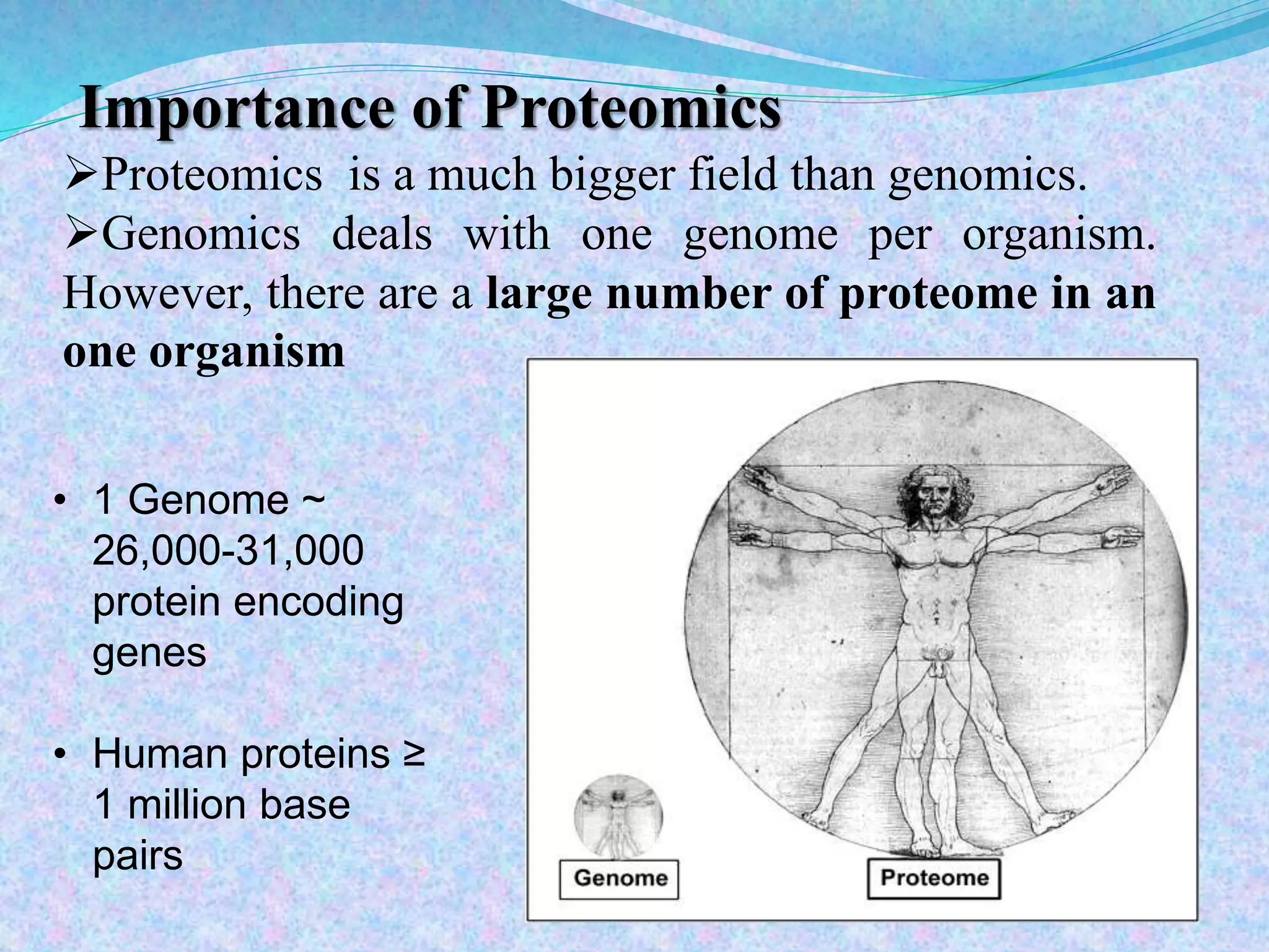
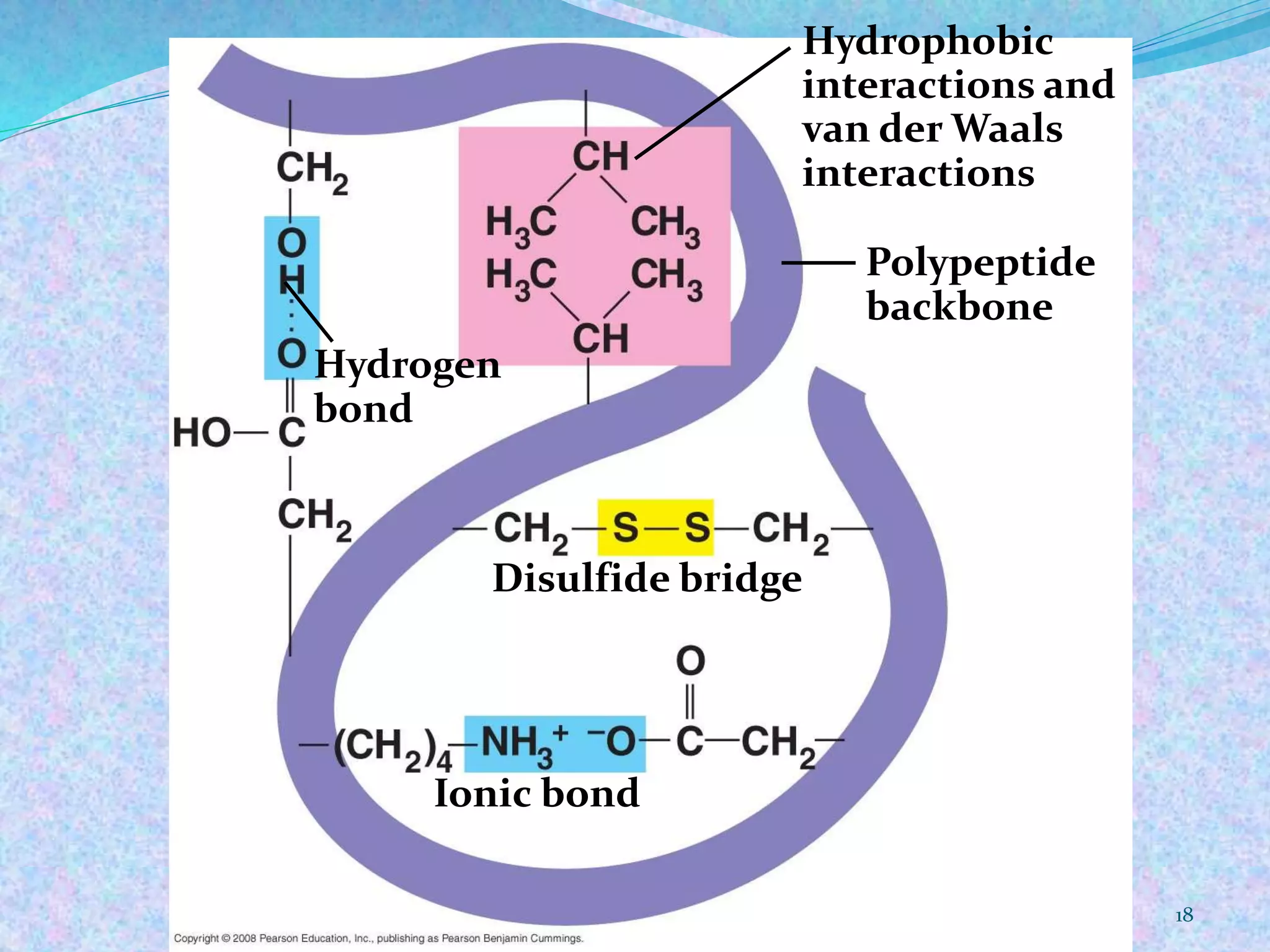
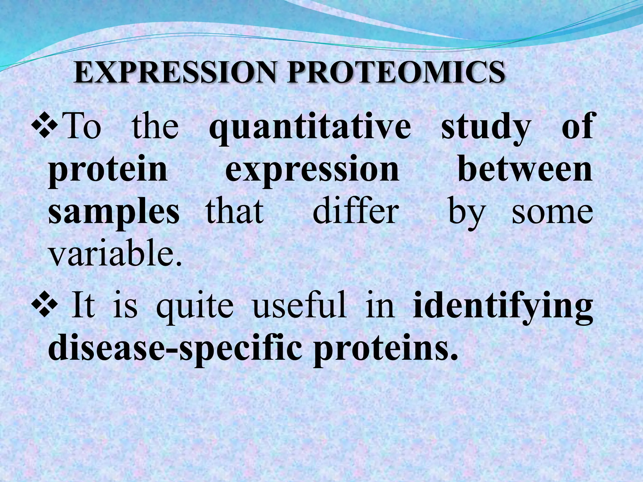
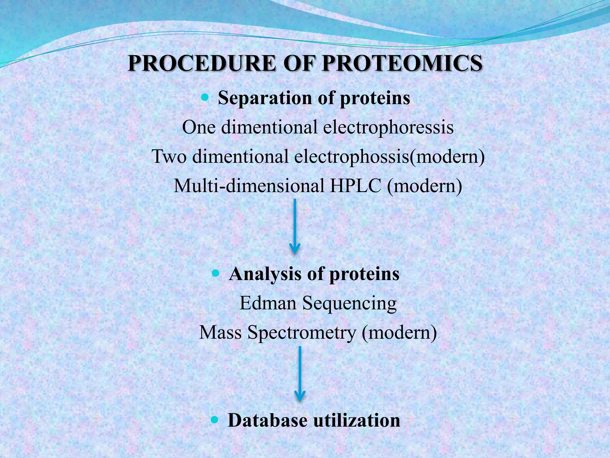
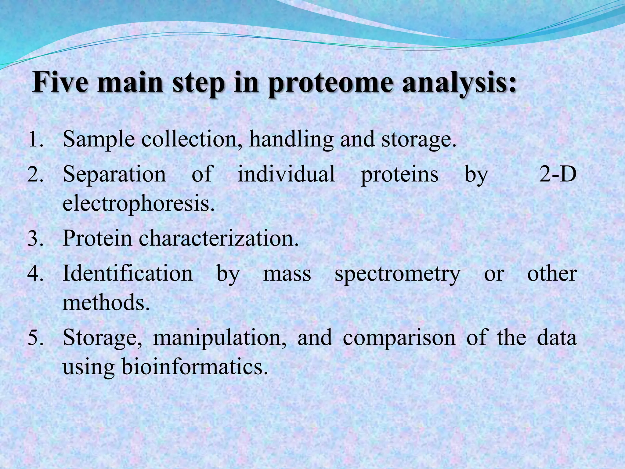
2. Submitted to
P.M. MISTRY
Assistant professor
Dept. of Genetics and Plant Breeding
N.M. College of agriculture
NAU, Navsari-396 450
Submitted by
Name: SENTHILKUMAR.V
Dept. of Genetics and Plant Breeding
M.sc.( Agri.) 1st semester
N.M. College of agriculture
NAU, Navsari-396 450
PROTEOMICS AND PROTEIN – PROTEIN INTERACTION
2
6. PROTEOMICS INTRODUCTION
PROTEOMICS is the study of the proteome, the full
protein complement of organisms e.g. plasma, cells
and tissue.
PROTEOMICS is the large scale study of proteins,
particularly their structures and functions
Understanding the proteome allows for:
Characterisation of proteins
Understanding protein interactions
Identification of disease biomarkers
7. HISTORY
“The Total Protein Complement Of A Genome.”
M. Wilkins Et Al. Electrophoresis 1995, 16, 1090-1094
“The Analysis Of The Entire Protein Complement
Expressed By A Genome, Or By A Cell Or Tissue Type.”
S. Fey, P.Mose-larsen, Centre For Proteome Analysis,
Odense, Denmark
Pharmaceutical Proteomics: “Proteome Approach To The
Interaction Of Drugs With Biological Systems.”
L. Anderson, Large Scale Biology, MA, USA
8. Importance of Proteomics
Proteomics is a much bigger field than genomics.
Genomics deals with one genome per organism.
However, there are a large number of proteome in an
one organism
• 1 Genome ~
26,000-31,000
protein encoding
genes
• Human proteins ≥
1 million base
pairs
11. DNA
Transcription Transcriptional control
RNA
Processing Post-transcriptional control
Translation Translational and degradation
Controls Translational frameshifting
mRNA
Protein > 200 known
post translation modifications
Fig. Protein Synthesis pathway.
FOCUS OF PROTEOMICS STUDIES
12. Aims of post translation
studies
Detect the protein expression by cell
to cell.
Store those information about
protein functions.
Compare the expression profiles
between a healthy cell vs diseased
cell.
Rational drug design.
15. FOUR LEVELS OF PROTEIN STRUCTURE
The primary structure of a protein is its unique
sequence of amino acids.
The Secondary structure, found in most proteins,
consists of coils and folds in the polypeptide chain.
The Tertiary structure is determined by interactions
among various side chains (R groups).
The Quaternary structure results when a protein
consists of multiple polypeptide chains.
STRUCTURE OF PROTEIN
20. TECHNIQUES INVOLVED TO STUDY OF PROTEIN
STRUCTURES
1) VIRTUAL LIGAND SCREENING
2) MALDI -TOF (Matrix Assisted Laser Desorption -
Time of Flight)
3) ESI -MS (Electro-spray ionization mass
spectrometry)
21. VIRTUAL LIGAND SCREENING
A computer technique, attempts to fit millions of
small molecules to 3D structure of protein.
The computer rates the quality of fit to various sites
in the protein with the goal of either enhancing or
disabling the function of protein in the cell.
22.
23. 1.Introduced in 1988.
2.It provides for the nondestructive vaporization
and ionization of both large and small
biomolecules.
3.Matrix play a key role.
MALDI-TOF TECHNIQUE
(Matrix-Assisted-Laser-Desorption-Ionization-
Time of Flight)
24. MALDI-TOF (MATRIX ASSISTED LASER DESORPTION-
Time Of Flight)
Protein placed on light absorbing material, and with a short
pulse of laser protein are ionized and desorbed in to
vacuum system known as MATRIX ASSISTED LASER
DESORPTION.
25. Time of Flight (TOF)
1.Accelerating a set of ions to a detector with the
same amount of energy.
2.Ions have the same energy, yet a different mass they
reach the detector at different time.
3.Arrival time of an ion at the detector is dependent
upon the mass, charge and kinetic energy of the ion.
26. Electro-spray Ionization Mass
Spectrometry (ESI MS)
Solution are forced directly from liquid
to gas phase.
A solution of analytes pass through
charged needle that is kept at high
electricpotential dispersed in to charged
microdroplets.
The solvent surrounding the
macromolecules rapidly evaporates, and
the resulting multiply charged
macromolecular ions are thus
introduced nondestructively in to a gas
is to be called as ESI MS.
Fenn's first electrospray
ionization source (top) coupled to
a single quadrupole mass
spectrometer
29. EXPRESSION PROTEOMICS
To the quantitative study of
protein expression between
samples that differ by some
variable.
It is quite useful in identifying
disease-specific proteins.
30. STRUCTURAL PROTEOMICS
To Concerns with mapping out the 3-D
structure and nature of protein complexes
present specifically in a particular
cell/organelle.
Aim of structural proteomics is to build a
body of structural information that will help
predict the probable structure and potential
function for almost any protein from a
knowledge of its coding sequence.
31. FUNCTIONAL PROTEOMICS
To study protein-protein interaction, cellular
localization and in order to understand the
physiological function of the whole set of proteome.
One of the recent successes of functional proteomics
is the identification and analysis of molecular
protein-network involved in the nuclear pore
complex (NPC) in yeast.
This success helps understand the translocation of
molecules from nucleus to the cytoplasm and vice-
versa.
35. Techniques of Structural Proteomics
High Throughput protein
structure determination
via
1) X-ray crystallography,
2) NMR spectroscopy or
3) comparative molecular
modeling
X-ray crystallography,
36. Techniques of Functional Proteomics
Silico method
Genome-wide Protein Tagging
Genome-wide Gene Deletion
Random Tagged Mutagenesis or Transposon Tagging
Yeast two-hybrid Methods(protein –protein interaction)
Protein Chips
38. PROTEOMICS ANAYSIS
AND STEPS
Sample prep.
• Immunoaffinity
depletion
• BCA protein assay
• Digestion of proteins
• Concentration
Sample analysis
• Spiking with internal
standard
• Blind run for protein
loading estimation
• Analysing samples in
triplicate
Bioinformatics
• Identification &
quantification using
• Expression analysis
39. PROCEDURE OF PROTEOMICS
Separation of proteins
One dimentional electrophoressis
Two dimentional electrophossis(modern)
Multi-dimensional HPLC (modern)
Analysis of proteins
Edman Sequencing
Mass Spectrometry (modern)
Database utilization
41. Five main step in proteome analysis:
1. Sample collection, handling and storage.
2. Separation of individual proteins by 2-D
electrophoresis.
3. Protein characterization.
4. Identification by mass spectrometry or other
methods.
5. Storage, manipulation, and comparison of the data
using bioinformatics.
42. FIRST STEP
Sample collection,
Handling and
Storage.
Sample prep.
•Immunoaffinity depletion
•BCA protein assay
•Digestion of proteins
•Concentration
44. 3.IDENTIFICATION AND
CHARACTERIZATION OF PROTEINS
.
•The primary method is electrophoresis or chromatography
coupled with mass spectrometry(MS).
•Defining the protein composition of a cell
A. mRNA splicing
B. covalent modification generate protein isoforms that
might contribute to important regulatory processes in the
cell.
C. Several approaches are being used to study post-
translational modifications on a proteome-wide scale.
46. Mass Spectrometry (MS)
• Mass spectrometry is used for protein identification.
•It is useful to obtain structural information like peptide
mass sequence.
•It is also useful in identifying type and location of
protein modification.
•A mass spectrometer separates proteins according to
their mass-to-charge(m/z) ratio. The molecule is first
ionized.
•The process of ionization of proteins forces them to move
towards the analyzer because of the charges on ions.
•MS can provide molecular weight and structural
information.
•MS always work with positive ions.
50. Protein Fingerprinting
• Protein fingerprinting , also called peptide mass fingerprinting
or peptide mapping.
•It is a technique for identification of proteins.
•Separated protein spots are obtained from the gel and then
identified using protein fingerprinting.
•The method is based on the use of a proteolytic enzyme to
digest the protein into a number of smaller peptides.
•Finger printing of the protein depends on the protease used, but is
always the same for each one.
•The most commonly used protease is trypsin, which cuts protein
at lysine and arginine positions.
•When the digestion in complete, a set of peptides are produced of
varying masses that are unique to that protein.
51.
52. Two –dimensional Gel Electrophoresis
• 2-D gel electrophoresis a method for the separation and
identification of proteins in a sample by displacement in 2
dimensions.
• First step is to separate based on charge or isoelectric point,
called isoelectric focusing.
•Then separate based on size (SDS-PAGE).
53. Isoelectric Focusing
•The isoelectric point is the pH at which the net
charge of the protein molecule is neutral.
•Different proteins have different isoelectric
points.
•Isoelectric point is found by drawing the sample
through a stable pH gradient.
•The range of the gradient determines the
resolution of the separation.
54. 5.Protein databases
UniProt
Protein Information Resource (PIR)
Swiss-Prot
Protein Data Bank (PDB)
National Center for Biotechnology Information
(NCBI)
Human Protein Reference Database
Proteomics Identifications Database (PRIDE)
55. POST-TRANSLATIONAL MODIFICATION
(PMTs)
• A protein can under go post-translational modification and hence
various proteins can be formed from a single gene.
•After transcription from DNA to RNA , the gene mRNA can be
spliced in different ways prior to translation into protein.
•Following translation, most proteins are chemically modified through
post-translation modification, mainly through the addition of
carbohydrate and phosphate groups.
•PTM events can affect nearly all properties of 3-D structure of a
protein: size, charge , hydrophobicity.
•A phosphorylation event would change the local environment
substantially, making likely changes in the 3-D structure.
•As proteomics is the elucidation of the totality of protein-related
events in the cell, it also includes PTM protein variants.
57. Protein – Protein interaction
Proteins often interact with each
other in very complex ways.
Proteins may act upon and be acted
upon by many other proteins in the
cell, or outside the cell.
That’s figuring out the function of
a gene’s protein product.
58. Individual proteins are rarely
able to perform their functions by
themselves.
Shown here, a protein
embedded in the membrane of the
cell is interacting with six other
proteins.
Protein-protein interaction
59. Types of Protein – Protein
interaction
Domain – domain
interactions
Domain – Peptide
interactions
Intramolecular protein-
protein interactions
60. Domain –Domain
Interactions
In domain –domain interactions, two independently
folded domains (completed protein)
It is interacted with more than one segments of
polypeptide
61. Domain – Peptide interactions
In interaction between a small unstructured proteins of
one structure and a folded domain protein.
64. Biochemical methods
1.Co-immuno precipitation
This method considered to be the gold standard
assay
Its isolated with specific antibody
2.Biomolecular fluroscence complementation
This method used for screening the protein –
protein interactions
3.Affinity electrophoresis
This method characterization of molecule
identify the glycan content and ligand binding
65. 4.Pulldown assay
Screening the protein-protein interactions
This methods combining from immuno
precipitation and affinity electro phoreosis
5.Tandam affinity purificaton
This method detect the transistant protein-
protein interactions
6.Strep –protein interaction
It is uses a combination of reversible link with
formaldehyde
66. Biophysical and theoretical
methods
Dual polarisation interferometry
DPI provides real time ,high resolution mesurements of molecular
size,density and mass based on kinetics principles
Static light shattering
This method used for rayleigh scattering of protein complexes
Fluorscence energy 2D FT –NMR spectro scopy
Basic method study in protein-protein interactions based on
fluorscence emitted.
Protein-protein docking
This method based on three dimensional protein structure from x-ray
diffraction of protein crystals
67. YEAST TWO-HYBRID SYSTEM FOR
PROTEIN-PROTEIN INTERACTION
•Most of the yeast two- hybrid systems utilize the reconstitution of an
active transcription factor to assay for protein-protein to make in
interactions.
•The Y2H system uses the trancription process to make the
prediction about protein interaction.
•The system requires that two yeast hybrids be prepared called
“bait-prey” system.
•The “bait” protein is fused to a transcrition factor DNA binding
domain.
•The other “prey” protein is fused to a transcription factor
activation domain.
68. •When expressed in a yeast cell containing the
appropriate reporter gene, interaction of the “bait”
with the “prey” brings the DNA binding domain and
the activation domain into close proximity, creating a
functional transcription factor.
•The ‘ bait prey’ nomenclature has applied to in
vitro method used to study protein analysis.
• In vitro method for protein interaction analysis
are often employed to confirm interaction
indicated by the Y2H method.
71. APPLICATION OF PROTEOMICS
To identify of potential of new drugs for
treatment of diseases.
This relies on identification of proteins which
associated with a disease , which computer
software can use as targets for drugs.
If a certain protein when implicated to a disease,
3D structure provides in turn to drug design to
interfere with the action of protein.
To identify unknown protein of interest.
Post translation modifaction.
Quantify protein and peptide.
73. 1. Proteomics is a composite study of a set of proteins .
2. The detailed protein studies will shed light on the role of protein
modification in protein function.
3. The development of proteomics renders us with a powerful tool to
examine biochemical processes at the molecular level and
identify sets of proteins.
4. During plant life some times in adverse conditions it can passes
through biotic/abiotic stresses, at a time proteomics can useful
to identify sets of proteins.