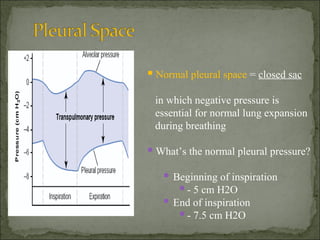
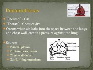
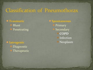
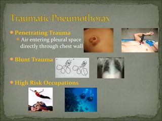
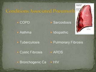
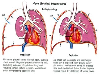
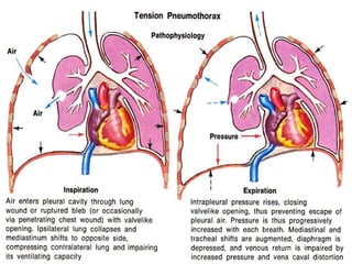
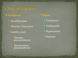
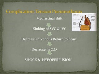
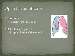
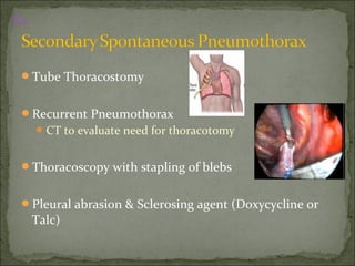
The document discusses pneumothorax, which is air in the pleural space between the lungs and chest wall. It notes that the normal pleural pressure is -5 cm H2O at the beginning of inspiration and -7.5 cm H2O at the end. Pneumothorax can be caused by trauma, medical procedures, or underlying lung conditions. Signs include shortness of breath, chest pain, and decreased breath sounds on examination. Treatment involves needle aspiration or chest tube placement to remove air and re-expand the lung. Recurrent or tension pneumothorax may require surgery.
- The document discusses pneumothorax, describing its classification, pathogenesis, clinical presentation, diagnosis, and treatment options.
- Key points include that tension pneumothorax can cause rapid deterioration and requires immediate treatment, while spontaneous pneumothorax is classified as primary or secondary depending on underlying lung health.
- Treatment involves observation for small primary pneumothoraces or procedures like aspiration, chest tube placement, or surgery depending on the size and symptoms.
Dr. Sagar Gandhi discusses pneumothorax in this document. Pneumothorax is defined as air in the pleural space between the lungs and chest wall. It can be primary or secondary and spontaneous or traumatic. Diagnosis is made through chest x-ray or CT scan. Treatment depends on size and includes observation, oxygen therapy, needle aspiration, catheter drainage, or chest tube placement. The goal is to promote lung re-expansion and prevent recurrence.
This document discusses pneumothorax, including causes, symptoms, diagnosis, and treatment. It defines pneumothorax as air in the pleural cavity, causing lung collapse. Common causes include spontaneous pneumothorax, trauma, and medical procedures. Symptoms range from minimal to severe dyspnea. Chest x-ray and CT scan are used to diagnose and estimate size. Treatment depends on severity but may include observation, needle aspiration, chest tube drainage, pleurodesis, or surgery. Recurrence risk varies from 36-83% without treatment to 0.6-2% after surgery.
A 5-day-old infant presented with shortness of breath, grunting, and poor feeding. Examination revealed reduced chest movements and diminished breath sounds on the left side. Chest X-ray showed pneumothorax. Pneumothorax is the presence of air in the pleural space, causing partial or complete lung collapse. It can be open, closed, or valvular. Causes include spontaneous (primary or secondary to lung disease) or traumatic (iatrogenic or non-iatrogenic) origins. Treatment options range from observation of small pneumothoraces to chest tube insertion or pleurodesis surgery for larger or recurrent cases.
This document provides information on pleural empyema, including its definition, etiology, stages, symptoms, investigations, and management. Pleural empyema, also known as pyothorax, is the accumulation of pus in the pleural cavity. It can develop as a complication of conditions like pneumonia or following trauma. Management involves treating the infection with antibiotics, draining the pus via procedures like chest tube insertion or VATS, and re-expanding the lung. Treatment may also include procedures like thoracocentesis, fibrinolytics, or open drainage if more invasive measures are needed.
PowerPoint presentation on Intercostal drainage (ICD) or Chest tube drainage. In this this presentation I have included different methods by which a chest tube can be inserted to drain fluid, pus, air from the Pleural cavity. please do mail me your feedback on this presentation at tinkujoseph2010@gmail.com.
This document provides an overview of chest x-ray interpretation from JSS Medical College in Mysuru, India. It begins with an introduction to radiographic densities and viewing chest x-rays. It then details an "ABCDEFGHI" approach to interpretation, covering the airway, bones, cardiomediastinal silhouette, diaphragm, effusions, lung fields, gastric bubble, hila, and impressions. Common abnormalities such as pneumonia, effusions, pneumothorax, and masses are described. The presentation emphasizes reviewing patient details, comparing to prior films, and examining all structures visible on a chest x-ray.
The document discusses pneumothorax, including its definition, pathophysiology, etiology, clinical manifestations, investigations, and management. Pneumothorax is defined as the presence of air in the pleural space. It can occur spontaneously due to ruptured blebs or bullae, or due to trauma. Clinical manifestations include dyspnea, chest pain, and decreased breath sounds on examination. Chest x-ray and CT scan are used to diagnose and characterize pneumothorax. Management involves oxygen therapy, needle aspiration, chest tube drainage, and chemical pleurodesis to promote lung re-expansion and prevent recurrence.
Pneumothorax is the presence of air or gas in the pleural space between the lung and chest wall. It occurs when there is a communication between an alveolus or air space and the pleural space, allowing air to enter the pleural space. Pneumothoraces are classified as spontaneous (primary or secondary), traumatic, or iatrogenic. Treatment depends on the type and severity but may include observation, oxygen therapy, needle aspiration, chest tube drainage, chemical pleurodesis, or surgery. Complications can include infection, bleeding, or mediastinal and subcutaneous emphysema.
The document discusses pleural effusion, which is the accumulation of fluid in the pleural space. Common causes include pneumonia, tuberculosis, pulmonary infarction, and malignant diseases. The fluid can be classified as a transudate or exudate based on biochemical analysis. Diagnosis involves chest x-ray, ultrasound, or CT scan to detect fluid levels. Diagnostic thoracentesis allows classification and identification of causative organisms if infected. Treatment focuses on draining large fluid volumes and addressing the underlying cause.
This document discusses the management of chest trauma. It describes various types of chest injuries including rib fractures, flail chest, pneumothorax, hemothorax, and tension pneumothorax. For each injury, it outlines the signs and symptoms and recommended treatment approaches. It emphasizes the importance of assessing airway, breathing, and circulation when treating chest trauma patients. It also provides details on procedures for needle chest decompression to treat tension pneumothorax.
Reexpansion pulmonary edema is a serious complication after sudden expansion of collapsed lung.Re-expansion pulmonary edema is an uncommon complication following drainage of a pneumothorax , pleural effusion or removal of any space occupying lesion.
The incidence referred is less than 1%, andmortality can reach up to 20%.
Dr. Jakeer Hussain discusses pneumothorax, beginning with an introduction and definition. He then covers the classification of pneumothorax as either spontaneous, traumatic, or iatrogenic. Spontaneous pneumothorax is further classified as primary or secondary. The document discusses signs, symptoms, investigations including x-ray and CT scan findings, differential diagnosis, quantification methods, and various treatment options including observation, oxygen supplementation, needle aspiration, tube thoracostomy, medical or VATS pleurodesis, and open thoracotomy.
LVRS involves surgically removing portions of emphysematous lung to allow the remaining lung tissue to expand. The NETT trial found LVRS benefits patients with upper lobe-predominant emphysema and low exercise capacity by improving lung function, exercise ability, and quality of life. Candidates for LVRS have severe emphysema, poor exercise capacity, marked lung hyperinflation, and meet criteria for pulmonary function tests, exercise testing, and cardiac/pulmonary evaluations. The procedure aims to improve ventilation/perfusion matching, reduce airway resistance, and allow the chest wall and diaphragm to resume a more normal position.
1. The document discusses a 60-year-old man who presents with a 3-month history of cough, sputum, and intermittent hemoptysis and is a chronic smoker. 2. On examination, tracheal shift to the right is noted along with decreased breath sounds in certain areas. 3. Differential diagnoses include bronchogenic carcinoma, neurogenic tumors, and other lung and mediastinal abnormalities. 4. The document then discusses various radiographic signs seen on chest x-rays that can help localize lesions and abnormalities, including the silhouette sign and Golden S sign indicative of lung collapse.
Pleural effusion may be defined figuratively as the juice, oozing from the leaky lingerie of the lung. However the text book definition is the abnormal accumulation of fluid in the pleural space due to disturbances in the forces that keep the pleural fluid economy in equilibrium...
A patient presented to the emergency department with sudden onset shortness of breath, cough, and chest pain. On examination, the patient's breathing sounds were decreased bilaterally with wheezing. Tests showed ischemic changes on ECG and type 1 respiratory failure on ABG. Chest X-ray revealed a secondary spontaneous pneumothorax. The patient was treated with intercostal drainage and discharged after two days with medications.
- ARDS is an acute respiratory condition characterized by diffuse lung inflammation and fluid buildup in the lungs, causing hypoxemia. Common causes include sepsis, aspiration, and pneumonia.
- The document discusses the definition, pathogenesis, clinical presentation, diagnosis, and management of ARDS. The primary goals of management are treating the underlying cause, maintaining oxygenation levels through ventilation strategies like low tidal volumes, and preventing further lung injury.
- Low tidal volume ventilation, which aims to limit overexpansion of alveoli, is the best proven strategy to improve survival based on current evidence. Other adjuncts like prone positioning and PEEP may also help optimize oxygenation in some cases.
Updates in Parapneumonic Effusion and EmpyemaGamal Agmy
The document discusses parapneumonic effusion and empyema. It classifies pneumonias and defines related terms like pleurisy, parapneumonic effusion, and empyema. It describes the pathophysiology of effusions in stages from pleuritis sicca to organization. Clinical presentation and differential diagnosis are also covered. Laboratory studies and imaging studies like chest radiography are important for diagnosing complicated parapneumonic effusions or empyemas to allow for optimal management.
A pleural effusion is a collection of fluid in the pleural space between the lungs and chest wall. It can be caused by conditions that increase hydrostatic pressure or decrease oncotic pressure, allowing fluid to accumulate. Diagnosis is typically made initially through chest x-ray or ultrasound imaging. Treatment depends on the underlying cause but may involve thoracentesis to drain fluid if it is causing breathing difficulties or pleurodesis to fuse the lung layers together to prevent further effusions.
This document provides an overview of pulmonary embolism (PE), including its definition, risk factors, types, natural history, symptoms, signs, investigations, diagnosis, and management. PE is defined as obstruction of the pulmonary artery or its branches by material such as thrombus. It discusses diagnostic tests like CT, VQ scan, echocardiogram and their role in determining pretest probability. Management involves anticoagulation with drugs like heparin, warfarin, rivaroxaban. Thrombolysis may be used for massive PE while inferior vena cava filters can be placed in patients who cannot receive anticoagulation.
CTEPH is a deadly disease that causes pulmonary hypertension. It is caused by blood clots in the lungs that do not fully resolve, leading to blockages in the pulmonary arteries. While medical therapies exist, they have mostly been tested in advanced cases. Without treatment, CTEPH progresses rapidly once symptoms appear. The disease is insidious in onset and can be misdiagnosed for years due to non-specific symptoms like dyspnea and exercise intolerance. This delays correct treatment and leads to right heart failure and death if left untreated.
This document discusses bronchiectasis, including its definition, etiology, clinical features, diagnosis, management, and complications. Some key points:
- Bronchiectasis is irreversible dilation of the airways caused by infection or other insults that damages the airways and impairs mucus clearance.
- It has various etiologies including infection, immunodeficiency, genetic disorders, and aspiration. Recurrent infections lead to a vicious cycle of inflammation and further airway damage.
- Symptoms include chronic productive cough and sputum. Investigations include chest CT, which shows characteristic findings like airway dilation.
- Management focuses on airway clearance, antibiotics for infections, and
The document discusses pleural effusions and empyema. It defines pleural effusions as excess fluid in the pleural space, which can be transudative or exudative based on its cause. Empyema is defined as pus or microorganisms present in the pleural fluid. Empyema progresses through exudative, fibrinopurulent and organizational stages. Treatment of empyema involves antibiotics, chest tube drainage, and sometimes surgery.
Acute respiratory distress syndrome (ARDS) is a sudden and progressive form of acute respiratory failure in which the alveolar capillary membrane becomes damaged and more permeable to intravascular fluid resulting in severe dyspnoea, hypoxemia and diffuse pulmonary infiltrates.
1. Pneumothorax is the presence of air in the pleural space causing lung collapse, and pneumomediastinum is the presence of air in the mediastinum.
2. Pneumothorax can be spontaneous, traumatic, or iatrogenic and is classified as primary or secondary depending on underlying lung conditions. Tension pneumothorax is a life-threatening form caused by trapped air that displaces mediastinal structures.
3. Chest x-ray is used to diagnose pneumothorax by identifying the visceral pleural line and lung collapse. Features of tension pneumothorax on x-ray include mediastinal shift and tracheal deviation. Pneum
This document provides information on empyema, including its definition, etiology, stages, symptoms, investigations, and management. Empyema is defined as infection of the pleural space resulting in pus accumulation. It is usually caused by bacteria spreading from a pneumonia or other infection. It progresses through exudative, fibrinopurulent, and organizing stages. Symptoms include fever, cough, chest pain, and shortness of breath. Diagnosis involves imaging like chest X-ray or CT scan and thoracentesis. Management includes antibiotics, chest tube drainage, fibrinolytics, VATS, and sometimes open drainage or decortication surgery. The goal is to treat infection, drain pus, and re-expand
This document discusses pneumothorax in neonates. It begins with an introduction that defines pneumothorax and discusses its prevalence. It then describes typical x-ray findings that can indicate pneumothorax such as increased lucency, decreased vascular markings, and flattened diaphragms. The document presents a case study of a premature infant born at 27 weeks who was intubated and transferred to the NICU with suspected pneumothorax. Example x-ray images are shown and diagnosed as demonstrating right and bilateral pneumothoraces. The conclusion emphasizes the importance of comparing current and past images and obtaining lateral views to properly diagnose pneumothorax.
This document discusses the anatomy and diseases of the upper and lower airways. It begins with an overview of the anatomy of the upper airways including the nose, pharynx and larynx. It then describes the anatomy of the lower airways including the trachea, bronchi and lungs. Key details about the histology and embryological development of the lungs are also provided. The document concludes with short descriptions of some common airway diseases such as asthma, COPD and cystic fibrosis that can affect the conducting airways and lungs.
For a leading US pharmaceutical consulting company, SmithStreetSolutions provided a comprehensive market analysis research paper that included current technology and marketing information of existing drugs in the market for a specific disease. The research also included results of multiple clinical trials measuring their safety, efficacy and their marketing mix. Based on the in-depth research SmithStreetSolutions provided , our client formulated a thorough marketing campaign recommendation including roll out timelines for the end client’s brand entry into the China market.
This document discusses the management of pregnant patients in oral surgery. It covers the stages of pregnancy, anatomical and physiological changes that occur, positioning considerations, and guidelines for medications and radiography. The first trimester is the most vulnerable period for fetal development. The second trimester is generally safest for dental treatment. Local anesthesia is considered safe in pregnancy, while certain antibiotics, analgesics and sedatives should be avoided or used cautiously. Proper positioning helps address issues like supine hypotension. Emergency situations like syncope, morning sickness and seizures require specific management to protect mother and fetus.
This document discusses air leak syndrome, which refers to the escape of air from the lungs into other tissues. It can include pulmonary interstitial emphysema, pneumothorax, pneumomediastinum, pneumopericardium, pneumoperitoneum, and subcutaneous emphysema. Pneumothorax, or air in the pleural space between the lung and chest wall, is the most common type. Risk factors include prematurity, mechanical ventilation, and diseases that damage lung tissue. Symptoms depend on the location and amount of air, and treatment ranges from supportive care to needle aspiration or chest tube insertion for more severe cases.
The document discusses one lung ventilation (OLV), which involves separately ventilating each lung during thoracic surgery. It covers the respiratory physiology of OLV, including how factors like anesthesia, paralysis, chest opening, and positioning impact ventilation and perfusion in the dependent versus non-dependent lungs. Specifically, it notes that while blood flow favors the dependent lung, ventilation is altered to favor the non-dependent lung due to changes in lung compliance. This can lead to ventilation-perfusion mismatches and hypoxemia.
This document discusses the diagnosis and treatment of primary and secondary spontaneous pneumothorax. Primary pneumothorax typically occurs in young, healthy individuals and is usually treated initially with observation or simple aspiration. Secondary pneumothorax occurs in the context of underlying lung disease and may require chest tube drainage. Treatment depends on factors such as pneumothorax size and severity of symptoms. Surgical intervention is recommended for recurrent or persistent cases.
This document discusses deep vein thrombosis (DVT) prophylaxis. It defines DVT as clot formation in the deep veins of the legs, with an annual incidence of 1-2 per 1000 people in the US. Risk factors include surgery, injury, prolonged bed rest, estrogen use, and inherited clotting disorders. Signs and symptoms include leg pain, swelling, and difficulty breathing. Diagnosis involves duplex ultrasound and other imaging tests. Complications include pulmonary embolism and post-thrombotic syndrome. Prophylaxis includes mechanical methods, aspirin, anticoagulants, and stratified prophylaxis based on patient risk factors. The goal of prophylaxis is to prevent DVT and its
Presentation1.pptx, radiological imaging of neonatal lung disease.Abdellah Nazeer
This document discusses several common neonatal lung diseases seen on chest radiographs. It describes the pathology and typical radiographic findings of transient tachypnea of the newborn (TTN), meconium aspiration syndrome (MAS), neonatal respiratory distress syndrome (NRDS), bronchopulmonary dysplasia (BPD), and pulmonary interstitial emphysema (PIE). For each condition, it provides details on risk factors, clinical presentation, effects on lung tissue, and characteristic imaging patterns seen on chest x-rays. Examples of chest radiographs demonstrating the radiographic manifestations of each disease are also included.
This document discusses respiratory distress in newborns, listing common medical and surgical causes such as transient tachypnea of the newborn (TTNB), respiratory distress syndrome (RDS), meconium aspiration syndrome, and pneumothorax. It then focuses on the pathophysiology, clinical presentation, diagnosis, and management of TTNB, RDS, and meconium aspiration syndrome. TTNB is usually mild and self-limited, resolving within 3 days with supportive care. RDS is caused by surfactant deficiency and presents with progressive respiratory distress, responding well to surfactant replacement therapy. Meconium aspiration syndrome involves airway obstruction and inflammation from aspirated meconium, often
The document discusses various techniques for invasive and non-invasive neonatal ventilation. It describes conventional mechanical ventilation modes like CMV, IMV, SIMV and newer modes like pressure support ventilation and proportional assist ventilation. It also covers high frequency ventilation, CPAP and newer non-invasive techniques like NIPPV and SNIPPV which aim to provide respiratory support without intubation. The goals, mechanisms, settings and potential complications of different ventilation strategies are outlined.
This document summarizes various types of thoracic trauma. It covers epidemiology, injuries to the chest wall including rib fractures and flail chest. It also discusses pulmonary injuries such as pulmonary contusion, pneumothorax, hemothorax, and tracheobronchial injuries. Cardiovascular injuries addressed include myocardial contusion, myocardial rupture, penetrating cardiac injury, and acute pericardial tamponade. Management strategies are provided for each type of injury.
This document provides information about mechanical ventilation in neonates from the NICU at Al Shifaa Hospital in Gaza. It discusses [1] the goals and indications for mechanical ventilation in neonates, [2] procedures for intubation and setting appropriate ventilator settings, and [3] concepts of lung physiology and mechanics relevant to neonatal ventilation. The document is intended to guide clinicians on best practices for mechanically ventilating neonates.
The document discusses control of breathing and neonatal respiratory diseases. It covers the respiratory center in the medulla, chemoreceptors, respiratory reflexes, lung mechanics, definitions of terms like tidal volume and compliance. It then summarizes several neonatal respiratory diseases like hyaline membrane disease, transient tachypnea of the newborn, pneumothorax, pulmonary interstitial emphysema, persistent pulmonary hypertension of the newborn, meconium aspiration syndrome, and chronic lung disease. For each disease it discusses clinical features, etiology, treatments and methods of prevention.
This document discusses neonatal respiratory distress, including signs, symptoms, and common etiologies. The main pulmonary causes discussed are transient tachypnea of newborn, respiratory distress syndrome, meconium aspiration syndrome, pneumonia, and air leak syndromes. For each cause, risk factors, pathophysiology, clinical manifestations, diagnostic findings, and management approaches are summarized. The document provides an overview of evaluation and treatment of neonatal respiratory distress.
This document provides an overview of pneumothorax, including:
- Classification as spontaneous (primary or secondary), traumatic, or iatrogenic
- Risk factors like smoking, COPD, and connective tissue diseases for secondary spontaneous pneumothorax
- Pathophysiology involving bleb/bullae rupture and air migration into the pleural space
- Clinical features like chest pain and shortness of breath, and radiological findings on CXR and CT scans
- Management approaches like chest tube insertion, pleurodesis, and VATS for recurrent or large pneumothoraces.
The document discusses pneumothorax, describing its history, classification, pathogenesis, pathophysiology, clinical presentation, diagnosis, and treatment. Pneumothorax is classified as primary, secondary, traumatic, or iatrogenic. It results from a communication between the lung and pleural space, eliminating the normal negative pressure. Clinical signs depend on factors like underlying lung disease and pneumothorax size. Chest x-ray is used to diagnose, while CT scanning can estimate size. Treatment depends on classification, severity, and recurrence risk.
The document discusses pneumothorax, beginning with a brief history and definitions. It then covers the classification, pathogenesis, pathophysiology, clinical presentation, diagnosis using imaging like chest X-rays and CT scans, and various treatment options including observation, aspiration, chest tube drainage and systems. Pneumothorax can be primary spontaneous, secondary, or iatrogenic and is typically diagnosed and monitored using chest imaging. Treatment depends on factors like size, symptoms and underlying lung condition, ranging from observation to tube drainage.
Pneumothorax is the presence of air in the pleural space and can be spontaneous, due to trauma, or iatrogenic. It is classified as primary spontaneous which occurs without lung disease usually in young males, secondary spontaneous which occurs with underlying lung pathology, or traumatic. Types include closed which seals off, open with a bronchopleural fistula, and tension which increases pressure. Clinical features include chest pain and shortness of breath. Diagnosis is made with chest x-ray showing increased radiolucency. Small primary pneumothoraces may resolve on their own while secondary pneumothoraces and those with symptoms require tube thoracostomy drainage. Recurrent cases require pleurodesis or surgery.
there is the introduction part of the torso trauma,
check out my next ppts for further more about torso trauma.
contents are in following order...
introduction
mechanism of injury
junctional zones of torso
tension pneumothorax
cardiac temponade
massive hemothorax
etc.
check out all slides
This document discusses pneumothorax, which is the accumulation of air in the pleural space causing lung collapse. It defines pneumothorax as air in the pleural space that can occur spontaneously or from trauma. The types are described as spontaneous, open, or tension pneumothorax. Clinical manifestations include shortness of breath, chest pain, and decreased breath sounds on the affected side. Diagnosis involves history, exam, chest x-ray and blood gas analysis. Management depends on the type but may include observation, needle aspiration, chest tube placement, or surgery to repair the lung. Nursing care focuses on dressing wounds, positioning, monitoring chest tube drainage, and watching for complications like respiratory failure.
This document discusses pneumothorax and hemothorax. It defines pneumothorax as a collection of air in the pleural space, which can be spontaneous or traumatic. Tension pneumothorax is a life-threatening condition where air builds up pressure in the pleural space. Hemothorax is defined as a collection of blood in the pleural space. The document covers causes, pathogenesis, clinical presentations, and treatment approaches for pneumothorax and hemothorax.
This document discusses pneumothorax, which is the presence of air in the pleural space outside the lung. It describes different types of pneumothorax including primary spontaneous, secondary spontaneous, closed, open, and tension pneumothorax. Risk factors, clinical features, diagnosis using chest x-ray, treatment options including chest tube insertion, and postoperative management of chest drains are covered. Surgical intervention is indicated for recurrent pneumothorax or when chest drainage fails.
Lung compression or chest trauma can cause several complications including pneumothorax, hemothorax, and flail chest. Pneumothorax involves air in the pleural space collapsing part or all of the lung. Hemothorax is a collection of blood in the pleural cavity. Flail chest occurs when multiple ribs are broken, detaching a segment of the chest wall. Nursing management focuses on stabilizing the patient, draining fluid or air from the chest, controlling pain, and supporting ventilation as needed.
Pneumothorax is the presence of air in the pleural cavity, which occurs when there is a breach in the parietal or visceral pleura. It can be classified as primary or secondary, and also as acute, chronic, small, large, partial or complete. Primary spontaneous pneumothorax typically occurs in young, tall, thin males and results from a rupture of a pulmonary bleb or bulla. Management depends on factors such as size, symptoms and underlying cause, and may include observation, aspiration, tube thoracostomy, chemical or surgical pleurodesis, or thoracoscopic procedures. Complications can include failure to reexpand, recurrence, infection or respiratory failure.
This document discusses pneumothorax, which is the presence of air in the pleural space. It can be spontaneous, due to underlying lung disease or trauma, or iatrogenic due to medical procedures. The types are described as closed, open, or tension pneumothorax. Clinical features include chest pain and breathlessness. Diagnosis is made through chest x-ray or CT scan. Treatment depends on the type and severity, ranging from needle aspiration for minor cases to chest tube drainage or surgery for more severe or recurrent cases.
This document discusses pneumothorax, which is the presence of air in the pleural space. It can be spontaneous, due to trauma, or iatrogenic. Spontaneous pneumothorax is classified as primary or secondary. Primary occurs without lung disease in young males and secondary occurs with underlying lung conditions. Types include closed, open, and tension. Clinical features, investigations like CXR and CT, and treatment approaches are described depending on the type and severity of pneumothorax. Needle aspiration, tube thoracostomy, surgery, and pleurodesis are common management steps.
The document discusses various life-threatening chest injuries that may occur from thoracic trauma. It identifies injuries like tension pneumothorax, open pneumothorax, flail chest, massive hemothorax, and cardiac tamponade as immediately life-threatening injuries that must be addressed during the primary survey. It also discusses potentially life-threatening injuries like tracheobronchial injuries, pulmonary contusions, hemothorax, and blunt cardiac injuries that require identification and treatment. Physical exams, chest x-rays, and other tests can help identify these injuries, which then often require simple procedures or surgeries to treat. The overall goals are to restore normal breathing and blood flow.
Pneumothorax is the presence of air in the pleural space. It can be classified as closed, open, or tension pneumothorax. The annual incidence is around 9 per 100,000 people. Risk factors include being a tall, thin male aged 20-40 who smokes cigarettes. Symptoms include chest pain and breathlessness. Chest x-ray is used for diagnosis and can classify pneumothorax as small or large based on rim size. Needle decompression is immediately needed for tension pneumothorax. Oxygen, aspiration, chest drain insertion, and surgery are treatment options depending on the severity of the case.
This document provides information on respiratory emergencies including causes, types of chest injuries, signs and symptoms, assessments, and treatments. It discusses specific chest injuries like rib fractures, flail chest, pneumothorax, hemothorax, and cardiac tamponade. For each injury, it describes definitions, risk factors, manifestations, diagnostic procedures, nursing care, and complications. The overall document aims to define respiratory emergencies, list causes, describe patient presentations, discuss assessments, and examine various chest injuries in detail.
Neonatal pneumothorax is the accumulation of air in the pleural cavity, which can collapse the lung. It occurs most commonly in preterm infants and those with underlying lung conditions requiring ventilation support. Symptoms range from none in mild cases to respiratory distress and hypotension in severe cases. Diagnosis is confirmed by chest x-ray showing hyperlucent lung fields. Small pneumothoraces may be observed but symptomatic or tension pneumothoraces require needle aspiration or chest tube placement to re-expand the lung. Persistent pneumothoraces lasting over a week sometimes require additional interventions like HFOV. Prognosis depends on the underlying condition but early and effective treatment prevents complications.
This document provides an overview of chest trauma. It begins with the anatomy of the thorax and then discusses various types of chest injuries including pneumothorax, hemothorax, flail chest, cardiac tamponade, and traumatic aortic rupture. For each type of injury, the document describes the mechanism of injury, signs and symptoms, and treatment approaches. It emphasizes the life-threatening nature of many chest injuries and stresses the importance of rapid diagnosis and management.
Pneumothorax refers to the presence of air in the pleural space and can occur spontaneously due to ruptured blebs or as a result of trauma or medical procedures. It presents clinically as reduced breath sounds, hyperresonance to percussion, and mediastinal shift. Chest x-ray or CT scan are used for diagnosis and show hypertranslucency. Treatment involves supplemental oxygen, aspiration, or chest tube placement. Physiotherapy focuses on improving ventilation and exercise tolerance. Recurrence can be prevented through procedures like pleurodesis or thoracotomy along with smoking cessation.
This document provides an overview of pneumothorax. It begins by defining pneumothorax as air in the pleural space between the lung and chest wall. The pathophysiology section explains how a pneumothorax occurs when air leaks into the pleural space, eliminating the normal negative pressure. This can impair breathing and circulation.
The document then classifies pneumothoraces as primary spontaneous, secondary spontaneous, or traumatic. Clinical features include dyspnea and chest pain. Diagnosis involves chest x-ray or CT scan to evaluate lung collapse and estimate size. Treatment depends on pneumothorax type and size but may include observation, oxygen therapy, needle aspiration, chest tube drainage, chemical pleuro
ABDOMINAL TRAUMA in pediatrics part one.drhasanrajab
Abdominal trauma in pediatrics refers to injuries or damage to the abdominal organs in children. It can occur due to various causes such as falls, motor vehicle accidents, sports-related injuries, and physical abuse. Children are more vulnerable to abdominal trauma due to their unique anatomical and physiological characteristics. Signs and symptoms include abdominal pain, tenderness, distension, vomiting, and signs of shock. Diagnosis involves physical examination, imaging studies, and laboratory tests. Management depends on the severity and may involve conservative treatment or surgical intervention. Prevention is crucial in reducing the incidence of abdominal trauma in children.
Muktapishti is a traditional Ayurvedic preparation made from Shoditha Mukta (Purified Pearl), is believed to help regulate thyroid function and reduce symptoms of hyperthyroidism due to its cooling and balancing properties. Clinical evidence on its efficacy remains limited, necessitating further research to validate its therapeutic benefits.
Adhd Medication Shortage Uk - trinexpharmacy.comreignlana06
The UK is currently facing a Adhd Medication Shortage Uk, which has left many patients and their families grappling with uncertainty and frustration. ADHD, or Attention Deficit Hyperactivity Disorder, is a chronic condition that requires consistent medication to manage effectively. This shortage has highlighted the critical role these medications play in the daily lives of those affected by ADHD. Contact : +1 (747) 209 – 3649 E-mail : sales@trinexpharmacy.com
Basavarajeeyam is an important text for ayurvedic physician belonging to andhra pradehs. It is a popular compendium in various parts of our country as well as in andhra pradesh. The content of the text was presented in sanskrit and telugu language (Bilingual). One of the most famous book in ayurvedic pharmaceutics and therapeutics. This book contains 25 chapters called as prakaranas. Many rasaoushadis were explained, pioneer of dhatu druti, nadi pareeksha, mutra pareeksha etc. Belongs to the period of 15-16 century. New diseases like upadamsha, phiranga rogas are explained.
8 Surprising Reasons To Meditate 40 Minutes A Day That Can Change Your Life.pptxHolistified Wellness
We’re talking about Vedic Meditation, a form of meditation that has been around for at least 5,000 years. Back then, the people who lived in the Indus Valley, now known as India and Pakistan, practised meditation as a fundamental part of daily life. This knowledge that has given us yoga and Ayurveda, was known as Veda, hence the name Vedic. And though there are some written records, the practice has been passed down verbally from generation to generation.
Local Advanced Lung Cancer: Artificial Intelligence, Synergetics, Complex Sys...Oleg Kshivets
Overall life span (LS) was 1671.7±1721.6 days and cumulative 5YS reached 62.4%, 10 years – 50.4%, 20 years – 44.6%. 94 LCP lived more than 5 years without cancer (LS=2958.6±1723.6 days), 22 – more than 10 years (LS=5571±1841.8 days). 67 LCP died because of LC (LS=471.9±344 days). AT significantly improved 5YS (68% vs. 53.7%) (P=0.028 by log-rank test). Cox modeling displayed that 5YS of LCP significantly depended on: N0-N12, T3-4, blood cell circuit, cell ratio factors (ratio between cancer cells-CC and blood cells subpopulations), LC cell dynamics, recalcification time, heparin tolerance, prothrombin index, protein, AT, procedure type (P=0.000-0.031). Neural networks, genetic algorithm selection and bootstrap simulation revealed relationships between 5YS and N0-12 (rank=1), thrombocytes/CC (rank=2), segmented neutrophils/CC (3), eosinophils/CC (4), erythrocytes/CC (5), healthy cells/CC (6), lymphocytes/CC (7), stick neutrophils/CC (8), leucocytes/CC (9), monocytes/CC (10). Correct prediction of 5YS was 100% by neural networks computing (error=0.000; area under ROC curve=1.0).
Recomendações da OMS sobre cuidados maternos e neonatais para uma experiência pós-natal positiva.
Em consonância com os ODS – Objetivos do Desenvolvimento Sustentável e a Estratégia Global para a Saúde das Mulheres, Crianças e Adolescentes, e aplicando uma abordagem baseada nos direitos humanos, os esforços de cuidados pós-natais devem expandir-se para além da cobertura e da simples sobrevivência, de modo a incluir cuidados de qualidade.
Estas diretrizes visam melhorar a qualidade dos cuidados pós-natais essenciais e de rotina prestados às mulheres e aos recém-nascidos, com o objetivo final de melhorar a saúde e o bem-estar materno e neonatal.
Uma “experiência pós-natal positiva” é um resultado importante para todas as mulheres que dão à luz e para os seus recém-nascidos, estabelecendo as bases para a melhoria da saúde e do bem-estar a curto e longo prazo. Uma experiência pós-natal positiva é definida como aquela em que as mulheres, pessoas que gestam, os recém-nascidos, os casais, os pais, os cuidadores e as famílias recebem informação consistente, garantia e apoio de profissionais de saúde motivados; e onde um sistema de saúde flexível e com recursos reconheça as necessidades das mulheres e dos bebês e respeite o seu contexto cultural.
Estas diretrizes consolidadas apresentam algumas recomendações novas e já bem fundamentadas sobre cuidados pós-natais de rotina para mulheres e neonatos que recebem cuidados no pós-parto em unidades de saúde ou na comunidade, independentemente dos recursos disponíveis.
É fornecido um conjunto abrangente de recomendações para cuidados durante o período puerperal, com ênfase nos cuidados essenciais que todas as mulheres e recém-nascidos devem receber, e com a devida atenção à qualidade dos cuidados; isto é, a entrega e a experiência do cuidado recebido. Estas diretrizes atualizam e ampliam as recomendações da OMS de 2014 sobre cuidados pós-natais da mãe e do recém-nascido e complementam as atuais diretrizes da OMS sobre a gestão de complicações pós-natais.
O estabelecimento da amamentação e o manejo das principais intercorrências é contemplada.
Recomendamos muito.
Vamos discutir essas recomendações no nosso curso de pós-graduação em Aleitamento no Instituto Ciclos.
Esta publicação só está disponível em inglês até o momento.
Prof. Marcus Renato de Carvalho
www.agostodourado.com
Osteoporosis - Definition , Evaluation and Management .pdfJim Jacob Roy
Osteoporosis is an increasing cause of morbidity among the elderly.
In this document , a brief outline of osteoporosis is given , including the risk factors of osteoporosis fractures , the indications for testing bone mineral density and the management of osteoporosis
2. Normal pleural space = closed sac
in which negative pressure is
essential for normal lung expansion
during breathing
What’s the normal pleural pressure?
Beginning of inspiration
- 5 cm H2O
End of inspiration
- 7.5 cm H2O
3. “Pneumo” – Gas
“Thorax” – Chest cavity
Occurs when air leaks into the space between the lungs
and chest wall, creating pressure against the lung
Sources
Visceral pleura
Ruptured esophagus
Chest wall defect
Gas-forming organisms
17. CXR
Inspiratory & expiratory images
Underlying Pul. disease
Harder to detect
CT
PSP – Blebs & Bullae
Small pneumothorax
Assess the need for
thoracotomy
18.
19.
20.
21. Absence of “lung sliding” as assessed on the time-
motion view
Demonstration of a "lung point" on the time-motion
view
Absence of vertical comet-tail artifacts
USGUSG
95% sensitivity95% sensitivity
100% specificity100% specificity
26. Immediate decompression via
chest tube or needle
thoracostomy
If a tension pneumothorax is
present, a “hiss of air” may be
heard escaping from the chest
cavity
Remove the needle,
leave the catheter in
place
Rx
31. Oxygen
Pneumothorax is smaller than 15%
Patient is asymptomatic
Needle aspiration
Pneumothorax is smaller than 15%
Symptomatic & hemodynamically stable
Pigtail catheter
Pneumothorax is greater than 15%
Rx
Normal pleural space = closed sac in which negative pressure is essential for normal lung expansion during breathing (simplified: space b/w lungs and chest wall)
*Remember from physics: Areas of high pressure will move into areas of lower pressure (I.e. when we exhale, we force air out of lungs, creating large area of negative pressure in pleural space, which entices air to rush back into lungs when we open mouth; inhalation = opposite, lots of positive pressure introduced into lungs, smaller negative pressure in pleural space around lungs).
“OPEN” vs. “CLOSED” chest injuries: open injuries have significant potential to compromise efficacy of pleural space and therefore respiratory function. Also potential for infection of exposed vital organs/systems, plus cavity area can pool blood quickly (internal bleeding).
Primary-smokers suggestive of subclinical disease
----------------------
primary spontaneous (no obvious underlying lung disease), secondary spontaneous (underlying lung disease), and iatrogenic pneumothoraces (which are traumatic but typically are smaller and more easily managed)
Primary spontaneous pneumothorax seems to result from rupture of a subpleural bleb, usually in an upper lobe.5 These blebs are usually multiple and have increased wall tension, allowing distention and eventual rupture. The mechanism of bleb formation remains unknown, but higher upper lobe transpulmonary pressure, local ischemia from decreased upper lobe blood flow, and subclinical emphysema-like changes have been postulated
-------------------
Primary spontaneous pneumothorax
Air in the intrapleural space without preceding trauma and without underlying clinical or radiologic evidence of lung disease
Typically in patients who are between 18 and 40 years of age
Primary spontaneous pneumothorax occurs in individuals without known lung disease and accounts for two-thirds of spontaneous pneumothoraces
Secondary spontaneous pneumothorax
Occurs in patients with underlying pulmonary structural pathology
Air enters the pleural space via distended, damaged, or compromised alveoli
May present with more serious clinical symptoms and sequelae due to comorbidity
Iatrogenic pneumothorax
Medical procedure resulting in traumatic pneumothorax (usually from a small-bore hollow needle)
Iatrogenic pneumothorax occurs secondary to a diagnostic or therapeutic procedure and is really a subset of penetrating traumatic pneumothorax.
------------------------------
Primary spontaneous pneumothorax
Spontaneous pneumothorax is heavily associated with smoking, with 80-90% of primary spontaneous pneumothorax cases occurring in smokers.
Physical height: It has been noted that typical patients tend to have a tall and thin body habitus. Whether height affects development of subpleural blebs or whether more negative apical pleural pressures cause preexisting blebs to rupture is unclear.
Valsalva results in increased intrathoracic pressure. However, contrary to popular belief, most spontaneous pneumothoraces occur while the patient is at rest.
Changes in atmospheric pressure, proximity to loud music, and low frequency noises have also been reported to be associated with pneumothorax
Familial associations have been noted in more than 10% of patients. Some are due to rare connective tissue diseases, but recently, mutations in the gene encoding folliculin (FLCN) have been described. These patients may represent an incomplete penetrance of a genetic disorder. Birt-Hogg-Dube syndrome is characterized by benign skin growths, pulmonary cysts, and renal cancers and is caused by mutations in the FLCN gene.
Secondary spontaneous pneumothorax
COPD or emphysema
Asthma
Cystic fibrosis
Interstitial lung disease
Tuberculosis
Bronchogenic or metastatic carcinoma
Pneumonia (fungal, caseating, HIV)
Collagen vascular disease including Marfan syndrome
Catamenial pneumothorax
---------------------------
The most common underlying abnormality in secondary spontaneous pneumothorax is COPD
-----------------------------------------
Regardless of the inciting event, once there is a break in the pleura, air travels down a pressure gradient into the intrapleural space until pressure equilibrium occurs with partial or total lung collapse. Altered ventilation perfusion relationships and decreased vital capacity then contribute to dyspnea and hypoxemia
flying
Iatrogenic pneumothorax occurs secondary to a diagnostic or therapeutic procedure and is really a subset of penetrating traumatic pneumothorax.
Spontaneous pneumothoraces in most patients occur from the rupture of blebs and bullae. While primary pneumothorax is defined as a lack of underlying pulmonary disease, these patients have asymptomatic blebs and bullae detected on CT scans or upon thoracotomy. Until a bleb ruptures and causes a pneumothorax, no clinical signs or symptoms are present.The pleural space has a negative pressure, with the chest wall tending to spring outward and the lung's elastic recoil tending to collapse. If the pleural space is invaded by gas from a ruptured bleb, the lung collapses until equilibrium is achieved or the rupture is sealed. As the pneumothorax enlarges, the lung becomes smaller.
The main physiologic consequence of this process is a decrease in vital capacity and partial pressure of oxygen. Young and otherwise healthy patients can tolerate these changes fairly well, with minimal changes in vital signs and symptoms, but those with underlying lung disease may have respiratory distress.
Equilibrium between intrathoracic and atmospheric pressure is immediate..
Spontaneous pneumothorax
Tachycardia (most common)
Tachypnea
Hypoxia
-----------------------
Symptoms are related to the size of the pneumothorax, rate of development, and underlying clinical status of the patient.
----------------------------------------------
Acute pleuritic chest pain occurs in 95 percent of patients and localizes to the side of the pneumothorax in >90 percent of cases. Dyspnea occurs in 80 percent of patients, and predicts a larger pneumothorax. Patients with COPD who develop a spontaneous pneumothorax may acutely decompensate, with 1 to 17 percent mortality.
Decreased breath sounds occur over the affected lung 85 percent of the time, but only 5 percent have tachypnea of more than 24 breaths/min or tachycardia of more than 120 beats/min. Hyperresonance occurs in fewer than one-third. Tracheal deviation and hemodynamic compromise are the hallmarks of tension pneumothorax that demand immediate treatment
Short-term complications of spontaneous pneumothorax include tension pneumothorax, failure to reexpand, persistent air leak, and complications related to the removal of intrapleural air, such as infection, technical errors, and reexpansion pulmonary edema
---------------------
Recurrence of pneumothorax is the major long-term complication and the reason that all patients with spontaneous pneumothorax require referral for potential definitive therapy. Primary spontaneous pneumothorax recurs in approximately 20 to 30 percent of patients and is independent of the method chosen to remove the intrapleural air
----------------
Secondary spontaneous pneumothorax recurs in 40 to 50 percent of patients
Chest radiography for evaluation of pneumothorax
Although expiratory images are thought to better depict subtle pneumothoraces (the volume of the pneumothorax is constant and hence proportionally higher on expiratory images), a randomized controlled trial revealed no difference in the ability of radiologists to detect pneumothoraces on inspiratory and expiratory images.
In patients with underlying pulmonary disease, the classic visceral pleural line may be harder to detect because the lung is hyperlucent, and little difference exists in the radiographic density between the pneumothorax and the emphysematous lung.
Ratio of lung size to hemithorax size to estimate pneumothorax size avoids the subjective underestimation of pneumothorax expressed as a percentage of previous lung volume
The size of a pneumothorax may be estimated by using the ratio of the lung diameter cubed to the hemithorax diameter cubed.
This formula assumes a constant shape of the lung when it collapses and is invalid if pleural adhesions are present.
A simple approach to classification of the pneumothorax as small or large involves measuring the distance from the apex of the lung to the top margin of the visceral pleura (thoracic cupola) on the upright chest radiograph.
Small pneumothorax: <3 cm distance to the apex
Large pneumothorax: >3 cm distance to the apex
A supine chest radiograph may depict the deep sulcus sign (very dark and deep costophrenic angle). The anterior costophrenic recess becomes the highest point in the hemithorax, resulting in an unusually sharp definition of the anterior diaphragmatic surface due to gas collection and a depressed costophrenic angle.
--------------------------------------------
CT of the thorax
When performed on primary spontaneous pneumothorax patients, CT detects multiple blebs and bullae in the setting of negative chest radiographic findings. This may not impact management, as there has been no correlation between number of blebs and recurrence.
CT can detect occult pneumothorax in patients who will require mechanical ventilation in trauma and emergency surgery settings.
CT has also been shown to be more sensitive than radiography for hemothorax and pulmonary contusion.
While the role of CT in trauma patients is evolving, it is controversial whether it significantly alters management and is not indicated in primary spontaneous pneumothorax.
CT may have a role in secondary spontaneous pneumothorax, especially to differentiate from giant bullous emphysema.
-------------------------------------------------------------------------------------
Ultrasonography
Ultrasonography can be used as a possible bedside technique to detect pneumothorax.
It may be useful in unstable patients who cannot undergo radiologic studies outside of the emergency department.
Many trauma centers are incorporating chest ultrasonography as an adjunct to the Focused Assessment with Sonography in Trauma (FAST) examination used for trauma patient screening.
Ultrasonography is operator dependent.
-------------------------------
Skin fold mimicking pneumothorax. A: AP supine chest radiograph shows opacification of the right medial lung outlined by a sharp edge (skin fold; arrows). Note that the lung peripheral to this edge is not hyperlucent, a clue that there is no pneumothorax. B: AP upright chest radiograph obtained 1 hour later no longer shows the skin fold. Redundant skin can result in skin folds on the chest radiograph, especially when the patient is supine. Changing patient positioning is often useful in differentiating a skin fold from a pneumothorax.
Pseudo-pneumothorax caused by a skin fold, scapular border, or tubing, is differentiated from true pneumothorax by looking for vascular markings inside the confines of the radiolucent area and the blending of these lines into the chest wall, rather than following the borders of a collapsed lung. Large bullae have been mistaken for pneumothoraces, but bullae and cysts have concave inner margins and rounded edges.
severe bullous COPD may mimic pneumothorax, and careful review of the CXR is needed with confirmatory CT, if the patient is stable. A thoracostomy with the chest tube inserted into a bulla mistaken for a pneumothorax results in a large pneumothorax, associated bronchopulmonary fistula, and its complications
---------------------------------------
Chest tube track mimicking pneumothorax. A: PA chest radiograph, coned to the left upper hemithorax, shows a thin curvilinear opacity paralleling the chest wall (arrows). B: AP chest radiograph obtained 1 day earlier shows a chest tube following the course of the opacity seen in (A).
Pneumothorax in a supine patient. AP supine chest radiograph shows the “deep sulcus†sign of pneumothorax on the right (curved arrow) and a basilar pneumothorax on the left (straight arrows)
---------------------------------
Pneumothorax is much more difficult to detect on a supine anteroposterior radiograph. On the anteroposterior view, a deep sulcus sign, representing a deep lateral costophrenic angle, sometimes is a clue to a pneumothorax.
The Lung Point, a Sign Specific to Pneumothorax
A patient with hard-to-detect or absent lung sliding and absence of interstitial syndrome will have an ultrasound profile of pneumothorax, i.e., a false-positive image. we can build a specific sign: with immediate and fleeting visualization at a precise location of the chest wall and along a definite line, at a precise moment of the respiratory cycle, usually inspiration, with the probe strictly motionless, the operator finds either lung sliding, lung rockets, or alteration of A lines, in an area previously observed with no lung sliding and the A-line sign, i.e., patterns that were barely suggestive of pneumothorax This sign has been called the lung point sensitivity of 66% and a specificity of 100% This sign can be explained if one considers that any lung, at the wall or not, in spontaneous or mechanical ventilation,
will slightly increase its volume on inspiration. Therefore, a lung sign will appear at the boundary area, at the precise line where the lung reaches the wall, since the lung surface in contact with the wall will increase .The poor sensitivity of ultrasound is easily explained: major, completely retracted pneumothorax will never touch the wall.
Emergency department evaluation and management of pneumothorax centers on immediate management of complications such as tension pneumothorax or ventilatory failure, and then on eliminating the intrapleural air and optimizing pleural healing.
Clinical diagnosis
Don’t wait for radiological diagnosis
Needle coverts it into simple pneumo…
All sides-tension pneumothorax
There is general agreement that a tension pneumothorax must be treated as an emergency by inserting a wide-bore needle or intercostal catheter (Davies, 1969), which can be connected subsequently to an underwater seal or to a valve (Knight, 1967). There is, however, considerable controversy about the treatment of cases not under tension.
--------------------------
Primary - rupture of apical pleural blebs, small cystic spaces that lie within or immediately under the visceral pleura
Nearly all patients with secondary pneumothorax should be treated with tube thoracostomy because of the lack of pulmonary reserve in these patients
----------------------------
Primary spontaneous pneumothorax
If the pneumothorax is smaller than 15% (or estimated as small, see Imaging Studies) and the patient is symptomatic but hemodynamically stable, needle aspiration is the treatment of choice.
If the pneumothorax is smaller than 15% and if the patient is asymptomatic, many consider observation to be the treatment of choice. (If the patient is admitted, administer oxygen, since this has been shown to speed resolution of the pneumothorax.)
If the pneumothorax is greater than 15% (or estimated as large, see Imaging Studies), aspiration using a pigtail catheter left to low suction or water seal is recommended.
Secondary spontaneous pneumothorax
Tube thoracostomy is the procedure of choice.
----------------------------------------
It is clearly important that treatment with high concentration oxygen should be avoided in patients with respiratory failure and in those with a tension pneumothorax. This method of treatment should probably be limited to the common primary type of spontaneous pneumothorax occurring in the absence of any generalized lung disease.
Computed tomography of the patient showing the pigtail
------------------------------------------------
The technique involves placing a small catheter either into the second anterior intercostal space in the midclavicular line or laterally at the fourth or fifth intercostal space in the anterior axillary line after local anesthesia and sterile preparation. A three-way stopcock is applied, and a 60-mL syringe is used to aspirate the pleural space until resistance is met and the patient coughs. The stopcock is closed, the tube is secured, and a chest radiograph is obtained to assure reexpansion. Aspiration of more than 4 L suggests continued air leak and failure of simple aspiration. Failure to fully expand warrants another aspiration attempt, addition of a Heimlich valve, or formal tube thoracostomy. If the procedure is successful, patients should be observed for 6 h and, if no recurrence is seen, the catheter is removed and the patient discharged with close follow-up within 24 h.
Secondary spontaneous pneumothorax
Tube thoracostomy is the procedure of choice.
Iatrogenic pneumothorax: Aspiration is the technique of choice for iatrogenic pneumothoraces because recurrence usually is not a factor. Tube thoracostomy is reserved for very symptomatic patients.