Downloaded 252 times

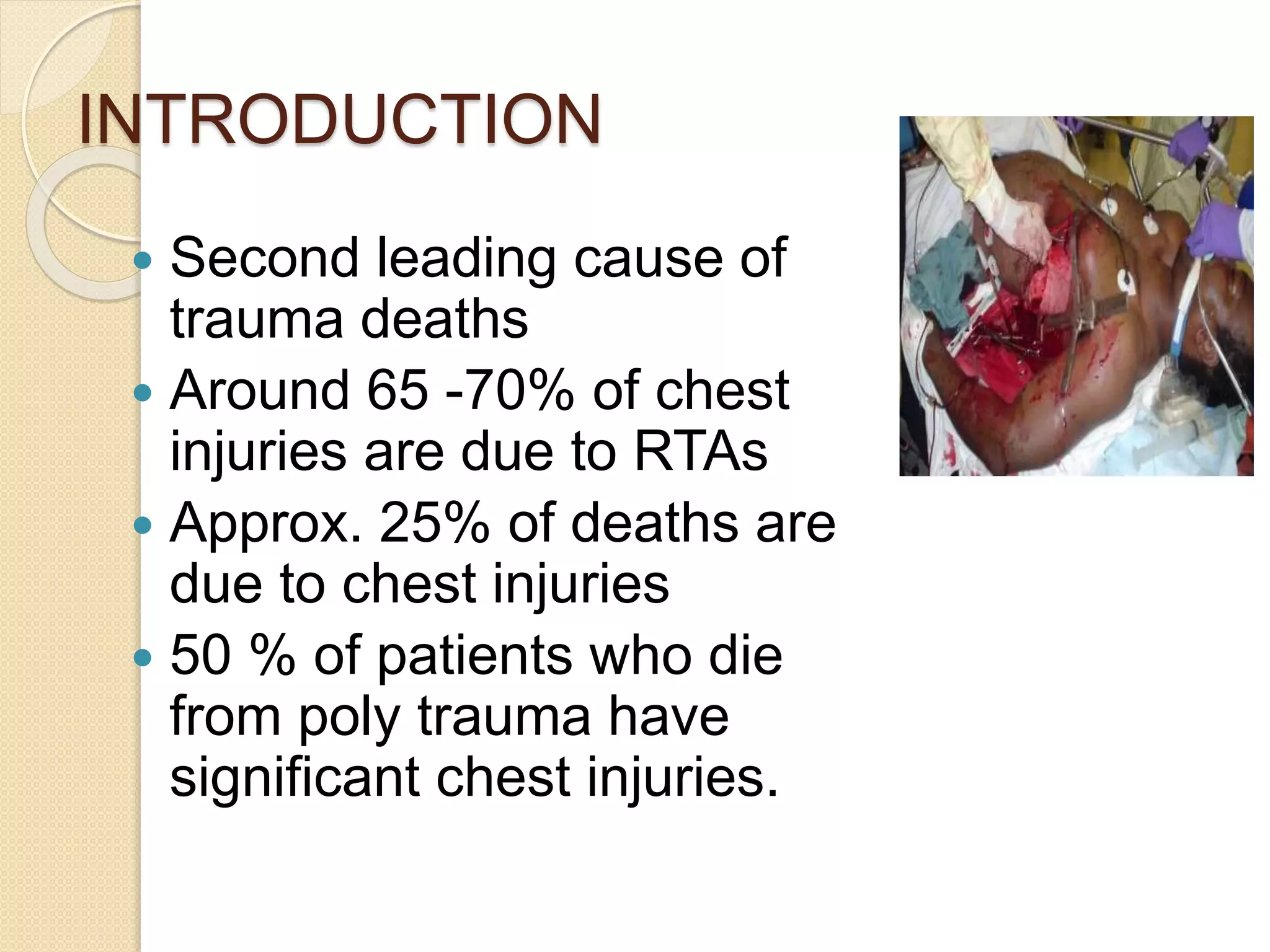


This document provides an overview of chest trauma, including the leading causes, types of injuries, clinical presentation, investigations, and management strategies. It discusses specific injuries such as pneumothorax, hemothorax, flail chest, cardiac tamponade, and aortic rupture. The summary emphasizes that chest trauma is a leading cause of trauma deaths, often involves multiple injuries, and requires rapid diagnosis and treatment of life-threatening conditions like tension pneumothorax to prevent death.