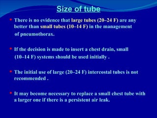
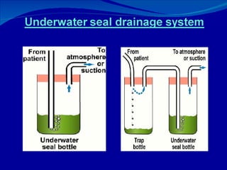
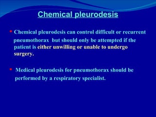
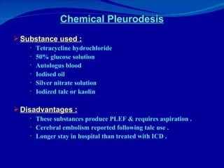
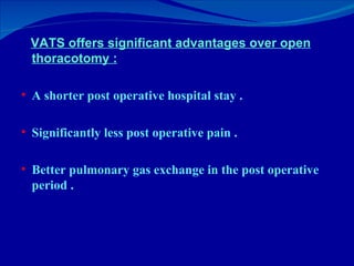
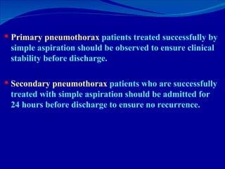
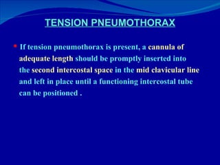
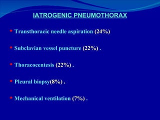
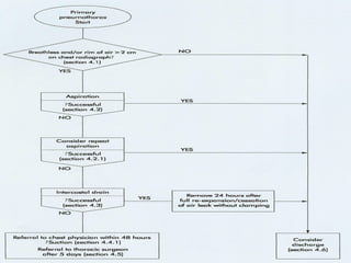
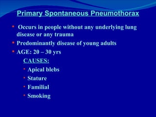
DR K . Mohammed Areef Specialist in pulmonology B.G.H
Spontaneous pneumothorax Primary Secondary
Primary Spontaneous Pneumothorax Occurs in people without any underlying lung disease or any trauma Predominantly disease of young adults AGE: 20 – 30 yrs CAUSES: Apical blebs Stature Familial Smoking


Thoracoscopic image of apical blebs in patient with spontaneous pneumothorax
Secondary Spontaneous Pneumothorax Occurs in the presence of lung diseases CAUSES: Chronic bronchitis & empysema Cavitating active pulmonary tuberculosis Bronchial asthma Suppurative diseases of lung & pleura Cystic fibrosis Pneumocystis carnii pneumonia contd
Secondary Spontaneous Pneumothorax RARE CAUSES: Cryptogenic fibrosing alveolitis Occupational lung diseases – Acute silicosis Granulomatous disease: Sarcoidosis late and fibrotic stage Histiocytosis x Marfanoid hypermobility syndrome Hereditary disorders : Neuro fibromatosis Tuberous sclerosis Coccidioidomycosis Parasitic infection of lung – Hydatid disease contd
Secondary spontaneous Pneumothorax Intra thoracic tumors : Metastatic sarcoma Germ cell tumor Primary bronchial carcinoma Cavitating pulmonary infarction Rhematoid arthritis Systemic sclerosis Haemosiderosis Pulmonary alveolar proteinosis Lymphangioleiomyomatosis Catamenial pneumothorax
CLINICAL EVALUATION AND IMAGING Clinical history and physical examination usually suggest the presence of a pneumothorax . The clinical symptoms associated with secondary pneumothorax are  more severe than  primary pneumothorax . Many patients particularly those with primary pneumothorax, do not seek medical advice for several days . This feature is important because the occurrence of  re-expansion   pulmonary edema (RPO)  after re-inflation may be related to the length of time the lung has been collapsed .
Pulmonary function tests  are weakly sensitive measures of the presence or size of pneumothorax and are not recommended   . Arterial blood gas measurements  are frequently abnormal in patients with pneumothorax with the arterial oxygen tension (PaO2) being less than 10.9 kPa (80 mm Hg) . In both primary and secondary spontaneous pneumothorax the diagnosis is normally established by  plain chest x ray . Expiratory chest radiographs  are not recommended for the routine diagnosis of pneumothorax   .
When a pneumothorax is suspected but not confirmed by  standard posteroanterior (PA)  chest radiographs,  lateral   radiographs  provide added information in up to 14% of cases.  The  lateral decubitus radiograph  is superior to the erect or supine chest radiograph and is felt to be  as sensitive as CT scanning  in pneumothorax detection.
CT scanning is recommended : When differentiating a  pneumothorax from complex bullous lung disease. When aberrant tube placement is suspected . When the plain chest radiograph is obscured by surgical emphysema .
 
SIZE OF PNEUMOTHORAX The size of a pneumothorax, in terms of volume, is difficult to assess accurately from a chest radiograph which is a two dimensional image.  In the 1993 guidelines pneumothorax were classified into three groups: •  “ Small” :  defined as a “small rim of air around the lung” . •  “ Moderate” :  defined as lung “collapsed halfway towards the heart border” •  “ Complete” :  defined as “airless lung, separate from the  diaphragm .
The volume of a pneumothorax approximates to the  ratio   of the cube of the of the lung diameter to the hemithorax diameter . A pneumothorax of  1 cm  on the PA chest radiograph occupies about  27%  of the hemithorax volume if the lung is 9 cm in diameter and the hemithorax 10 cm:  (10 3  – 9 3 )/10 3  = 27%.  A  2 cm  radiographic pneumothorax occupies  49%  of the hemithorax on the same basis .
Volume of pneumothorax (12 3  -  9.5 3 ) /12 3  = 50% Tx suggested  :  Intercostal tube drainage 9.5 cm 12 cm
The previous classification of the size of a pneumothorax  tends to underestimate its volume.  In these new guidelines the size of a pneumothorax is divided into  “small” or “large”  depending on the presence of a visible rim of  <2 cm or >2 cm  between the lung margin and the chest wall .
TREATMENT OPTIONS  FOR SPONTANEOUS PNEUMOTHORAX
Observation Observation should be the treatment of choice for small closed pneumothorax without significant breathlessness.  Patients with small  (<2 cm) primary pneumothorax  not associated with breathlessness should be considered for discharge with early outpatient review.  Observation alone is only recommended in patients with  small secondary pneumothorax of  < 1 cm  depth or isolated apical pneumothorax in asymptomatic patients.
If a patient with a pneumothorax is admitted overnight for observation,  high flow (10 l/min)  oxygen should be administered, with appropriate caution in patients with COPD who may be sensitive to higher concentrations of oxygen.  Breathless patients should not be left without intervention regardless of the size of the pneumothorax on a chest radiograph.
Simple aspiration Simple aspiration is recommended as first line treatment for all  primary pneumothorax  requiring intervention.  Simple aspiration  in  secondary  pneumothorax   is recommended as an initial treatment only in  small (<2 cm) pneumothorax  in minimally breathless patients under the age of 50 years.  Patients with  secondary pneumothorax  treated  successfully with simple aspiration should be admitted to hospital and observed for at least 24 hours  before discharge.
Advantages of simple aspiration Patients treated with simple aspiration were less likely to be hospitalized . Less likely to suffer a recurrence of the pneumothorax over the next 12 months.  Reduction in total pain scores during hospitalization  . Shorter hospital stays .
Simple aspiration Kit
Repeat aspiration and CSAP Repeated aspiration is reasonable for  primary pneumothorax  when the first aspiration has been unsuccessful (i.e. patient still symptomatic) and a  volume  of <2.5 l  has been aspirated on the first attempt.  Catheter aspiration of pneumothorax (CASP) can be used where the equipment and experience is available. Catheter aspiration kits with an integral one way valve system may reduce the need for repeat aspiration.
CASP Catheter aspiration of simple pneumothorax (CASP) involves a small (8 F) catheter being passed over a guide wire into the pleural space.  A three way stopcock is attached and air may be aspirated via a 50 ml syringe.   Addition of a Heimlich valve and suction may improve success rates further.
The Heimlich valve is a one-way, rubber flutter valve.  The proximal end attaches to the chest tube .  The distal end connects to a suction device or is left open to the atmosphere.  It allows outpatient treatment of a pneumothorax.
Intercostal tube drainage If simple aspiration or catheter aspiration drainage of  any pneumothorax  is unsuccessful in controlling symptoms, then an intercostal tube should be inserted.  Intercostal tube drainage is recommended in  secondary   pneumothorax  except in patients who are not breathless and have a very small (<1 cm or apical) pneumothorax.  A bubbling chest tube should never be clamped.
A chest tube which is not bubbling should not usually be clamped.  If a chest tube for pneumothorax is clamped, this should be under the supervision of a respiratory physician or thoracic surgeon . If a patient with a clamped drain becomes breathless or develops subcutaneous emphysema, the drain must be immediately unclamped and medical advice sought.
 
Complications of ICD Penetration of the major organs such as lung, stomach, spleen, liver, heart and great vessels, and are potentially fatal. Pleural infection  [ Empyema ] Surgical emphysema  [ Malpositioned, kinked, blocked or clamped tube , small tube with large leak ]
Size of tube There is no evidence that  large tubes (20–24 F)  are any better than  small tubes (10–14 F)  in the management of pneumothorax.  If the decision is made to insert a chest drain, small (10–14 F) systems should be used initially . The initial use of large (20–24 F) intercostal tubes is not recommended . It may become necessary to replace a small chest tube with a larger one if there is a persistent air leak.
Factors which might predispose to small tube failure, thus favoring the choice of a larger tube . The presence of pleural fluid . The presence of a large air leak which exceeds the capacity of the smaller tubes .
Referral to respiratory specialists Pneumothorax which  fail to respond within 48 hours  to treatment should be referred to a respiratory physician.  May require sustained chest drainage with complex  drain management  (suction, chest drain repositioning)   and thoracic surgery decisions   .
Chest drain suction Suction to an intercostal tube should not be applied directly after tube insertion, but can be added  after 48 hours  for  persistent air leak  or  failure of a pneumothorax to re-expand .  High volume, low pressure  (–10 to –20 cm H2O) suction systems are recommended.  Patients requiring suction should only be managed on lung units where there is specialist medical and nursing experience.
 
Chemical pleurodesis Chemical pleurodesis can control difficult or recurrent pneumothorax  but should only be attempted if the patient is  either unwilling or unable to undergo surgery.  Medical pleurodesis for pneumothorax should be performed by a respiratory specialist.
Chemical Pleurodesis Substance used : Tetracycline hydrochloride 50% glucose solution Autologus blood Iodised oil Silver nitrate solution Iodized talc or kaolin Disadvantages : These substances produce PLEF & requires aspiration . Cerebral embolism reported following talc use . Longer stay in hospital than treated with ICD .
Referral to thoracic surgeons In cases of  persistent air leak or failure of the lung to re-expand , the managing respiratory specialist should seek an early  (3–5 days)  thoracic surgical opinion.  Open thoracotomy  and  pleurectomy  remains the  procedure with the lowest recurrence rate for difficult or recurrent pneumothorax.  Minimally invasive procedures, thoracoscopy (VATS), pleural abrasion, and surgical talc pleurodesis  are all effective alternative strategies.
Indications for operative intervention Second ipsilateral pneumothorax First contralateral pneumothorax Bilateral spontaneous pneumothorax Persistent air leak  (>5–7 days of tube drainage; air leak or failure to completely re-expand) Professions at risk (e.g. pilots, divers)
Surgical strategies Two objectives in the surgical management of a pneumothorax.  The first widely accepted objective is  resection of blebs  or  the suture of apical perforations  to treat the underlying defect .  The second objective is to create a  pleural symphysis  to prevent recurrences.
Open thoracotomy Pleural abrasion  . Parietal pleurectomy  for recurrent pneumothorax.  Both of these techniques are designed  To obliterate the pleural space by creating symphysis between the two pleural layers . Between the visceral pleura and subpleural plane, in the case of parietal pleurectomy .
Open thoracotomy yields the lowest postoperative recurrence results.  Bulla ligation/excision, thoracotomy with pleural abrasion , and either  apical or total parietal pleurectomy  all have failure rates under  0.5%. Open thoracotomy is generally performed using  Single lung ventilation . A limited posterolateral thoracotomy allowing parietal pleurectomy, excision, or stapling of bullae or pleural abrasion.
Video assisted thoracoscopic surgery (VATS) Bullectomy,  pleurectomy,  pleural abrasion, and surgical pleurodesis have all been shown to have reasonable success rates when carried out thoracoscopically . Recurrence rates of pneumothorax after VATS are  5 –10% which are higher than the 1% rates reported after open procedures .
VATS
Pleural Abrasion Metallic scrubber before mechanical pleural abrasion.  Before abrasion, the parietal pleura looks normal . The parietal pleura is reddened and bleeds slightly .
VATS offers significant advantages over open thoracotomy : A shorter post operative hospital stay . Significantly less post operative pain . Better pulmonary gas exchange in the post operative period .
Discharge and follow up Patients discharged without intervention should  avoid   air travel  until a chest radiograph has confirmed resolution of the pneumothorax.  Diving should be permanently avoided  after a pneumothorax, unless the patient has had bilateral surgical pleurectomy.
Primary pneumothorax  patients treated successfully by simple aspiration should be observed to ensure clinical stability before discharge.  Secondary pneumothorax  patients who are successfully treated with simple aspiration should be admitted for 24 hours before discharge to ensure no recurrence.
PNEUMOTHORAX AND CYSTIC FIBROSIS Early and aggressive treatment of pneumothorax in cystic fibrosis is recommended.  Surgical intervention should be considered after the first episode, provided the patient is fit for the procedure.
TENSION PNEUMOTHORAX If tension pneumothorax is present, a  cannula of adequate length  should be promptly inserted into the  second intercostal space  in the  mid clavicular line and left in place until a functioning intercostal tube can be positioned .
IATROGENIC PNEUMOTHORAX Transthoracic needle aspiration  (24%) Subclavian vessel puncture  (22%)  . Thoracocentesis  (22%)  . Pleural biopsy (8%) . Mechanical ventilation  (7%)  .
Pneumothorax Kit Supplied sterile in peel-open packages which includes:  Catheter introducer needle .  Heimlich Valve . Connecting tube, one-way stopcock, Syringe, molnar disc with pull tie .  Small roll of transparent tape.
Future potential areas for research Prospective randomised controlled trials comparing: •  Simple observation versus aspiration ± tube drainage  for primary pneumothoraces larger than 2 cm on the chest radiograph . •  Use of small catheter/Heimlich valve kits versus intercostal tube drainage following failed aspiration in primary pneumothoraces . •  Small catheter aspiration (CASP) versus conventional aspiration or tube drainage .
•  VATS versus open thoracotomy for the difficult pneumothorax. •  Use of suction with regard to its timing and optimal mode. •  Comparison of “clamping” and “ non-clamping” strategies after cessation of air leak.


Spontaneous Pneumothorax An Update

  • 1.
    DR K .Mohammed Areef Specialist in pulmonology B.G.H
  • 2.
  • 3.
    Primary Spontaneous PneumothoraxOccurs in people without any underlying lung disease or any trauma Predominantly disease of young adults AGE: 20 – 30 yrs CAUSES: Apical blebs Stature Familial Smoking
  • 4.
  • 5.
  • 6.
  • 7.
    Thoracoscopic image ofapical blebs in patient with spontaneous pneumothorax
  • 8.
    Secondary Spontaneous PneumothoraxOccurs in the presence of lung diseases CAUSES: Chronic bronchitis & empysema Cavitating active pulmonary tuberculosis Bronchial asthma Suppurative diseases of lung & pleura Cystic fibrosis Pneumocystis carnii pneumonia contd
  • 9.
    Secondary Spontaneous PneumothoraxRARE CAUSES: Cryptogenic fibrosing alveolitis Occupational lung diseases – Acute silicosis Granulomatous disease: Sarcoidosis late and fibrotic stage Histiocytosis x Marfanoid hypermobility syndrome Hereditary disorders : Neuro fibromatosis Tuberous sclerosis Coccidioidomycosis Parasitic infection of lung – Hydatid disease contd
  • 10.
    Secondary spontaneous PneumothoraxIntra thoracic tumors : Metastatic sarcoma Germ cell tumor Primary bronchial carcinoma Cavitating pulmonary infarction Rhematoid arthritis Systemic sclerosis Haemosiderosis Pulmonary alveolar proteinosis Lymphangioleiomyomatosis Catamenial pneumothorax
  • 11.
    CLINICAL EVALUATION ANDIMAGING Clinical history and physical examination usually suggest the presence of a pneumothorax . The clinical symptoms associated with secondary pneumothorax are more severe than primary pneumothorax . Many patients particularly those with primary pneumothorax, do not seek medical advice for several days . This feature is important because the occurrence of re-expansion pulmonary edema (RPO) after re-inflation may be related to the length of time the lung has been collapsed .
  • 12.
    Pulmonary function tests are weakly sensitive measures of the presence or size of pneumothorax and are not recommended . Arterial blood gas measurements are frequently abnormal in patients with pneumothorax with the arterial oxygen tension (PaO2) being less than 10.9 kPa (80 mm Hg) . In both primary and secondary spontaneous pneumothorax the diagnosis is normally established by plain chest x ray . Expiratory chest radiographs are not recommended for the routine diagnosis of pneumothorax .
  • 13.
    When a pneumothoraxis suspected but not confirmed by standard posteroanterior (PA) chest radiographs, lateral radiographs provide added information in up to 14% of cases. The lateral decubitus radiograph is superior to the erect or supine chest radiograph and is felt to be as sensitive as CT scanning in pneumothorax detection.
  • 14.
    CT scanning isrecommended : When differentiating a pneumothorax from complex bullous lung disease. When aberrant tube placement is suspected . When the plain chest radiograph is obscured by surgical emphysema .
  • 15.
  • 16.
    SIZE OF PNEUMOTHORAXThe size of a pneumothorax, in terms of volume, is difficult to assess accurately from a chest radiograph which is a two dimensional image. In the 1993 guidelines pneumothorax were classified into three groups: • “ Small” : defined as a “small rim of air around the lung” . • “ Moderate” : defined as lung “collapsed halfway towards the heart border” • “ Complete” : defined as “airless lung, separate from the diaphragm .
  • 17.
    The volume ofa pneumothorax approximates to the ratio of the cube of the of the lung diameter to the hemithorax diameter . A pneumothorax of 1 cm on the PA chest radiograph occupies about 27% of the hemithorax volume if the lung is 9 cm in diameter and the hemithorax 10 cm: (10 3 – 9 3 )/10 3 = 27%. A 2 cm radiographic pneumothorax occupies 49% of the hemithorax on the same basis .
  • 18.
    Volume of pneumothorax(12 3 - 9.5 3 ) /12 3 = 50% Tx suggested : Intercostal tube drainage 9.5 cm 12 cm
  • 19.
    The previous classificationof the size of a pneumothorax tends to underestimate its volume. In these new guidelines the size of a pneumothorax is divided into “small” or “large” depending on the presence of a visible rim of <2 cm or >2 cm between the lung margin and the chest wall .
  • 20.
    TREATMENT OPTIONS FOR SPONTANEOUS PNEUMOTHORAX
  • 21.
    Observation Observation shouldbe the treatment of choice for small closed pneumothorax without significant breathlessness. Patients with small (<2 cm) primary pneumothorax not associated with breathlessness should be considered for discharge with early outpatient review. Observation alone is only recommended in patients with small secondary pneumothorax of < 1 cm depth or isolated apical pneumothorax in asymptomatic patients.
  • 22.
    If a patientwith a pneumothorax is admitted overnight for observation, high flow (10 l/min) oxygen should be administered, with appropriate caution in patients with COPD who may be sensitive to higher concentrations of oxygen. Breathless patients should not be left without intervention regardless of the size of the pneumothorax on a chest radiograph.
  • 23.
    Simple aspiration Simpleaspiration is recommended as first line treatment for all primary pneumothorax requiring intervention. Simple aspiration in secondary pneumothorax is recommended as an initial treatment only in small (<2 cm) pneumothorax in minimally breathless patients under the age of 50 years. Patients with secondary pneumothorax treated successfully with simple aspiration should be admitted to hospital and observed for at least 24 hours before discharge.
  • 24.
    Advantages of simpleaspiration Patients treated with simple aspiration were less likely to be hospitalized . Less likely to suffer a recurrence of the pneumothorax over the next 12 months. Reduction in total pain scores during hospitalization . Shorter hospital stays .
  • 25.
  • 26.
    Repeat aspiration andCSAP Repeated aspiration is reasonable for primary pneumothorax when the first aspiration has been unsuccessful (i.e. patient still symptomatic) and a volume of <2.5 l has been aspirated on the first attempt. Catheter aspiration of pneumothorax (CASP) can be used where the equipment and experience is available. Catheter aspiration kits with an integral one way valve system may reduce the need for repeat aspiration.
  • 27.
    CASP Catheter aspirationof simple pneumothorax (CASP) involves a small (8 F) catheter being passed over a guide wire into the pleural space. A three way stopcock is attached and air may be aspirated via a 50 ml syringe. Addition of a Heimlich valve and suction may improve success rates further.
  • 28.
    The Heimlich valveis a one-way, rubber flutter valve. The proximal end attaches to the chest tube . The distal end connects to a suction device or is left open to the atmosphere. It allows outpatient treatment of a pneumothorax.
  • 29.
    Intercostal tube drainageIf simple aspiration or catheter aspiration drainage of any pneumothorax is unsuccessful in controlling symptoms, then an intercostal tube should be inserted. Intercostal tube drainage is recommended in secondary pneumothorax except in patients who are not breathless and have a very small (<1 cm or apical) pneumothorax. A bubbling chest tube should never be clamped.
  • 30.
    A chest tubewhich is not bubbling should not usually be clamped. If a chest tube for pneumothorax is clamped, this should be under the supervision of a respiratory physician or thoracic surgeon . If a patient with a clamped drain becomes breathless or develops subcutaneous emphysema, the drain must be immediately unclamped and medical advice sought.
  • 31.
  • 32.
    Complications of ICDPenetration of the major organs such as lung, stomach, spleen, liver, heart and great vessels, and are potentially fatal. Pleural infection [ Empyema ] Surgical emphysema [ Malpositioned, kinked, blocked or clamped tube , small tube with large leak ]
  • 33.
    Size of tubeThere is no evidence that large tubes (20–24 F) are any better than small tubes (10–14 F) in the management of pneumothorax. If the decision is made to insert a chest drain, small (10–14 F) systems should be used initially . The initial use of large (20–24 F) intercostal tubes is not recommended . It may become necessary to replace a small chest tube with a larger one if there is a persistent air leak.
  • 34.
    Factors which mightpredispose to small tube failure, thus favoring the choice of a larger tube . The presence of pleural fluid . The presence of a large air leak which exceeds the capacity of the smaller tubes .
  • 35.
    Referral to respiratoryspecialists Pneumothorax which fail to respond within 48 hours to treatment should be referred to a respiratory physician. May require sustained chest drainage with complex drain management (suction, chest drain repositioning) and thoracic surgery decisions .
  • 36.
    Chest drain suctionSuction to an intercostal tube should not be applied directly after tube insertion, but can be added after 48 hours for persistent air leak or failure of a pneumothorax to re-expand . High volume, low pressure (–10 to –20 cm H2O) suction systems are recommended. Patients requiring suction should only be managed on lung units where there is specialist medical and nursing experience.
  • 37.
  • 38.
    Chemical pleurodesis Chemicalpleurodesis can control difficult or recurrent pneumothorax but should only be attempted if the patient is either unwilling or unable to undergo surgery. Medical pleurodesis for pneumothorax should be performed by a respiratory specialist.
  • 39.
    Chemical Pleurodesis Substanceused : Tetracycline hydrochloride 50% glucose solution Autologus blood Iodised oil Silver nitrate solution Iodized talc or kaolin Disadvantages : These substances produce PLEF & requires aspiration . Cerebral embolism reported following talc use . Longer stay in hospital than treated with ICD .
  • 40.
    Referral to thoracicsurgeons In cases of persistent air leak or failure of the lung to re-expand , the managing respiratory specialist should seek an early (3–5 days) thoracic surgical opinion. Open thoracotomy and pleurectomy remains the procedure with the lowest recurrence rate for difficult or recurrent pneumothorax. Minimally invasive procedures, thoracoscopy (VATS), pleural abrasion, and surgical talc pleurodesis are all effective alternative strategies.
  • 41.
    Indications for operativeintervention Second ipsilateral pneumothorax First contralateral pneumothorax Bilateral spontaneous pneumothorax Persistent air leak (>5–7 days of tube drainage; air leak or failure to completely re-expand) Professions at risk (e.g. pilots, divers)
  • 42.
    Surgical strategies Twoobjectives in the surgical management of a pneumothorax. The first widely accepted objective is resection of blebs or the suture of apical perforations to treat the underlying defect . The second objective is to create a pleural symphysis to prevent recurrences.
  • 43.
    Open thoracotomy Pleuralabrasion . Parietal pleurectomy for recurrent pneumothorax. Both of these techniques are designed To obliterate the pleural space by creating symphysis between the two pleural layers . Between the visceral pleura and subpleural plane, in the case of parietal pleurectomy .
  • 44.
    Open thoracotomy yieldsthe lowest postoperative recurrence results. Bulla ligation/excision, thoracotomy with pleural abrasion , and either apical or total parietal pleurectomy all have failure rates under 0.5%. Open thoracotomy is generally performed using Single lung ventilation . A limited posterolateral thoracotomy allowing parietal pleurectomy, excision, or stapling of bullae or pleural abrasion.
  • 45.
    Video assisted thoracoscopicsurgery (VATS) Bullectomy, pleurectomy, pleural abrasion, and surgical pleurodesis have all been shown to have reasonable success rates when carried out thoracoscopically . Recurrence rates of pneumothorax after VATS are 5 –10% which are higher than the 1% rates reported after open procedures .
  • 46.
  • 47.
    Pleural Abrasion Metallicscrubber before mechanical pleural abrasion. Before abrasion, the parietal pleura looks normal . The parietal pleura is reddened and bleeds slightly .
  • 48.
    VATS offers significantadvantages over open thoracotomy : A shorter post operative hospital stay . Significantly less post operative pain . Better pulmonary gas exchange in the post operative period .
  • 49.
    Discharge and followup Patients discharged without intervention should avoid air travel until a chest radiograph has confirmed resolution of the pneumothorax. Diving should be permanently avoided after a pneumothorax, unless the patient has had bilateral surgical pleurectomy.
  • 50.
    Primary pneumothorax patients treated successfully by simple aspiration should be observed to ensure clinical stability before discharge. Secondary pneumothorax patients who are successfully treated with simple aspiration should be admitted for 24 hours before discharge to ensure no recurrence.
  • 51.
    PNEUMOTHORAX AND CYSTICFIBROSIS Early and aggressive treatment of pneumothorax in cystic fibrosis is recommended. Surgical intervention should be considered after the first episode, provided the patient is fit for the procedure.
  • 52.
    TENSION PNEUMOTHORAX Iftension pneumothorax is present, a cannula of adequate length should be promptly inserted into the second intercostal space in the mid clavicular line and left in place until a functioning intercostal tube can be positioned .
  • 53.
    IATROGENIC PNEUMOTHORAX Transthoracicneedle aspiration (24%) Subclavian vessel puncture (22%) . Thoracocentesis (22%) . Pleural biopsy (8%) . Mechanical ventilation (7%) .
  • 54.
    Pneumothorax Kit Suppliedsterile in peel-open packages which includes: Catheter introducer needle . Heimlich Valve . Connecting tube, one-way stopcock, Syringe, molnar disc with pull tie . Small roll of transparent tape.
  • 55.
    Future potential areasfor research Prospective randomised controlled trials comparing: • Simple observation versus aspiration ± tube drainage for primary pneumothoraces larger than 2 cm on the chest radiograph . • Use of small catheter/Heimlich valve kits versus intercostal tube drainage following failed aspiration in primary pneumothoraces . • Small catheter aspiration (CASP) versus conventional aspiration or tube drainage .
  • 56.
    • VATSversus open thoracotomy for the difficult pneumothorax. • Use of suction with regard to its timing and optimal mode. • Comparison of “clamping” and “ non-clamping” strategies after cessation of air leak.
  • 57.
  • 58.
  • 59.