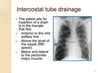
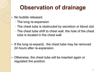
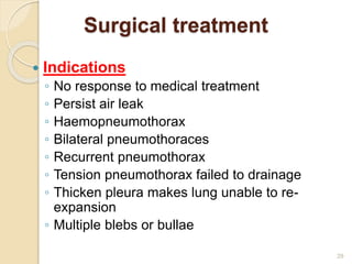
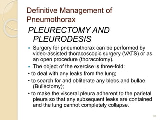
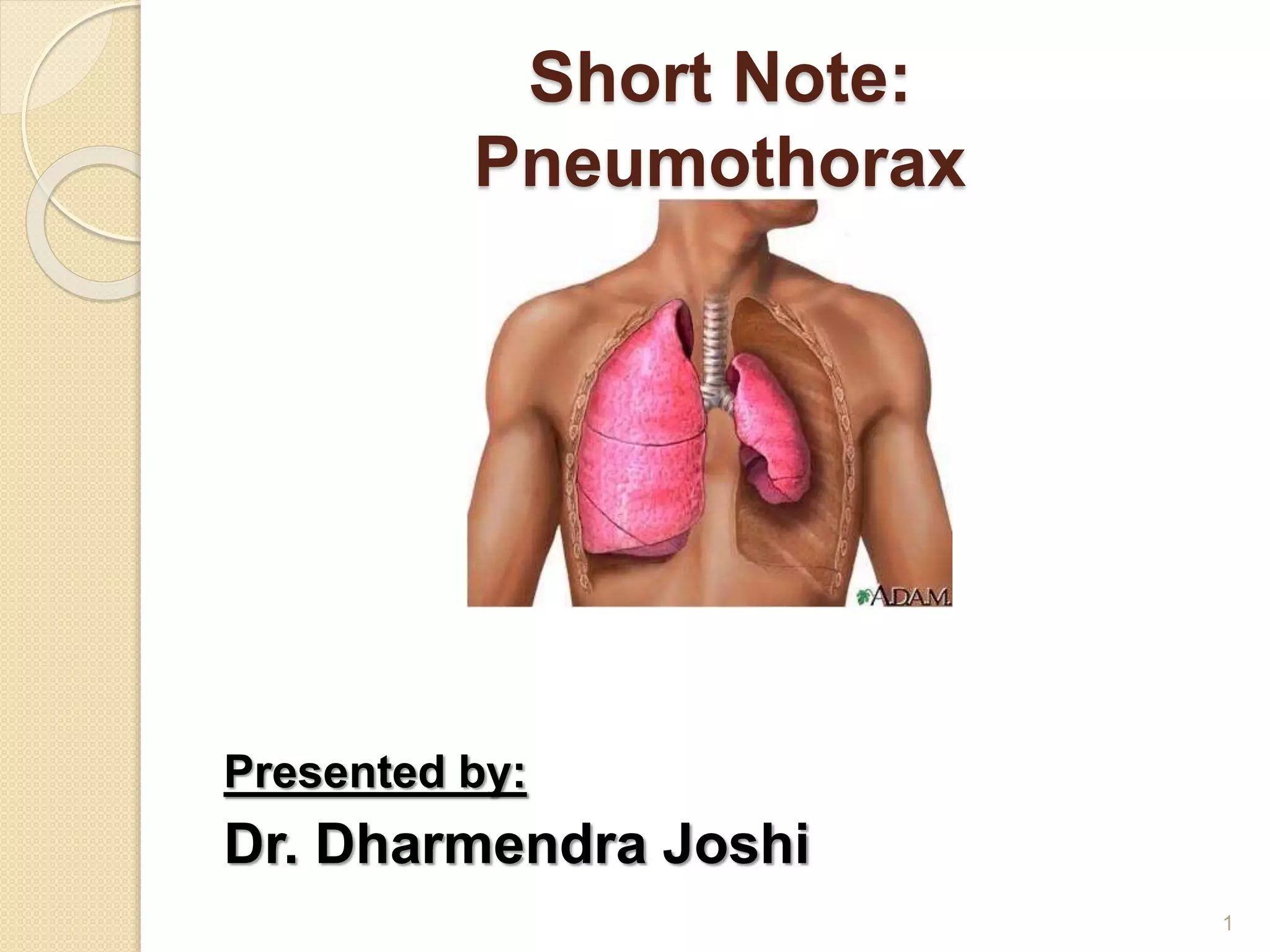
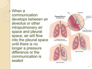
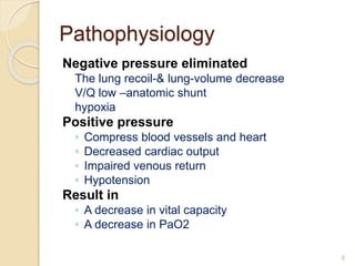
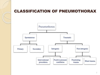
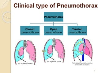
This document provides an overview of pneumothorax, including its definition, classification, mechanisms, clinical presentation, diagnosis, and management. Pneumothorax is defined as the presence of air in the pleural space, and can be spontaneous, traumatic, or iatrogenic. It presents with symptoms like dyspnea and chest pain. Diagnosis is typically made through chest x-ray or CT scan. Management depends on the type and severity, and may include oxygen therapy, needle aspiration, chest tube drainage, or surgery like pleurodesis for recurrent cases.
Thoracentesis (thor-a-sen-tee-sis) is a procedure that is done to remove a sample of fluid from around the lung.
The lung is covered with a tissue called the pleura. The inside of the chest is also lined with pleura.
The space between these two areas is called the pleural space.
This space normally contains just a thin layer of fluid, however, some conditions such as pneumonia, some types of cancer, or congestive heart failure may cause excessive fluid to develop (pleural effusion).
Thoracentesis, also known as pleural fluid analysis, is a procedure in which a needle is inserted through the back of the chest wall into the pleural space (a space that exists between the two lungs and the anterior chest wall) to remove fluid or air.
Pleural fluid analysis is the microscopic and chemical lab analysis of the fluid obtained during thoracentesis.
IndDiagnostic: determination of pleural effusion etiology (e.g. transudative versus exudative) usually requires the removal of 50 to 100mL of pleural fluid for laboratory studies. Most new effusions require diagnostic thoracentesis, an exception being a new effusion with a clear clinical diagnosis (e.g. CHF) with no evidence for superimposed pleural space infection
Therapeutic: reduce dyspnea and respiratory compromise in patients with large pleural effusions. This is typically achieved by removing a much larger volume of fluid compared to the diagnostic thoracentesis
ications
Despite modern anti-tuberculous chemotherapy, approximately 2% of all cases of pulmonary mycobacterial infection require surgical treatment.Therefore, surgical treatment of pulmonary mycobacterial disease is rarely necessary.Types of surgical procedures for PTB include: Collapse therapy, pulmonary resection, lung decortication, drainage procedures such as closed tube thoracostomy, rib resection and open window thoracotomy beside pulmonary resection+ collapse therapy (thoracoplasty). The decreasing morbidity and mortality of pulmonary resection for PTB is due to careful patient selection ( failure of chemotherapy, massive haemoptysis, BPF), improved anaesthetic techniques, stapling devices and better chemotherapy.The prognosis after successful resection is excellent ( 90% survive and remain disease free).
Respiratory Disorders
Disease Condition Pneumothorax, Causes, Sign and Symptoms, Pathophysiology, Types, Assessment and Dignostic Test, Management
By HIREN GEHLOTH For Nursing Students Medical Surgical Nursing
LAUGH A LOT IT CLEARS THE LUNGS
TEACHING IS ONE PROFESSION THAT CREATE ALL OTHER PROFESSION
Hemopneumothorax, or haemopneumothorax is the condition of having air in the chest cavity (pneumothorax) and blood in the chest cavity (hemothorax). A hemothorax, pneumothorax, or the combination of both can occur due to an injury to the lung or chest.
Bilateral Pulmonary Hydatid Cysts with Ruptured & Infected Hydatid Cyst of Le...Dharmendra Joshi
Bilateral Pulmonary Hydatid Cysts with Ruptured & Infected Hydatid Cyst of Left Lung - A Case Presentation
Operations:
First Operation:
VATS Enucleation of Hydatid Cyst of Lung (Right side)
Second Operation:
VATS followed by minimally invasive open Enucleation and Capitonnage of Hydatid Cyst of Lung (Left side)
Prix Galien International 2024 Forum ProgramLevi Shapiro
June 20, 2024, Prix Galien International and Jerusalem Ethics Forum in ROME. Detailed agenda including panels:
- ADVANCES IN CARDIOLOGY: A NEW PARADIGM IS COMING
- WOMEN’S HEALTH: FERTILITY PRESERVATION
- WHAT’S NEW IN THE TREATMENT OF INFECTIOUS,
ONCOLOGICAL AND INFLAMMATORY SKIN DISEASES?
- ARTIFICIAL INTELLIGENCE AND ETHICS
- GENE THERAPY
- BEYOND BORDERS: GLOBAL INITIATIVES FOR DEMOCRATIZING LIFE SCIENCE TECHNOLOGIES AND PROMOTING ACCESS TO HEALTHCARE
- ETHICAL CHALLENGES IN LIFE SCIENCES
- Prix Galien International Awards Ceremony
Ozempic: Preoperative Management of Patients on GLP-1 Receptor Agonists Saeid Safari
Preoperative Management of Patients on GLP-1 Receptor Agonists like Ozempic and Semiglutide
ASA GUIDELINE
NYSORA Guideline
2 Case Reports of Gastric Ultrasound
Couples presenting to the infertility clinic- Do they really have infertility...Sujoy Dasgupta
Dr Sujoy Dasgupta presented the study on "Couples presenting to the infertility clinic- Do they really have infertility? – The unexplored stories of non-consummation" in the 13th Congress of the Asia Pacific Initiative on Reproduction (ASPIRE 2024) at Manila on 24 May, 2024.
Title: Sense of Taste
Presenter: Dr. Faiza, Assistant Professor of Physiology
Qualifications:
MBBS (Best Graduate, AIMC Lahore)
FCPS Physiology
ICMT, CHPE, DHPE (STMU)
MPH (GC University, Faisalabad)
MBA (Virtual University of Pakistan)
Learning Objectives:
Describe the structure and function of taste buds.
Describe the relationship between the taste threshold and taste index of common substances.
Explain the chemical basis and signal transduction of taste perception for each type of primary taste sensation.
Recognize different abnormalities of taste perception and their causes.
Key Topics:
Significance of Taste Sensation:
Differentiation between pleasant and harmful food
Influence on behavior
Selection of food based on metabolic needs
Receptors of Taste:
Taste buds on the tongue
Influence of sense of smell, texture of food, and pain stimulation (e.g., by pepper)
Primary and Secondary Taste Sensations:
Primary taste sensations: Sweet, Sour, Salty, Bitter, Umami
Chemical basis and signal transduction mechanisms for each taste
Taste Threshold and Index:
Taste threshold values for Sweet (sucrose), Salty (NaCl), Sour (HCl), and Bitter (Quinine)
Taste index relationship: Inversely proportional to taste threshold
Taste Blindness:
Inability to taste certain substances, particularly thiourea compounds
Example: Phenylthiocarbamide
Structure and Function of Taste Buds:
Composition: Epithelial cells, Sustentacular/Supporting cells, Taste cells, Basal cells
Features: Taste pores, Taste hairs/microvilli, and Taste nerve fibers
Location of Taste Buds:
Found in papillae of the tongue (Fungiform, Circumvallate, Foliate)
Also present on the palate, tonsillar pillars, epiglottis, and proximal esophagus
Mechanism of Taste Stimulation:
Interaction of taste substances with receptors on microvilli
Signal transduction pathways for Umami, Sweet, Bitter, Sour, and Salty tastes
Taste Sensitivity and Adaptation:
Decrease in sensitivity with age
Rapid adaptation of taste sensation
Role of Saliva in Taste:
Dissolution of tastants to reach receptors
Washing away the stimulus
Taste Preferences and Aversions:
Mechanisms behind taste preference and aversion
Influence of receptors and neural pathways
Impact of Sensory Nerve Damage:
Degeneration of taste buds if the sensory nerve fiber is cut
Abnormalities of Taste Detection:
Conditions: Ageusia, Hypogeusia, Dysgeusia (parageusia)
Causes: Nerve damage, neurological disorders, infections, poor oral hygiene, adverse drug effects, deficiencies, aging, tobacco use, altered neurotransmitter levels
Neurotransmitters and Taste Threshold:
Effects of serotonin (5-HT) and norepinephrine (NE) on taste sensitivity
Supertasters:
25% of the population with heightened sensitivity to taste, especially bitterness
Increased number of fungiform papillae
The prostate is an exocrine gland of the male mammalian reproductive system
It is a walnut-sized gland that forms part of the male reproductive system and is located in front of the rectum and just below the urinary bladder
Function is to store and secrete a clear, slightly alkaline fluid that constitutes 10-30% of the volume of the seminal fluid that along with the spermatozoa, constitutes semen
A healthy human prostate measures (4cm-vertical, by 3cm-horizontal, 2cm ant-post ).
It surrounds the urethra just below the urinary bladder. It has anterior, median, posterior and two lateral lobes
It’s work is regulated by androgens which are responsible for male sex characteristics
Generalised disease of the prostate due to hormonal derangement which leads to non malignant enlargement of the gland (increase in the number of epithelial cells and stromal tissue)to cause compression of the urethra leading to symptoms (LUTS
Explore natural remedies for syphilis treatment in Singapore. Discover alternative therapies, herbal remedies, and lifestyle changes that may complement conventional treatments. Learn about holistic approaches to managing syphilis symptoms and supporting overall health.
TEST BANK for Operations Management, 14th Edition by William J. Stevenson, Ve...kevinkariuki227
TEST BANK for Operations Management, 14th Edition by William J. Stevenson, Verified Chapters 1 - 19, Complete Newest Version.pdf
- Video recording of this lecture in English language: https://youtu.be/lK81BzxMqdo
- Video recording of this lecture in Arabic language: https://youtu.be/Ve4P0COk9OI
- Link to download the book free: https://nephrotube.blogspot.com/p/nephrotube-nephrology-books.html
- Link to NephroTube website: www.NephroTube.com
- Link to NephroTube social media accounts: https://nephrotube.blogspot.com/p/join-nephrotube-on-social-media.html
micro teaching on communication m.sc nursing.pdfAnurag Sharma
Microteaching is a unique model of practice teaching. It is a viable instrument for the. desired change in the teaching behavior or the behavior potential which, in specified types of real. classroom situations, tends to facilitate the achievement of specified types of objectives.
Tom Selleck Health: A Comprehensive Look at the Iconic Actor’s Wellness Journeygreendigital
Tom Selleck, an enduring figure in Hollywood. has captivated audiences for decades with his rugged charm, iconic moustache. and memorable roles in television and film. From his breakout role as Thomas Magnum in Magnum P.I. to his current portrayal of Frank Reagan in Blue Bloods. Selleck's career has spanned over 50 years. But beyond his professional achievements. fans have often been curious about Tom Selleck Health. especially as he has aged in the public eye.
Follow us on: Pinterest
Introduction
Many have been interested in Tom Selleck health. not only because of his enduring presence on screen but also because of the challenges. and lifestyle choices he has faced and made over the years. This article delves into the various aspects of Tom Selleck health. exploring his fitness regimen, diet, mental health. and the challenges he has encountered as he ages. We'll look at how he maintains his well-being. the health issues he has faced, and his approach to ageing .
Early Life and Career
Childhood and Athletic Beginnings
Tom Selleck was born on January 29, 1945, in Detroit, Michigan, and grew up in Sherman Oaks, California. From an early age, he was involved in sports, particularly basketball. which played a significant role in his physical development. His athletic pursuits continued into college. where he attended the University of Southern California (USC) on a basketball scholarship. This early involvement in sports laid a strong foundation for his physical health and disciplined lifestyle.
Transition to Acting
Selleck's transition from an athlete to an actor came with its physical demands. His first significant role in "Magnum P.I." required him to perform various stunts and maintain a fit appearance. This role, which he played from 1980 to 1988. necessitated a rigorous fitness routine to meet the show's demands. setting the stage for his long-term commitment to health and wellness.
Fitness Regimen
Workout Routine
Tom Selleck health and fitness regimen has evolved. adapting to his changing roles and age. During his "Magnum, P.I." days. Selleck's workouts were intense and focused on building and maintaining muscle mass. His routine included weightlifting, cardiovascular exercises. and specific training for the stunts he performed on the show.
Selleck adjusted his fitness routine as he aged to suit his body's needs. Today, his workouts focus on maintaining flexibility, strength, and cardiovascular health. He incorporates low-impact exercises such as swimming, walking, and light weightlifting. This balanced approach helps him stay fit without putting undue strain on his joints and muscles.
Importance of Flexibility and Mobility
In recent years, Selleck has emphasized the importance of flexibility and mobility in his fitness regimen. Understanding the natural decline in muscle mass and joint flexibility with age. he includes stretching and yoga in his routine. These practices help prevent injuries, improve posture, and maintain mobilit
2. Introduction:
Laennec described the clinical picture of
Pneumothorax in 1819.
The modern description of primary
spontaneous pneumothorax occurring in
otherwise healthy people was provided
by Kjaergard in 1932.
Primary pneumothorax remains a
significant global problem.
Incidence is 18-28/100,000 per year for
men and 1.2-6/100,000 per year for
women.
2
3. Definition:
Pneumothorax is defined as
presence of air or gas in the
pleural space
(OR)
Presence of air outside the lung,
within the pleural space.
3
4. Mechanism:
In normal people, the pressure in
pleural space is negative during the
entire respiratory cycle.
Two opposite forces result in negative
pressure in pleural space.
(outward pull of the chest wall and
elastic recoil of the lung)
The negative pressure will
be disappeared if any communication
develops .
4
5. When a
communication
develops between an
alveolus or other
intrapulmonary air
space and pleural
space, air will flow
into the pleural space
until there is no
longer a pressure
difference or the
communication is
sealed
5
6. Pathophysiology
Negative pressure eliminated
The lung recoil-& lung-volume decrease
V/Q low –anatomic shunt
hypoxia
Positive pressure
◦ Compress blood vessels and heart
◦ Decreased cardiac output
◦ Impaired venous return
◦ Hypotension
Result in
◦ A decrease in vital capacity
◦ A decrease in PaO2
6
10. Clinical manifestation
Tension pneumothorax
◦ Distressed with rapid labored respiration
◦ Cyanosis
◦ Marked tachycardia
Patient who suddenly deteriorate
clinically,
be suspected in the patient with
◦ Mechanical ventilation
◦ Cardiopulmonary resuscitation
10
11. Physical examination:
◦ Depend on size of pneumothorax
◦ The vital signs usually normal
◦ Unilateral Chest movements
◦ The trachea may be shifted toward the
contralateral side if the pneumothorax is
large
◦ Tactile fremitus is absent
◦ The percussion note is hyper-resonant
◦ The breath sounds are reduced or absent
on the affected side
◦ The lower edge of the liver may be shifted
inferiorly with a right-side pneumothorax
11
17. CT scanning
It is recommended in difficult cases
such as patients in whom the lungs
are obscured by overlying surgical
emphysema
To differentiate a pneumothorax from
suspected bulla in complex cystic lung
disease
17
19. MANAGMENT
Goals
◦ To promote lung expansion
◦ To eliminate the pathogenesis
◦ To decrease pneumothorax recurrence
Treatment options according to
◦ Classification of pneumothorax
◦ Pathogenesis
◦ The extension of lung collapse
◦ Severity of disease
◦ Complication and concomitant underlying
diseases
19
21. 21
Inhalation of high concentration of oxygen
may reduce the total pressure of gases in
pleural capillaries by reducing the partial
pressure of nitrogen
This should increase the pressure gradient
between the pleural capillaries and the
pleural cavity
Thereby increasing absorption of air from
the pleural cavity
O2 TREATMENT: PSP or SSP
22. 22
The rate of resolution/re-absorption of
spontaneous pneumothorax is 1.25 –
1.8% of volume of hemi thorax every 24
hours
The addition of high flow oxygen
therapy has been shown to result in a
4-fold increase in the rate of
pneumothorax reabsorption during the
periods of oxygen supplementation
23. 23
Simple aspiration
Simple aspiration is recommended as first line
treatment for all PSP requiring intervention
Simple aspiration is less likely to succeed in
secondary pneumothoraces and in this situation,
is only recommended as an initial treatment in
small (<2 cm) pneumothoraces in minimally
breathless patients under the age of 50 years
Patients with secondary pneumothoraces
treated successfully with simple aspiration
should be admitted to hospital and observed for
at least 24 hours before discharge
24. 24
Repeated aspiration is reasonable for
primary pneumothorax when the first
aspiration has been unsuccessful
A volume of < 2.5 L has been aspirated on
the first attempt
The aspiration can be used by needle or
catheter
Catheter aspiration
25. Intercostal tube drainage
INDICATIONS
◦ Unstable
pneumothorax
◦ Severe dyspnea
◦ Large lung collapse
◦ Open or tension
pneumothorax
◦ Recurrent
pneumothorax
◦ Simple aspiration or
catheter aspiration
drainage is
unsuccessful in
controlling symptoms 25
26. 26
The safest site for
Insertion of a drain
is in the triangle
that lies:
a. Anterior to the mid
axillary line
b. Above the level of
the nipple (fifth
space)
c. Below and lateral
to the pectoralis
major muscle.
Intercostal tube drainage
27. 27
Intercostal tube drainage
Fix the catheter and cover with
gauze
Making a small incision
Using a forceps to extend the
hole
Inserting a catheter into
pleural cavity
28. 28
Observation of drainage
No bubble released:
◦ The lung re-expansion
◦ The chest tube is obstructed by secretion or blood clot
◦ The chest tube shift to chest wall, the hole of the chest
tube is located in the chest wall
If the lung re-expand, the chest tube may be removed
24 hours after re-expansion.
Otherwise, the chest tube will be inserted again or
regulated the position.
29. 29
Surgical treatment
Indications
◦ No response to medical treatment
◦ Persist air leak
◦ Haemopneumothorax
◦ Bilateral pneumothoraces
◦ Recurrent pneumothorax
◦ Tension pneumothorax failed to drainage
◦ Thicken pleura makes lung unable to re-
expansion
◦ Multiple blebs or bullae
30. Definitive Management of
Pneumothorax
PLEURECTOMY AND
PLEURODESIS
Surgery for pneumothorax can be performed by
video-assisted thoracoscopic surgery (VATS) or as
an open procedure (thoracotomy).
The object of the exercise is three-fold:
• to deal with any leaks from the lung;
• to search for and obliterate any blebs and bullae
(Bullectomy);
• to make the visceral pleura adherent to the parietal
pleura so that any subsequent leaks are contained
and the lung cannot completely collapse.
30
31. Pleural adhesion is achieved in
one of three ways:
• Pleurectomy: systematically strip the
parietal pleura from the chest wall.
• Pleural abrasion: a scourer is used to
scrape off the slick surface of the
parietal pleura.
• Chemical pleurodesis: usually talc is
used and is insufflated into the chest
cavity.
31
33. Recurrence:
The best estimates of recurrence rates
are:
• of patients who experience a first event,
only about one-third experience
recurrence;
• of those who have a second episode,
about one-half go on to experience a
third episode;
• those who have had three episodes will
probably go on to have repeated
recurrences.
33