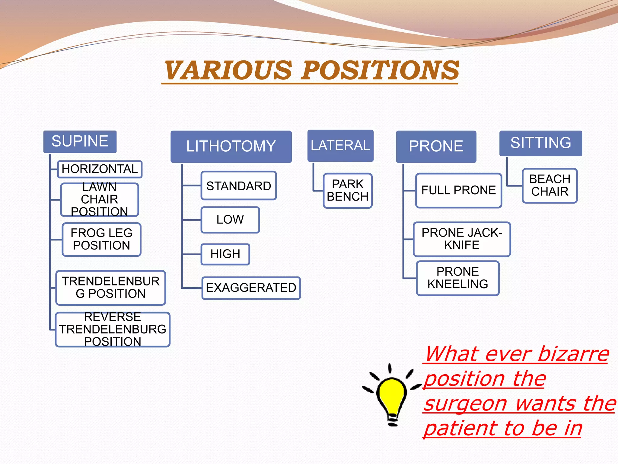
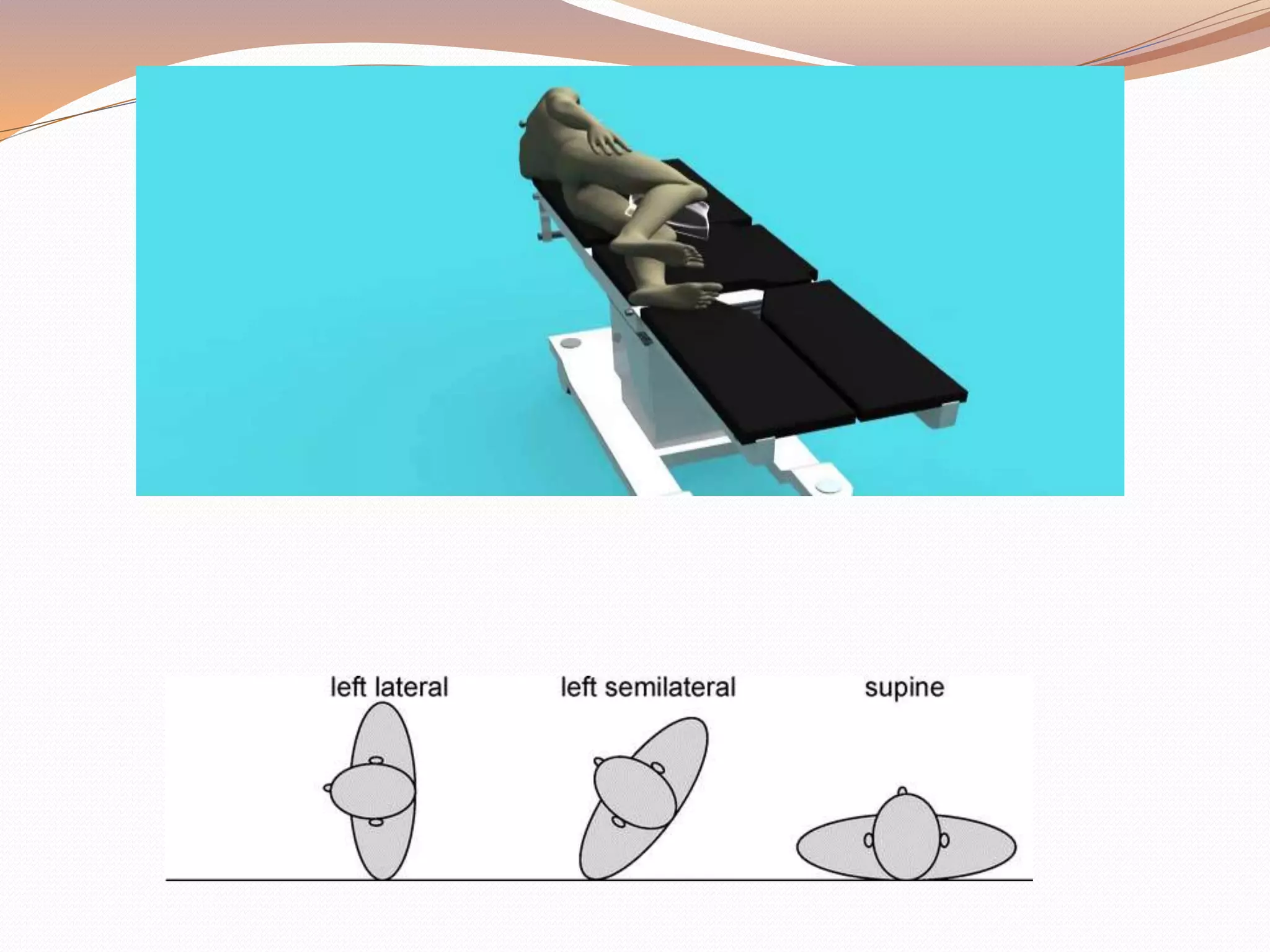
DR. IQRAA KHANUM
Patient positioning is a major responsibility that is
shared by the entire operating room team. A balance
between optimal surgical positioning and patient
well-being is sometimes required.
Patient’s position during anesthesia care should be
natural- one that would be well tolerated if the
patient were awake and unsedated.
 Anaesthesia blunts natural
compensatory mechanisms,
rendering surgical patients
vulnerable to positional changes.
 Positions deemed optimal for
surgery often result in undesirable
physiologic changes such as
hypotension from impaired venous
return to the heart or oxygen
desaturation as a result of
ventilation perfusion mismatching.
 Peripheral nerve injuries during
surgery remain a significant source
of perioperative morbidity.
PHYSIOLOGICAL CHANGES RELATED
TO CHANGE IN BODY POSITION
Most changes are related to gravitational effects
on cardiovascular system and respiratory
system.
Changes in position redistribute blood within the
venous, arterial, and pulmonary vasculature.
Pulmonary mechanics also change
with varying body positions.
CARDIOVASCULAR CONCERNS
Cardiac output ↑ on assuming supine position
Venous blood from lower body
↓ flows back
To heart
↓
Stretches atrial wall
↓ (Laplace’s law)
Stroke volume ↑
↓
↑ blood pressure
(clinically normal BP observed)
Baroreceptors in Aorta Baroreceptors in Carotid
↓ via ↓ via sinus
Vagus nerve Glossopharyngeal nerve
Medulla Oblongata
↓ efferent
↑ Parasympathetic activity
↓
↓ HR ↓SV ↓Contractility
↓
Little change in BP noted
General anaesthesia, muscle relaxation, positive
pressure ventilation, and neuraxial blockade all
interfere with venous return to the heart, arterial
tone, and autoregulatory mechanism.
Therefore, arterial blood pressure is often labile
immediately after the start of anesthesia and during
positioning.
PULMONARY CONCERNS
In an erect person
-Abdominal contents & diaphragm move caudally
-FRC ↑ TLC ↑
Anesthetized person who are spontaneously
breathing—
↓Tidal Volume
↓Functional residual capacity
↑Closing volume
Positive pressure ventilation with muscle relaxation
may ameliorate ventilation perfusion mismatches
under GA by maintaining adequate minute
ventilation.
Perfusion appears to follow a central-to-peripheral
spectrum in each lobe that is maintained with
changes in cardiac output.
Also, gravity affects the preferential perfusion of the
dependent portions of the lungs.
VARIOUS POSITIONS
SITTING
BEACH
CHAIR
SUPINE
HORIZONTAL
LAWN
CHAIR
POSITION
FROG LEG
POSITION
TRENDELENBUR
G POSITION
REVERSE
TRENDELENBURG
POSITION
LITHOTOMY
STANDARD
LOW
HIGH
EXAGGERATED
LATERAL
PARK
BENCH
PRONE
FULL PRONE
PRONE JACK-
KNIFE
PRONE
KNEELING
What ever bizarre
position the
surgeon wants the
patient to be in
SUPINE
 Most common with the least amount of harm
 Placed on back with legs extended and
uncrossed at the ankles
 Spinal column should be in alignment with legs
parallel to the OR bed
 Head in line with the spine and the face is
upward
 Hips are parallel to the spine
 Padding is placed under the head, arms, and
heels with a pillow placed under the knees
 Safety belt placed 2” above the knees while not
impeding circulation
Associated
arm position:
 Arms either on arm boards
abducted <90 degrees to
minimize the likelihood of
brachial plexus injury.
 When arms are adducted,
they are usually held
alongside the body with a
“draw sheet” that passes
under the body.
 The elbows and any
protruding objects, such as
intravenous line and
stopcocks are padded.
SUPINE CONCERNS
 Greatest concerns are circulation and pressure points
 Most Common Nerve Damage:
 Brachial Plexus: positioning the arm >90*
 Radial and Ulnar: compression against the OR bed,
metal attachments, or when team members lean
against the arms during the procedure
 Peroneal and Tibial: Crossing of feet and plantar
flexion of ankles and feet
 Vulnerable Bony Prominences:
(due to rubbing and sustained pressure)
 Occiput, spine, scapula, Olecranon, Sacrum,
Calcaneous
SUPINE POSITION- PRESSURE POINTS
SUPINE VARIATIONS
LAWN CHAIR POSITION
 Back of the bed is raised
 Legs below the knees are lowered
to an equivalent angle
 Slight trendelenburg tilt
ADVANTAGES:
 Better tolerated by awake
patient or under monitored
anesthesia care
 Venous drainage from lower
extremities enhanced
 Xiphoid to pubic distance reduced and
easing closure of laparotomy incisions
 FROG LEG POSITION
hips and knees are flexed and the hips are
externally rotated with soles of feet facing each other.
allows access to perineum, medial thighs,
genitalia and rectum
TRENDELENBURG POSITION
Tilting a supine patient head down, the Trendelenburg
position, is often used to increase venous return during
hypotension, to improve exposure during abdominal and
laparoscopic surgery, and to prevent air emboli and
facilitate cannulation during central line placement.
The Trendelenburg position has significant cardiovascular
and respiratory consequences.
 The head-down position
 Increases central venous, intracranial, and intraocular
pressures.
Prolonged head-down position also can lead to swelling of
the face, conjunctiva, larynx, and tongue with an
increased potential for postoperative upper airway
obstruction.
The cephalic movement of abdominal viscera against the
diaphragm also decreases functional residual capacity
and pulmonary compliance.
 In spontaneously ventilating patients, the work of
breathing increases.
 In mechanically ventilated patients, airway pressures
must be higher to ensure adequate ventilation.
The stomach also lies above the glottis. Endotracheal
intubation is often preferred to protect the airway from
pulmonary aspiration related to reflux and to reduce
atelectasis.
Because of the risk of edema to the trachea and mucosa
surrounding the airway during surgeries in which
patients have been in the Trendelenburg position for
prolonged periods, it may be prudent to verify an air leak
around the endotracheal tube or visualize the larynx
before extubation.
REVERSE TRENDELENBURG POSITION
Reverse Trendelenburg position (head-up tilt) is often
employed to facilitate upper abdominal surgery by
shifting the abdominal contents caudad.
This position is increasingly popular because of the
growing number of laparoscopic surgeries.
Caution is advised to prevent patients from slipping
on the table, and more frequent monitoring of arterial
blood pressure may be prudent to detect
hypotension owing to decreased venous return.
In addition, the position of the head above the heart
reduces perfusion pressure to the brain and should
be taken into consideration when determining
optimal blood pressure.
In all positions in which the head is at a different
level than the heart, the effect of the hydrostatic
gradient on cerebral arterial and venous pressures
should be carefully considered in terms of cerebral
perfusion pressure. Careful documentation of any
potential arterial pressure gradients is especially
prudent.
Trendelenburg position and reverse Trendelenburg
position. Shoulder braces should be avoided to
prevent brachial plexus compression injuries
COMPLICATIONS- SUPINE AND ITS
VARIANTS
Pressure alopecia:
Lumps, such as those caused by monitoring cable
connectors, should not be placed under head padding
because they may create focal areas of pressure.)
Backache :
as the normal lumbar lordotic curvature, particularly
the tone of the paraspinous musculature, is lost
during general anesthesia with muscle relaxation or a
neuraxial block.
Tissues overlying all bony prominences, such as the
heels and sacrum, must be padded to prevent soft
tissue ischemia owing to pressure, especially during
prolonged surgery.
Peripheral nerve injury :(Ulnar neuropathy is the
most common lesion.)
 Regardless of the position of the upper extremities,
maintaining the head in a relatively midline
position can help minimize the risk of stretch
injury to the brachial plexus.
 ASA practice Advisory recommends limiting arm
abductionin supine patient to less than 90 degrees
at the shoulder with the hand and forearm either
supinated or kept in neutral position
NB: When patients are very heavy, caution is advised
when placing them in reverse axis on the operating
room table.
LITHOTOMY
The classic lithotomy
position is frequently
used during
gynaecologic, rectal, and
urologic surgeries.
 The hips are flexed 80 to
100 degrees from the
trunk, and the legs are
abducted 30 to 45
degrees from the midline.
Initiation of the lithotomy position requires
coordinated positioning of the lower extremities by
two assistants to avoid torsion of the lumbar spine.
Both legs should be raised together, flexing the hips
and knees simultaneously.
After the surgery, the patient must be returned to the
supine position in a coordinated manner. The legs
should be removed from the holders simultaneously,
knees brought together in the midline, and the legs
slowly straightened and lowered onto the operating
room table.
Standard Lithotomy Position
Low Lithotomy Position
High Lithotomy Position
Hemi Lithotomy Position
Exaggerated lithotomy position
Various types of stirrups
 Candy cane
 Allen stirrups
 Knee cradles
Stirrups
COMPLICATIONS
When the legs are elevated, preload increases,
causing a transient increase in cardiac output and,
to a lesser extent, cerebral venous and intracranial
pressure in otherwise healthy patients.
In addition, the lithotomy position causes the
abdominal viscera to displace the diaphragm
cephalad, reducing lung compliance and potentially
resulting in a decreased tidal volume.
If obesity or a large abdominal mass is present
(tumor, gravid uterus), abdominal pressure may
increase significantly enough to obstruct venous
return to the heart.
Lastly, the normal lordotic curvature of the lumbar
spine is lost in the lithotomy position, potentially
aggravating any previous lower back pain.
Lower extremity compartment syndrome
NERVE INJURIES IN LITHOTOMY
POSITION
Improper position of
arms in lithotomy
position with fingers
at risk for
compression when
the lower section of
the bed is raised.
COMPARTMENT SYNDROME
Rare complication caused by inadequate tissue
perfusion that is associated with the lithotomy
position
Local arterial pressure decreases 0.78mmHg for
each cm the leg is raised above the right atrium
Decompression fasciotomy- tissue pressure
>30mmHg
Irreversible muscle damage- pressure > 50mm Hg
LATERAL DECUBITUS
The lateral decubitus position is used most
frequently for surgery involving the thorax,
retroperitoneal structures, or hip.
The patient’s head must be kept in a neutral position
to prevent excessive lateral rotation of the neck and
stretch injuries to the brachial plexus.
(Additional head support may be required )
The dependent ear should be checked to avoid
folding and undue pressure.
 It is advised to verify that the eyes are securely
taped before repositioning if the patient is asleep.
The dependent eye must be checked frequently for
external compression.
Watch for compression of the dependent axillary
structures.
(Regardless of the technique, the pulse should be
monitored in thedependent arm for early detection of
compression to axillary neurovascular structures.)
Vascular compression and venous engorgement in
the dependent arm may affect the pulse oximetry
reading, and a low saturation reading may be an
early warning of compromised circulation.
When a kidney rest is used, it must be properly placed
under the dependent iliac crest to prevent inadvertent
compression of the inferior vena cava.
Finally, a pillow or other padding is generally placed
between the knees with the dependent leg flexed to
minimize
excessive pressure on bony prominences and stretch of
low
extremity nerves.
The lateral decubitus position also is associated
with pulmonary compromise. In a patient who is
mechanically ventilated,
 lateral weight of the mediastinum
disproportionate cephalad pressure of
abdominal contents on the dependent lung
favors overventilation of the nondependent lung
pulmonary blood flow to the
underventilated,dependent lung increases
owing to the effect of gravity.
ventilation-perfusion matching worsens
PARK-BENCH POSITION (SEMI‐PRONE
POSITION)
 Modification of lat. position.
 Better access to posterior fossa.
 Upper arm positioned along lateral trunk &
upper shoulder is taped towards table.
 Patient looks like he is trying to look at the
floor
PRONE( VENTRAL DECUBITUS)
Used primarily for surgical access to the posterior
fossa of the skull, the posterior spine, the buttocks
and perirectal area, and the lower extremities.
As with the supine position, if the legs are in plane
with the torso, hemodynamic reserve is maintained
Pulmonary function may be superior to the supine or
lateral decubitus positions if there is no significant
abdominal pressure and the patient is properly
positioned.
The legs should be padded and flexed slightly at the
knees and hips. The head may be supported face-
down with its weight borne by the bony structures or
turned to the side.
Both arms may be positioned to the patient’s sides
and tucked in the neutral position or placed next to
the patient’s head on arm boards—sometimes called
the prone “superman” position.
Extra padding under the elbow is needed to prevent
compression of the ulnar nerve.
 The arms should not be abducted greater than 90
degrees to prevent excessive stretching of the
brachial plexus.
Finally, elastic stockings and active compression
devices are needed for the lower extremities to
minimize pooling of the blood, especially with any
flexion of the body.
When general anesthesia is planned, the patient is first
intubated on the stretcher, and all intravascular access is
obtained as needed.
The endotracheal tube is well secured to prevent
dislodgment and loosening of tape owing to drainage of
saliva when prone.
With the coordination of the entire operating room
staff(minimum of 5), the patient is turned prone onto the
operating room table, keeping the neck in line with the
spine during the move.
The anesthesiologist is primarily responsible for
coordinating the move and for repositioning of the head.
It is recommended to disconnect blood pressure cuffs and
arterial and venous lines that are on the side that rotates
furthest to avoid dislodgment.
Full monitoring should be reinstituted as rapidly as
possible.
Endotracheal tube position and adequate ventilation are
reassessed immediately after the move.
Because the abdominal wall is easily displaced,
external pressure on the abdomen may elevate intra-
abdominal pressure in the prone position.
External pressure on the abdomen may push the
diaphragm cephalad, decreasing functional residual
capacity and pulmonary compliance, and increasing
peak airway pressure.
Abdominal pressure also may impede venous return
through compression of the inferior vena cava
As such careful attention must be paid to the ability
of the abdomen to hang free and to move with
respiration.
The prone position presents special risks for
morbidly obese patients, whose respiration is
already compromised, and who may be difficult to
reposition quickly.
POSITIONING AIDS AND SUPPORT
1.Pin (Mayfield) head holder
2.Mirror system
3.Radiolucent pin head holder
4.Horseshoe head rest
5.Foam head support (e.g., Voss, O.S.I., Prone-View)
6.Vacuum mattress (“bean bag”)
7.Wilson-type frame
8.Andrews (“hinder binder”)-type frame
9.Relton -Hall (four-poster) frame
MIRROR
SYSTEM
HORSESHOE HEAD
REST
MAYFIELD HEAD
PINS
COMPLICATIONSAirway
• Accidental extubation
• Obstruction of ETT
bloody secretions/
sputum plugs
• Facial, Airway edema
 Prolonged head low
position, ↑ crystalloid
infusion
 Problems with
extubation
53
 Visual loss
Neck injury
Excessive lateral torsion or hyperflexion → Post-op
pain, cervical nerve root or vascular compression
Accentuation of pre-existing trauma
Multiple skeletal injuries may be further
exacerbated during positioning
54
JACK KNIFE POSITION
Used for anal surgeries, pilonidal sinus excision
Places patient prone with head & feet at a lower
level
KNEE CHEST POSITION
Further exaggeration of jack knife position
Used for sigmoidoscopies or lumbar laminectomies
Severe hypotension is seen due to pooling of blood in
the legs
PERIOPERATIVE EYE INJURY AND
VISUAL LOSS
Most common- corneal abrasions
Postoperative visual loss—
Ischemic optic neuropathy
central retinal artery occlusion
Risk factors – prolonged hypotension, long duration
of surgery, large blood loss, large crystalloid use,
anemia, hemodilution, increased IOP, venous pressure
from prone position,
ASA GUIDELINES FOR PREVENTION
OF PERIOPERATIVE VISUAL LOSS
Patient Positioning
 There is no pathophysiologic mechanism by which
facial edema can cause perioperative ION.
 There is no evidence that ocular compression causes
isolated perioperative anterior ION or posterior ION.
 – However, direct pressure on the eye should be
avoided to prevent central retinal artery occlusion
(CRAO).
 The high‐risk patient should be positioned so that the
head is level with or higher than the heart when
possible.
 The high‐risk patient's head should be maintained in a
neutral forward position (e.g., without significant neck
flexion, extension, lateral flexion, or rotation) when
possible.
 Avoid external compression of the abdomen or chest
SITTING POSITION
The sitting position ( although infrequently used
because of the perception of risk from venous and
paradoxical air embolism, offers advantages to the
surgeon in approaching the posterior cervical spine
and the posterior fossa.)
The main advantages of the sitting position over the
prone position for neurosurgical and cervical spine
surgeries are;
 excellent surgical exposure
 decreased blood in the operative field
 reduced perioperative blood loss.
 superior access to the airway, reduced facial
swelling, and improved ventilation, particularly in
obese patients.(to the anesthesiologist)
The head may be fixed in pins for neurosurgery or
taped in place with adequate support for other
surgeries.
Arms must be supported to the point of slight
elevation of the shoulders to avoid traction on the
shoulder muscles and potential stretching of upper
extremity neurovascular structures.
The knees are usually slightly flexed for balance and
to reduce stretching of the sciatic nerve, and the feet
are supported and padded.
Because of the pooling of blood into the lower body
under general anesthesia patients are particularly
prone to hypotensive episodes.
Head and neck position has been associated with
complications during surgery to the posterior spine or
skull in the sitting position.
Excessive cervical flexion has numerous adverse
consequences.
 It can impede arterial and venous blood flow,
causing hypoperfusion or venous congestion of the
brain.
 It may impede normal respiratory excursion.
 Excessive flexion also can obstruct the
endotracheal tube and place significant pressure
on the tongue, leading to macroglossia.
Because of the elevation of the surgical field above
the heart, and the inability of the dural venous
sinuses to collapse because of their bony
attachments, the risk of venous air embolism is a
constant concern.
 Arrhythmia, desaturation, pulmonary hypertension,
circulatory compromise, or cardiac arrest may occur
if sufficient quantities are entrained.
Potential complications from sitting position
 Venous air emboli.
 Need to take measures to detect and extract
VAE
 Hypotension.
 Brainstem manipulations resulting in
hemodynamic changes.
 Risk of airway obstruction.
 Macroglossia.
 Pneumocephalus
 Quadriplegia.
BEACH CHAIR POSITION
 Used for shoulder
surgeries including
arthoscopies.
 Superior access to
shoulder
 Associated with
neurologic injury,
cervical neurapraxia,
and hypotensive
bradycardia
(epinephrine
containing
interscalene block)
Excessive cervical flexion
It can impede arterial and venous blood flow,
causing hypoperfusion or venous congestion of the
brain.
Obstruct the ET tube and place significant pressure
on the tongue, leading to edema.
 Generally, maintaining at least two fingers' distance
between the mandible and the sternum is
recommended.
If TEE is used for air embolism monitoring because
the oesophageal probe lies between the flexed spine
and the airway, adding potential for compression of
laryngeal structures and the tongue.
Pneumocephalus
Air enters into the supratentorial
space, much as air enters an
inverted bottle.
Tension pneumocephalus is one of
the causes of delayed awakening
or nonawakening after posterior
fossa and supratentorial
procedures.
The diagnosis of pneumocephalus
is confirmed by a brow-up lateral
x-ray or CT scan.
The treatment is a twist-drill hole
followed by needle puncture of the
dura.
Venous air embolism
VAE is detectable by precordial Doppler in
approximately 40% of patients and by TEE in 76% of
patients.
The rate of VAE is apparently lower with cervical
laminectomy (25% using TEE in the sitting position
versus 76% for posterior fossa procedures.
Transverse and sigmoid sinuses
Monitoring
The monitors employed for the detection of VAE should
provide
(1) a high level of sensitivity
(2) a high level of specificity
(3) a rapid response
(4) a quantitative measure of the VAE event
(5) an indication of the course of recovery from the VAE
event.
The combination of a precordial Doppler and expired CO2
monitoring meet these criteria and are the current
standard of care.
TEE is more sensitive than precordial Doppler to VAE
and offers the advantage of identifying right-to-left
shunting of air.
VENOUS AIR EMBOLISM‐MONITOR WARNINGS
Mandatory monitoring: EKG, SPO2,
ETCO2, BP,DOPPLER,CVP,PAP
Signs and Symptoms
 No physiologic change – TEE, Doppler
 Modest physiologic change – EtCO2, PAP
 Clinically apparent changes – CO, CVP
 Cardiovascular Collapse – BP, ECG
Management
Prevent further air entry
Notify surgeon (flood or pack surgical field)
Jugular compression
Lower the head
Treat intravascular Air
Aspirate right heart catheter
Discontinue N2O
FIO2: 1.0
Pressors/ inotropes
Chest compression
NERVE INJURIES
Peripheral nerve injury, although rare, accounted for
18% of cases in the 1990-1994 ASA Closed Claims
Database, second only to death.
Peripheral nerve injury is often a result of patient
positioning. The mechanisms of injury are stretching,
compression, and ischemia.
 Ulnar neuropathy is the most common postoperative
nerve injury, followed by injury to the brachial
plexus, lumbosacral nerve roots, and spinal cord.
Ulnar Nerve Injury
 Most common nerve injury in anesthetized
patient.
 Often injured when compressed between the
posterior aspect of medial epicondyle of elbow and
arm board or bed.
 More likely with elbow flexed or forearm pronated.
 Symptoms include loss of sensation of lateral
portion of hand and inability to abduct or oppose
the fifth finger (claw hand).
Brachial plexus nerve injury
 Second most common type of nerve injury
 Injury occurs often when plexus is stretched or compressed
between the clavicle and first rib
 Seen in prone and supine procedures where head rotated
and laterally flexed to the same side and/or arm is extended
posteriorly past the plane of the torso
Manifestations depend on which nerves are injured in the
plexus:
 Median – “Ape hand” deformity, inability to oppose thumb
 Axillary – inability to abduct the arm
 Ulnar – “Claw hand” deformity
 Musculocutaneous – inability to flex forearm
 Radial – wrist drop
ASA GUIDELINES TO PREVENT
PERIOPERATIVE PERIPHERAL
NEUROPATHY
POSITIONING OF THE UPPER EXTREMITY Iin supine position , shoulder abduction
should be limited to 90. reduce the
pressure at the ulnar nerve canal
When the arms are placed to the sides,
forearm should be placed on neutral
position Whenever the arms in
abduction and supported on boards the
forearm shd be in supine or neutral
position
External pressure on radial n at
humeral groove shall be avoided
Positioning of the lowerextremity The lithotomy position that stretches the
hamstring gp beyond a comfortable range
may pull the sciaticnerve
Extended pressure at the head of the
fibula shall beavoided
Neither hip extension or flexion inc
risk of femoral neuropathy
padded protection Padded armrests may reduce the
potential for upper limbneuropathy
Elbow and head of the fibula padding
reduce the risk of peripheral
neuropathies
Post surgicalevaluation The postsurgical evaluation of the limb
nerve function shd lead to an early
diagnosis of a peripheral neuropathy
THANK YOU…

Patient positioning and anaesthetic consideration

  • 1.
  • 2.
    Patient positioning isa major responsibility that is shared by the entire operating room team. A balance between optimal surgical positioning and patient well-being is sometimes required. Patient’s position during anesthesia care should be natural- one that would be well tolerated if the patient were awake and unsedated.
  • 3.
     Anaesthesia bluntsnatural compensatory mechanisms, rendering surgical patients vulnerable to positional changes.  Positions deemed optimal for surgery often result in undesirable physiologic changes such as hypotension from impaired venous return to the heart or oxygen desaturation as a result of ventilation perfusion mismatching.  Peripheral nerve injuries during surgery remain a significant source of perioperative morbidity.
  • 4.
    PHYSIOLOGICAL CHANGES RELATED TOCHANGE IN BODY POSITION Most changes are related to gravitational effects on cardiovascular system and respiratory system. Changes in position redistribute blood within the venous, arterial, and pulmonary vasculature. Pulmonary mechanics also change with varying body positions.
  • 5.
    CARDIOVASCULAR CONCERNS Cardiac output↑ on assuming supine position Venous blood from lower body ↓ flows back To heart ↓ Stretches atrial wall ↓ (Laplace’s law) Stroke volume ↑ ↓ ↑ blood pressure (clinically normal BP observed)
  • 6.
    Baroreceptors in AortaBaroreceptors in Carotid ↓ via ↓ via sinus Vagus nerve Glossopharyngeal nerve Medulla Oblongata ↓ efferent ↑ Parasympathetic activity ↓ ↓ HR ↓SV ↓Contractility ↓ Little change in BP noted
  • 7.
    General anaesthesia, musclerelaxation, positive pressure ventilation, and neuraxial blockade all interfere with venous return to the heart, arterial tone, and autoregulatory mechanism. Therefore, arterial blood pressure is often labile immediately after the start of anesthesia and during positioning.
  • 8.
    PULMONARY CONCERNS In anerect person -Abdominal contents & diaphragm move caudally -FRC ↑ TLC ↑
  • 9.
    Anesthetized person whoare spontaneously breathing— ↓Tidal Volume ↓Functional residual capacity ↑Closing volume Positive pressure ventilation with muscle relaxation may ameliorate ventilation perfusion mismatches under GA by maintaining adequate minute ventilation. Perfusion appears to follow a central-to-peripheral spectrum in each lobe that is maintained with changes in cardiac output. Also, gravity affects the preferential perfusion of the dependent portions of the lungs.
  • 10.
    VARIOUS POSITIONS SITTING BEACH CHAIR SUPINE HORIZONTAL LAWN CHAIR POSITION FROG LEG POSITION TRENDELENBUR GPOSITION REVERSE TRENDELENBURG POSITION LITHOTOMY STANDARD LOW HIGH EXAGGERATED LATERAL PARK BENCH PRONE FULL PRONE PRONE JACK- KNIFE PRONE KNEELING What ever bizarre position the surgeon wants the patient to be in
  • 11.
  • 12.
     Most commonwith the least amount of harm  Placed on back with legs extended and uncrossed at the ankles  Spinal column should be in alignment with legs parallel to the OR bed  Head in line with the spine and the face is upward  Hips are parallel to the spine  Padding is placed under the head, arms, and heels with a pillow placed under the knees  Safety belt placed 2” above the knees while not impeding circulation
  • 13.
    Associated arm position:  Armseither on arm boards abducted <90 degrees to minimize the likelihood of brachial plexus injury.  When arms are adducted, they are usually held alongside the body with a “draw sheet” that passes under the body.  The elbows and any protruding objects, such as intravenous line and stopcocks are padded.
  • 14.
    SUPINE CONCERNS  Greatestconcerns are circulation and pressure points  Most Common Nerve Damage:  Brachial Plexus: positioning the arm >90*  Radial and Ulnar: compression against the OR bed, metal attachments, or when team members lean against the arms during the procedure  Peroneal and Tibial: Crossing of feet and plantar flexion of ankles and feet  Vulnerable Bony Prominences: (due to rubbing and sustained pressure)  Occiput, spine, scapula, Olecranon, Sacrum, Calcaneous
  • 15.
  • 16.
    SUPINE VARIATIONS LAWN CHAIRPOSITION  Back of the bed is raised  Legs below the knees are lowered to an equivalent angle  Slight trendelenburg tilt ADVANTAGES:  Better tolerated by awake patient or under monitored anesthesia care  Venous drainage from lower extremities enhanced  Xiphoid to pubic distance reduced and easing closure of laparotomy incisions
  • 17.
     FROG LEGPOSITION hips and knees are flexed and the hips are externally rotated with soles of feet facing each other. allows access to perineum, medial thighs, genitalia and rectum
  • 18.
    TRENDELENBURG POSITION Tilting asupine patient head down, the Trendelenburg position, is often used to increase venous return during hypotension, to improve exposure during abdominal and laparoscopic surgery, and to prevent air emboli and facilitate cannulation during central line placement. The Trendelenburg position has significant cardiovascular and respiratory consequences.  The head-down position  Increases central venous, intracranial, and intraocular pressures. Prolonged head-down position also can lead to swelling of the face, conjunctiva, larynx, and tongue with an increased potential for postoperative upper airway obstruction.
  • 19.
    The cephalic movementof abdominal viscera against the diaphragm also decreases functional residual capacity and pulmonary compliance.  In spontaneously ventilating patients, the work of breathing increases.  In mechanically ventilated patients, airway pressures must be higher to ensure adequate ventilation. The stomach also lies above the glottis. Endotracheal intubation is often preferred to protect the airway from pulmonary aspiration related to reflux and to reduce atelectasis. Because of the risk of edema to the trachea and mucosa surrounding the airway during surgeries in which patients have been in the Trendelenburg position for prolonged periods, it may be prudent to verify an air leak around the endotracheal tube or visualize the larynx before extubation.
  • 20.
    REVERSE TRENDELENBURG POSITION ReverseTrendelenburg position (head-up tilt) is often employed to facilitate upper abdominal surgery by shifting the abdominal contents caudad. This position is increasingly popular because of the growing number of laparoscopic surgeries. Caution is advised to prevent patients from slipping on the table, and more frequent monitoring of arterial blood pressure may be prudent to detect hypotension owing to decreased venous return. In addition, the position of the head above the heart reduces perfusion pressure to the brain and should be taken into consideration when determining optimal blood pressure.
  • 21.
    In all positionsin which the head is at a different level than the heart, the effect of the hydrostatic gradient on cerebral arterial and venous pressures should be carefully considered in terms of cerebral perfusion pressure. Careful documentation of any potential arterial pressure gradients is especially prudent.
  • 22.
    Trendelenburg position andreverse Trendelenburg position. Shoulder braces should be avoided to prevent brachial plexus compression injuries
  • 23.
    COMPLICATIONS- SUPINE ANDITS VARIANTS Pressure alopecia: Lumps, such as those caused by monitoring cable connectors, should not be placed under head padding because they may create focal areas of pressure.) Backache : as the normal lumbar lordotic curvature, particularly the tone of the paraspinous musculature, is lost during general anesthesia with muscle relaxation or a neuraxial block. Tissues overlying all bony prominences, such as the heels and sacrum, must be padded to prevent soft tissue ischemia owing to pressure, especially during prolonged surgery.
  • 24.
    Peripheral nerve injury:(Ulnar neuropathy is the most common lesion.)  Regardless of the position of the upper extremities, maintaining the head in a relatively midline position can help minimize the risk of stretch injury to the brachial plexus.  ASA practice Advisory recommends limiting arm abductionin supine patient to less than 90 degrees at the shoulder with the hand and forearm either supinated or kept in neutral position NB: When patients are very heavy, caution is advised when placing them in reverse axis on the operating room table.
  • 25.
    LITHOTOMY The classic lithotomy positionis frequently used during gynaecologic, rectal, and urologic surgeries.  The hips are flexed 80 to 100 degrees from the trunk, and the legs are abducted 30 to 45 degrees from the midline.
  • 26.
    Initiation of thelithotomy position requires coordinated positioning of the lower extremities by two assistants to avoid torsion of the lumbar spine. Both legs should be raised together, flexing the hips and knees simultaneously. After the surgery, the patient must be returned to the supine position in a coordinated manner. The legs should be removed from the holders simultaneously, knees brought together in the midline, and the legs slowly straightened and lowered onto the operating room table.
  • 27.
  • 28.
  • 29.
  • 30.
  • 31.
  • 32.
    Various types ofstirrups  Candy cane  Allen stirrups  Knee cradles
  • 33.
  • 34.
    COMPLICATIONS When the legsare elevated, preload increases, causing a transient increase in cardiac output and, to a lesser extent, cerebral venous and intracranial pressure in otherwise healthy patients. In addition, the lithotomy position causes the abdominal viscera to displace the diaphragm cephalad, reducing lung compliance and potentially resulting in a decreased tidal volume.
  • 35.
    If obesity ora large abdominal mass is present (tumor, gravid uterus), abdominal pressure may increase significantly enough to obstruct venous return to the heart. Lastly, the normal lordotic curvature of the lumbar spine is lost in the lithotomy position, potentially aggravating any previous lower back pain. Lower extremity compartment syndrome
  • 36.
    NERVE INJURIES INLITHOTOMY POSITION
  • 37.
    Improper position of armsin lithotomy position with fingers at risk for compression when the lower section of the bed is raised.
  • 38.
    COMPARTMENT SYNDROME Rare complicationcaused by inadequate tissue perfusion that is associated with the lithotomy position Local arterial pressure decreases 0.78mmHg for each cm the leg is raised above the right atrium Decompression fasciotomy- tissue pressure >30mmHg Irreversible muscle damage- pressure > 50mm Hg
  • 39.
  • 40.
    The lateral decubitusposition is used most frequently for surgery involving the thorax, retroperitoneal structures, or hip. The patient’s head must be kept in a neutral position to prevent excessive lateral rotation of the neck and stretch injuries to the brachial plexus. (Additional head support may be required ) The dependent ear should be checked to avoid folding and undue pressure.  It is advised to verify that the eyes are securely taped before repositioning if the patient is asleep.
  • 41.
    The dependent eyemust be checked frequently for external compression. Watch for compression of the dependent axillary structures. (Regardless of the technique, the pulse should be monitored in thedependent arm for early detection of compression to axillary neurovascular structures.) Vascular compression and venous engorgement in the dependent arm may affect the pulse oximetry reading, and a low saturation reading may be an early warning of compromised circulation.
  • 42.
    When a kidneyrest is used, it must be properly placed under the dependent iliac crest to prevent inadvertent compression of the inferior vena cava. Finally, a pillow or other padding is generally placed between the knees with the dependent leg flexed to minimize excessive pressure on bony prominences and stretch of low extremity nerves.
  • 43.
    The lateral decubitusposition also is associated with pulmonary compromise. In a patient who is mechanically ventilated,  lateral weight of the mediastinum disproportionate cephalad pressure of abdominal contents on the dependent lung favors overventilation of the nondependent lung pulmonary blood flow to the underventilated,dependent lung increases owing to the effect of gravity. ventilation-perfusion matching worsens
  • 44.
    PARK-BENCH POSITION (SEMI‐PRONE POSITION) Modification of lat. position.  Better access to posterior fossa.  Upper arm positioned along lateral trunk & upper shoulder is taped towards table.  Patient looks like he is trying to look at the floor
  • 46.
  • 47.
    Used primarily forsurgical access to the posterior fossa of the skull, the posterior spine, the buttocks and perirectal area, and the lower extremities. As with the supine position, if the legs are in plane with the torso, hemodynamic reserve is maintained Pulmonary function may be superior to the supine or lateral decubitus positions if there is no significant abdominal pressure and the patient is properly positioned.
  • 48.
    The legs shouldbe padded and flexed slightly at the knees and hips. The head may be supported face- down with its weight borne by the bony structures or turned to the side. Both arms may be positioned to the patient’s sides and tucked in the neutral position or placed next to the patient’s head on arm boards—sometimes called the prone “superman” position. Extra padding under the elbow is needed to prevent compression of the ulnar nerve.  The arms should not be abducted greater than 90 degrees to prevent excessive stretching of the brachial plexus. Finally, elastic stockings and active compression devices are needed for the lower extremities to minimize pooling of the blood, especially with any flexion of the body.
  • 49.
    When general anesthesiais planned, the patient is first intubated on the stretcher, and all intravascular access is obtained as needed. The endotracheal tube is well secured to prevent dislodgment and loosening of tape owing to drainage of saliva when prone. With the coordination of the entire operating room staff(minimum of 5), the patient is turned prone onto the operating room table, keeping the neck in line with the spine during the move. The anesthesiologist is primarily responsible for coordinating the move and for repositioning of the head. It is recommended to disconnect blood pressure cuffs and arterial and venous lines that are on the side that rotates furthest to avoid dislodgment. Full monitoring should be reinstituted as rapidly as possible. Endotracheal tube position and adequate ventilation are reassessed immediately after the move.
  • 50.
    Because the abdominalwall is easily displaced, external pressure on the abdomen may elevate intra- abdominal pressure in the prone position. External pressure on the abdomen may push the diaphragm cephalad, decreasing functional residual capacity and pulmonary compliance, and increasing peak airway pressure. Abdominal pressure also may impede venous return through compression of the inferior vena cava As such careful attention must be paid to the ability of the abdomen to hang free and to move with respiration. The prone position presents special risks for morbidly obese patients, whose respiration is already compromised, and who may be difficult to reposition quickly.
  • 51.
    POSITIONING AIDS ANDSUPPORT 1.Pin (Mayfield) head holder 2.Mirror system 3.Radiolucent pin head holder 4.Horseshoe head rest 5.Foam head support (e.g., Voss, O.S.I., Prone-View) 6.Vacuum mattress (“bean bag”) 7.Wilson-type frame 8.Andrews (“hinder binder”)-type frame 9.Relton -Hall (four-poster) frame
  • 52.
  • 53.
    COMPLICATIONSAirway • Accidental extubation •Obstruction of ETT bloody secretions/ sputum plugs • Facial, Airway edema  Prolonged head low position, ↑ crystalloid infusion  Problems with extubation 53
  • 54.
     Visual loss Neckinjury Excessive lateral torsion or hyperflexion → Post-op pain, cervical nerve root or vascular compression Accentuation of pre-existing trauma Multiple skeletal injuries may be further exacerbated during positioning 54
  • 55.
    JACK KNIFE POSITION Usedfor anal surgeries, pilonidal sinus excision Places patient prone with head & feet at a lower level
  • 56.
    KNEE CHEST POSITION Furtherexaggeration of jack knife position Used for sigmoidoscopies or lumbar laminectomies Severe hypotension is seen due to pooling of blood in the legs
  • 57.
    PERIOPERATIVE EYE INJURYAND VISUAL LOSS Most common- corneal abrasions Postoperative visual loss— Ischemic optic neuropathy central retinal artery occlusion Risk factors – prolonged hypotension, long duration of surgery, large blood loss, large crystalloid use, anemia, hemodilution, increased IOP, venous pressure from prone position,
  • 58.
    ASA GUIDELINES FORPREVENTION OF PERIOPERATIVE VISUAL LOSS Patient Positioning  There is no pathophysiologic mechanism by which facial edema can cause perioperative ION.  There is no evidence that ocular compression causes isolated perioperative anterior ION or posterior ION.  – However, direct pressure on the eye should be avoided to prevent central retinal artery occlusion (CRAO).  The high‐risk patient should be positioned so that the head is level with or higher than the heart when possible.  The high‐risk patient's head should be maintained in a neutral forward position (e.g., without significant neck flexion, extension, lateral flexion, or rotation) when possible.  Avoid external compression of the abdomen or chest
  • 59.
  • 60.
    The sitting position( although infrequently used because of the perception of risk from venous and paradoxical air embolism, offers advantages to the surgeon in approaching the posterior cervical spine and the posterior fossa.) The main advantages of the sitting position over the prone position for neurosurgical and cervical spine surgeries are;  excellent surgical exposure  decreased blood in the operative field  reduced perioperative blood loss.  superior access to the airway, reduced facial swelling, and improved ventilation, particularly in obese patients.(to the anesthesiologist)
  • 61.
    The head maybe fixed in pins for neurosurgery or taped in place with adequate support for other surgeries. Arms must be supported to the point of slight elevation of the shoulders to avoid traction on the shoulder muscles and potential stretching of upper extremity neurovascular structures. The knees are usually slightly flexed for balance and to reduce stretching of the sciatic nerve, and the feet are supported and padded. Because of the pooling of blood into the lower body under general anesthesia patients are particularly prone to hypotensive episodes.
  • 62.
    Head and neckposition has been associated with complications during surgery to the posterior spine or skull in the sitting position. Excessive cervical flexion has numerous adverse consequences.  It can impede arterial and venous blood flow, causing hypoperfusion or venous congestion of the brain.  It may impede normal respiratory excursion.  Excessive flexion also can obstruct the endotracheal tube and place significant pressure on the tongue, leading to macroglossia.
  • 63.
    Because of theelevation of the surgical field above the heart, and the inability of the dural venous sinuses to collapse because of their bony attachments, the risk of venous air embolism is a constant concern.  Arrhythmia, desaturation, pulmonary hypertension, circulatory compromise, or cardiac arrest may occur if sufficient quantities are entrained.
  • 64.
    Potential complications fromsitting position  Venous air emboli.  Need to take measures to detect and extract VAE  Hypotension.  Brainstem manipulations resulting in hemodynamic changes.  Risk of airway obstruction.  Macroglossia.  Pneumocephalus  Quadriplegia.
  • 65.
    BEACH CHAIR POSITION Used for shoulder surgeries including arthoscopies.  Superior access to shoulder  Associated with neurologic injury, cervical neurapraxia, and hypotensive bradycardia (epinephrine containing interscalene block)
  • 66.
    Excessive cervical flexion Itcan impede arterial and venous blood flow, causing hypoperfusion or venous congestion of the brain. Obstruct the ET tube and place significant pressure on the tongue, leading to edema.  Generally, maintaining at least two fingers' distance between the mandible and the sternum is recommended. If TEE is used for air embolism monitoring because the oesophageal probe lies between the flexed spine and the airway, adding potential for compression of laryngeal structures and the tongue.
  • 67.
    Pneumocephalus Air enters intothe supratentorial space, much as air enters an inverted bottle. Tension pneumocephalus is one of the causes of delayed awakening or nonawakening after posterior fossa and supratentorial procedures. The diagnosis of pneumocephalus is confirmed by a brow-up lateral x-ray or CT scan. The treatment is a twist-drill hole followed by needle puncture of the dura.
  • 68.
    Venous air embolism VAEis detectable by precordial Doppler in approximately 40% of patients and by TEE in 76% of patients. The rate of VAE is apparently lower with cervical laminectomy (25% using TEE in the sitting position versus 76% for posterior fossa procedures. Transverse and sigmoid sinuses
  • 69.
    Monitoring The monitors employedfor the detection of VAE should provide (1) a high level of sensitivity (2) a high level of specificity (3) a rapid response (4) a quantitative measure of the VAE event (5) an indication of the course of recovery from the VAE event. The combination of a precordial Doppler and expired CO2 monitoring meet these criteria and are the current standard of care. TEE is more sensitive than precordial Doppler to VAE and offers the advantage of identifying right-to-left shunting of air.
  • 70.
    VENOUS AIR EMBOLISM‐MONITORWARNINGS Mandatory monitoring: EKG, SPO2, ETCO2, BP,DOPPLER,CVP,PAP
  • 71.
    Signs and Symptoms No physiologic change – TEE, Doppler  Modest physiologic change – EtCO2, PAP  Clinically apparent changes – CO, CVP  Cardiovascular Collapse – BP, ECG
  • 72.
    Management Prevent further airentry Notify surgeon (flood or pack surgical field) Jugular compression Lower the head Treat intravascular Air Aspirate right heart catheter Discontinue N2O FIO2: 1.0 Pressors/ inotropes Chest compression
  • 73.
    NERVE INJURIES Peripheral nerveinjury, although rare, accounted for 18% of cases in the 1990-1994 ASA Closed Claims Database, second only to death. Peripheral nerve injury is often a result of patient positioning. The mechanisms of injury are stretching, compression, and ischemia.  Ulnar neuropathy is the most common postoperative nerve injury, followed by injury to the brachial plexus, lumbosacral nerve roots, and spinal cord.
  • 74.
    Ulnar Nerve Injury Most common nerve injury in anesthetized patient.  Often injured when compressed between the posterior aspect of medial epicondyle of elbow and arm board or bed.  More likely with elbow flexed or forearm pronated.  Symptoms include loss of sensation of lateral portion of hand and inability to abduct or oppose the fifth finger (claw hand).
  • 75.
    Brachial plexus nerveinjury  Second most common type of nerve injury  Injury occurs often when plexus is stretched or compressed between the clavicle and first rib  Seen in prone and supine procedures where head rotated and laterally flexed to the same side and/or arm is extended posteriorly past the plane of the torso Manifestations depend on which nerves are injured in the plexus:  Median – “Ape hand” deformity, inability to oppose thumb  Axillary – inability to abduct the arm  Ulnar – “Claw hand” deformity  Musculocutaneous – inability to flex forearm  Radial – wrist drop
  • 77.
    ASA GUIDELINES TOPREVENT PERIOPERATIVE PERIPHERAL NEUROPATHY
  • 78.
    POSITIONING OF THEUPPER EXTREMITY Iin supine position , shoulder abduction should be limited to 90. reduce the pressure at the ulnar nerve canal When the arms are placed to the sides, forearm should be placed on neutral position Whenever the arms in abduction and supported on boards the forearm shd be in supine or neutral position External pressure on radial n at humeral groove shall be avoided Positioning of the lowerextremity The lithotomy position that stretches the hamstring gp beyond a comfortable range may pull the sciaticnerve Extended pressure at the head of the fibula shall beavoided Neither hip extension or flexion inc risk of femoral neuropathy padded protection Padded armrests may reduce the potential for upper limbneuropathy Elbow and head of the fibula padding reduce the risk of peripheral neuropathies Post surgicalevaluation The postsurgical evaluation of the limb nerve function shd lead to an early diagnosis of a peripheral neuropathy
  • 79.