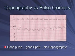
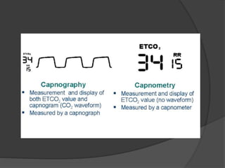
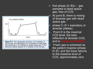
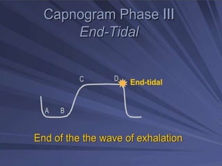
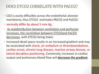
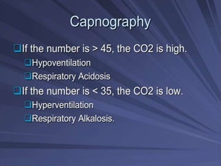
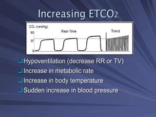
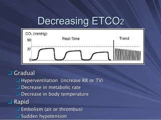
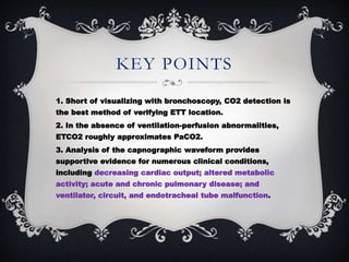
3. WHAT IS THE DIFFERENCE
BETWEEN CAPNOMETRY AND
CAPNOGRAPHY? WHICH IS
BETTER? Capnometry is defined as the numeric
measurement and display of the level
of CO2.
It is not nearly as valuable as
capnography, in which the expired
CO2 level is graphically displayed as a
function of time and concentration
4.
5.
6.
7.
8.
9. IMPORTANCE OF MEASURING
CO2
best method of verifying correct endotracheal
tube (ETT) placement.
provides valuable information about important
physiologic factors, including ventilation,
cardiac output, and metabolic activity, and
proper ventilator functioning.
used to predict outcome of resuscitation.
A prospective observation
study of 150 out-of-hospital cardiac arrests
found that an ETCO2level less than 10 mm Hg
after 20 minutes of standard advanced cardiac
life support was 100% predictive of failure to
resuscitate
10. CAPNOGRAPHICWAVE
FORM
The important features include
I. baseline level,
II. the extent and rate of rise of CO2, and
III. the contour of the capnograph.
11. first phase (A–B)n - gas
sampled is dead space
gas, free of CO2.
At point B, there is mixing
of alveolar gas with dead
space gas.
phase C–D = expiratory or
alveolar plateau.
Point D is the maximal
CO2 level, the best
reflection of alveolar CO2,
and is known as ETCO2.
Fresh gas is entrained as
the patient inspires (phase
D–E), and the trace returns
to the baseline level of
CO2, approximately zero
12.
13. DOESETCO2 CORRELATEWITH PACO2?
• CO2 is easily diffusible across the endothelial-alveolar
membrane; thus ETCO2 -estimates PACO2 and PaCO2.
• normally differ by about 5 mm Hg .
• As maldistribution between ventilation and perfusion
increases, the correlation between ETCO2and PaCO2
decreases, with ETCO2 being lower.
• Increased dead space results in an increased gradient and may
be associated with shock, air embolism or thromboembolism,
cardiac arrest, chronic lung disease, reactive airway disease, or
lateral decubitus positioning. Conversely, increases in cardiac
output and pulmonary blood flow will decrease the gradient.
14.
15.
16.
17.
18.
19.
20.
21.
22.
23. KEY POINTS
1. Short of visualizing with bronchoscopy, CO2 detection is
the best method of verifying ETT location.
2. In the absence of ventilation-perfusion abnormalities,
ETCO2 roughly approximates PaCO2.
3. Analysis of the capnographic waveform provides
supportive evidence for numerous clinical conditions,
including decreasing cardiac output; altered metabolic
activity; acute and chronic pulmonary disease; and
ventilator, circuit, and endotracheal tube malfunction.