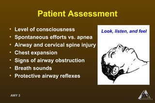
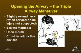
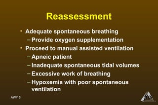
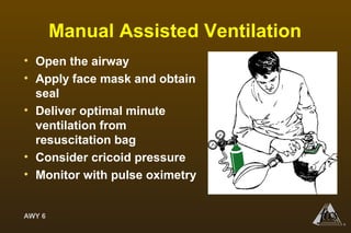
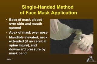
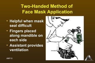
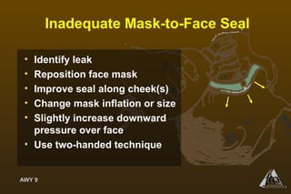
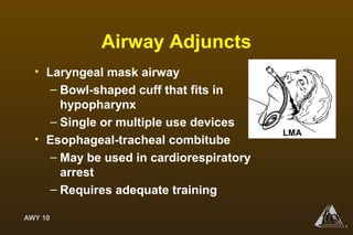
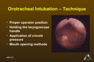
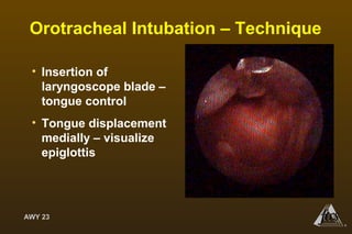
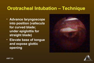
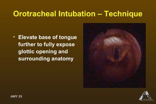
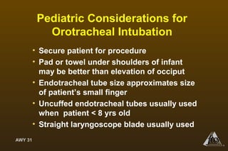
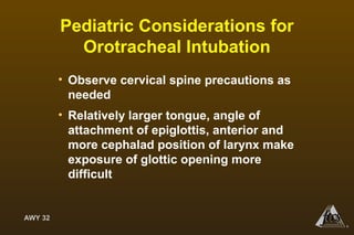
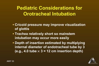
AWY 1
®
Airway Management
AWY 1
®
AWY 2
®
Objectives
• Recognize signs of a threatened airway
• Describe techniques for establishing an airway
and for mask ventilation
• Be familiar with airway adjuncts
• Describe proper preparation for endotracheal
intubation
• Decribe alternative methods to establish an
airway when intubation is not possible
AWY 2
®
AWY 3
®
Patient Assessment
• Level of consciousness
• Spontaneous efforts vs. apnea
• Airway and cervical spine injury
• Chest expansion
• Signs of airway obstruction
• Breath sounds
• Protective airway reflexes
Look, listen, and feel
AWY 4
®
Opening the Airway – the Triple
Airway Maneuver
• Slightly extend neck
(when cervical spine
injury not suspected)
• Elevate mandible
• Open mouth
• Consider adjunctive
devices
AWY 5
®
Reassessment
• Adequate spontaneous breathing
– Provide oxygen supplementation
• Proceed to manual assisted ventilation
– Apneic patient
– Inadequate spontaneous tidal volumes
– Excessive work of breathing
– Hypoxemia with poor spontaneous
ventilation
AWY 6
®
Manual Assisted Ventilation
• Open the airway
• Apply face mask and obtain
seal
• Deliver optimal minute
ventilation from
resuscitation bag
• Consider cricoid pressure
• Monitor with pulse oximetry
AWY 7
®
Single-Handed Method
of Face Mask Application
• Base of mask placed
over chin and mouth
opened
• Apex of mask over nose
• Mandible elevated, neck
extended (if no cervical
spine injury), and
downward pressure by
mask hand
AWY 8
®
Two-Handed Method of
Face Mask Application
• Helpful when mask
seal difficult
• Fingers placed
along mandible on
each side
• Assistant provides
ventilation
AWY 9
®
Inadequate Mask-to-Face Seal
• Identify leak
• Reposition face mask
• Improve seal along cheek(s)
• Change mask inflation or size
• Slightly increase downward
pressure over face
• Use two-handed technique
AWY 10
®
Airway Adjuncts
• Laryngeal mask airway
– Bowl-shaped cuff that fits in
hypopharynx
– Single or multiple use devices
• Esophageal-tracheal combitube
– May be used in cardiorespiratory
arrest
– Requires adequate training
LMA
AWY 11
®
Indications for Endotracheal
Intubation
• Airway protection
• Relief of obstruction
• Need for mechanical ventilation/O2 therapy
• Respiratory failure
• Shock
• Need for hyperventilation
• Reduce the work of breathing
• Facilitate suctioning/pulmonary toilet
AWY 12
®
Preparation for Intubation
• Assess degree of difficulty for intubation
• Assure optimal ventilation and
oxygenation
• Consider gastric decompression
• Analgesia, sedation, amnesia,
neuromuscular blockade as needed
AWY 13
®
Degree of Difficulty Assessment
• Neck mobility
• External face
• Mouth
• Tongue and pharynx
• Jaw
• Consider options for obtaining an
airway that maintain ventilation
• Obtain expert assistance
AWY 14
®
Options for Airway Management
• Awake intubation
• Flexible fiberoptic intubation
• Awake tracheostomy
• Laryngeal mask airway or esophageal-
tracheal combitube
• Needle cricothyrotomy
• Surgical cricothyrotomy
AWY 15
®
Difficult Airway
AWY 16
®
Analgesia, Sedation, Amnesia,
Neuromuscular Blockade
• Analgesia – topical, nerve blocks, sedation
• Sedation/amnesia – rapid acting, short
duration, reversible
– Fentanyl: 25–100 µg iv, titrated to effect
– Midazolam: 1 mg iv, titrated to effect
– Etomidate: 0.3–0.4 mg/kg iv, titrated to effect
– Lidocaine: 1-1.5 mg/kg iv
AWY 17
®
Analgesia, Sedation, Amnesia,
Neuromuscular Blockade
• Assess need for neuromuscular blockers
– Succinylcholine: 1–1.5 mg/kg iv bolus
– Vecuronium: 0.1–0.3 mg/kg iv bolus
– Rocuronium: 0.6-1.0 mg/kg iv bolus
– Cisatracurium: 0.1-0.2 mg/kg iv bolus
AWY 18
®
Early Complications
• Hemodynamic alterations
–Hypertension
–Tachycardia
–Hypotension
• Consider effects of sedative agents
AWY 19
®
Key Points
AWY 20
®
Orotracheal Intubation –
Preparation
• Appropriate monitoring – oximetry, ECG, BP
– Assemble equipment
– Laryngoscope – test light, select blade
– Endotracheal tube – test cuff, lubricate
– Stylet – insert, angulate
– Suction – test
– Magill forceps
AWY 21
®
Orotracheal Intubation –
Preparation
• Don protective garb
• Elevate occiput with pad if no cervical
spine injury suspected
• Provide anesthesia, sedation, amnesia,
and neuromuscular blockade as required
AWY 22
®
Orotracheal Intubation – Technique
• Proper operator position
• Holding the laryngoscope
handle
• Application of cricoid
pressure
• Mouth opening methods
AWY 23
®
Orotracheal Intubation – Technique
• Insertion of
laryngoscope blade –
tongue control
• Tongue displacement
medially – visualize
epiglottis
AWY 24
®
Orotracheal Intubation – Technique
• Advance laryngoscope
into position (vallecula
for curved blade;
under epiglottis for
straight blade)
• Elevate base of tongue
and expose glottic
opening
AWY 25
®
Orotracheal Intubation – Technique
• Elevate base of tongue
further to fully expose
glottic opening and
surrounding anatomy
AWY 26
®
Orotracheal Intubation – Technique
• Insert endotracheal tube under direct vision
to 23–25 cm at lip
• Remove stylet and laryngoscope, inflate
tube cuff
• Confirm tube position – breath sounds, CO2
detector
• Secure endotracheal tube
• Obtain chest radiograph
AWY 27
®
Orotracheal Intubation – Technique
• Straight blade
position, elevating
the epiglottis
• Be aware of
laryngospasm when
epiglottis is touched
AWY 28
®
Pediatric Considerations
• Infections commonly cause airway
obstruction in young children
• Because infants are obligate “nose
breathers” until ~ age 6 months, suctioning
nares may establish an open airway
• When possible, allow child to assume
position of comfort in early respiratory
compromise
AWY 29
®
Pediatric Considerations
• Face mask may agitate child – several
delivery devices should be available
• If obtunded or unable to assume a
comfortable position, sniffing position is
preferred in infants and young children to
minimize airway obstruction from soft tissues
(when no cervical spine injury is suspected)
• Overextension of neck may cause airway
obstruction
AWY 30
®
Pediatric Considerations
• Positive pressure during bag-mask
ventilation may cause gastric distention;
a nasogastric tube may be needed
• Tongue in infants and children up to ~ age 2
yrs occupies relatively large portion of oral
cavity and is likely to cause obstruction
during spontaneous breathing and manually
assisted ventilation
AWY 31
®
Pediatric Considerations for
Orotracheal Intubation
• Secure patient for procedure
• Pad or towel under shoulders of infant
may be better than elevation of occiput
• Endotracheal tube size approximates size
of patient’s small finger
• Uncuffed endotracheal tubes usually used
when patient < 8 yrs old
• Straight laryngoscope blade usually used
AWY 32
®
Pediatric Considerations for
Orotracheal Intubation
• Observe cervical spine precautions as
needed
• Relatively larger tongue, angle of
attachment of epiglottis, anterior and
more cephalad position of larynx make
exposure of glottic opening more
difficult
AWY 33
®
Pediatric Considerations for
Orotracheal Intubation
• Cricoid pressure may improve visualization
of glottis
• Trachea relatively short so mainstem
intubation may occur more easily
• Depth of insertion estimated by multiplying
internal diameter of endotracheal tube by 3
(e.g., 4.0 tube Ă— 3 = 12 cm insertion depth)

Airway management

  • 1.
  • 2.
    AWY 2 ® Objectives • Recognizesigns of a threatened airway • Describe techniques for establishing an airway and for mask ventilation • Be familiar with airway adjuncts • Describe proper preparation for endotracheal intubation • Decribe alternative methods to establish an airway when intubation is not possible AWY 2 ®
  • 3.
    AWY 3 ® Patient Assessment •Level of consciousness • Spontaneous efforts vs. apnea • Airway and cervical spine injury • Chest expansion • Signs of airway obstruction • Breath sounds • Protective airway reflexes Look, listen, and feel
  • 4.
    AWY 4 ® Opening theAirway – the Triple Airway Maneuver • Slightly extend neck (when cervical spine injury not suspected) • Elevate mandible • Open mouth • Consider adjunctive devices
  • 5.
    AWY 5 ® Reassessment • Adequatespontaneous breathing – Provide oxygen supplementation • Proceed to manual assisted ventilation – Apneic patient – Inadequate spontaneous tidal volumes – Excessive work of breathing – Hypoxemia with poor spontaneous ventilation
  • 6.
    AWY 6 ® Manual AssistedVentilation • Open the airway • Apply face mask and obtain seal • Deliver optimal minute ventilation from resuscitation bag • Consider cricoid pressure • Monitor with pulse oximetry
  • 7.
    AWY 7 ® Single-Handed Method ofFace Mask Application • Base of mask placed over chin and mouth opened • Apex of mask over nose • Mandible elevated, neck extended (if no cervical spine injury), and downward pressure by mask hand
  • 8.
    AWY 8 ® Two-Handed Methodof Face Mask Application • Helpful when mask seal difficult • Fingers placed along mandible on each side • Assistant provides ventilation
  • 9.
    AWY 9 ® Inadequate Mask-to-FaceSeal • Identify leak • Reposition face mask • Improve seal along cheek(s) • Change mask inflation or size • Slightly increase downward pressure over face • Use two-handed technique
  • 10.
    AWY 10 ® Airway Adjuncts •Laryngeal mask airway – Bowl-shaped cuff that fits in hypopharynx – Single or multiple use devices • Esophageal-tracheal combitube – May be used in cardiorespiratory arrest – Requires adequate training LMA
  • 11.
    AWY 11 ® Indications forEndotracheal Intubation • Airway protection • Relief of obstruction • Need for mechanical ventilation/O2 therapy • Respiratory failure • Shock • Need for hyperventilation • Reduce the work of breathing • Facilitate suctioning/pulmonary toilet
  • 12.
    AWY 12 ® Preparation forIntubation • Assess degree of difficulty for intubation • Assure optimal ventilation and oxygenation • Consider gastric decompression • Analgesia, sedation, amnesia, neuromuscular blockade as needed
  • 13.
    AWY 13 ® Degree ofDifficulty Assessment • Neck mobility • External face • Mouth • Tongue and pharynx • Jaw • Consider options for obtaining an airway that maintain ventilation • Obtain expert assistance
  • 14.
    AWY 14 ® Options forAirway Management • Awake intubation • Flexible fiberoptic intubation • Awake tracheostomy • Laryngeal mask airway or esophageal- tracheal combitube • Needle cricothyrotomy • Surgical cricothyrotomy
  • 15.
  • 16.
    AWY 16 ® Analgesia, Sedation,Amnesia, Neuromuscular Blockade • Analgesia – topical, nerve blocks, sedation • Sedation/amnesia – rapid acting, short duration, reversible – Fentanyl: 25–100 µg iv, titrated to effect – Midazolam: 1 mg iv, titrated to effect – Etomidate: 0.3–0.4 mg/kg iv, titrated to effect – Lidocaine: 1-1.5 mg/kg iv
  • 17.
    AWY 17 ® Analgesia, Sedation,Amnesia, Neuromuscular Blockade • Assess need for neuromuscular blockers – Succinylcholine: 1–1.5 mg/kg iv bolus – Vecuronium: 0.1–0.3 mg/kg iv bolus – Rocuronium: 0.6-1.0 mg/kg iv bolus – Cisatracurium: 0.1-0.2 mg/kg iv bolus
  • 18.
    AWY 18 ® Early Complications •Hemodynamic alterations –Hypertension –Tachycardia –Hypotension • Consider effects of sedative agents
  • 19.
  • 20.
    AWY 20 ® Orotracheal Intubation– Preparation • Appropriate monitoring – oximetry, ECG, BP – Assemble equipment – Laryngoscope – test light, select blade – Endotracheal tube – test cuff, lubricate – Stylet – insert, angulate – Suction – test – Magill forceps
  • 21.
    AWY 21 ® Orotracheal Intubation– Preparation • Don protective garb • Elevate occiput with pad if no cervical spine injury suspected • Provide anesthesia, sedation, amnesia, and neuromuscular blockade as required
  • 22.
    AWY 22 ® Orotracheal Intubation– Technique • Proper operator position • Holding the laryngoscope handle • Application of cricoid pressure • Mouth opening methods
  • 23.
    AWY 23 ® Orotracheal Intubation– Technique • Insertion of laryngoscope blade – tongue control • Tongue displacement medially – visualize epiglottis
  • 24.
    AWY 24 ® Orotracheal Intubation– Technique • Advance laryngoscope into position (vallecula for curved blade; under epiglottis for straight blade) • Elevate base of tongue and expose glottic opening
  • 25.
    AWY 25 ® Orotracheal Intubation– Technique • Elevate base of tongue further to fully expose glottic opening and surrounding anatomy
  • 26.
    AWY 26 ® Orotracheal Intubation– Technique • Insert endotracheal tube under direct vision to 23–25 cm at lip • Remove stylet and laryngoscope, inflate tube cuff • Confirm tube position – breath sounds, CO2 detector • Secure endotracheal tube • Obtain chest radiograph
  • 27.
    AWY 27 ® Orotracheal Intubation– Technique • Straight blade position, elevating the epiglottis • Be aware of laryngospasm when epiglottis is touched
  • 28.
    AWY 28 ® Pediatric Considerations •Infections commonly cause airway obstruction in young children • Because infants are obligate “nose breathers” until ~ age 6 months, suctioning nares may establish an open airway • When possible, allow child to assume position of comfort in early respiratory compromise
  • 29.
    AWY 29 ® Pediatric Considerations •Face mask may agitate child – several delivery devices should be available • If obtunded or unable to assume a comfortable position, sniffing position is preferred in infants and young children to minimize airway obstruction from soft tissues (when no cervical spine injury is suspected) • Overextension of neck may cause airway obstruction
  • 30.
    AWY 30 ® Pediatric Considerations •Positive pressure during bag-mask ventilation may cause gastric distention; a nasogastric tube may be needed • Tongue in infants and children up to ~ age 2 yrs occupies relatively large portion of oral cavity and is likely to cause obstruction during spontaneous breathing and manually assisted ventilation
  • 31.
    AWY 31 ® Pediatric Considerationsfor Orotracheal Intubation • Secure patient for procedure • Pad or towel under shoulders of infant may be better than elevation of occiput • Endotracheal tube size approximates size of patient’s small finger • Uncuffed endotracheal tubes usually used when patient < 8 yrs old • Straight laryngoscope blade usually used
  • 32.
    AWY 32 ® Pediatric Considerationsfor Orotracheal Intubation • Observe cervical spine precautions as needed • Relatively larger tongue, angle of attachment of epiglottis, anterior and more cephalad position of larynx make exposure of glottic opening more difficult
  • 33.
    AWY 33 ® Pediatric Considerationsfor Orotracheal Intubation • Cricoid pressure may improve visualization of glottis • Trachea relatively short so mainstem intubation may occur more easily • Depth of insertion estimated by multiplying internal diameter of endotracheal tube by 3 (e.g., 4.0 tube × 3 = 12 cm insertion depth)