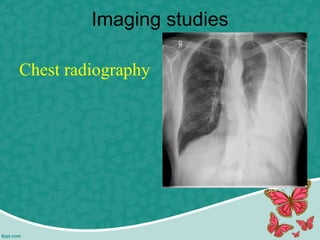
Normally, the pleural space contains a small amount of fluid (5 to 15 mL), which acts as a lubricant that allows the pleural surfaces to move without friction.
But if fluid builds up from either increased production or inadequate removal pleural effusion results.
Pleural effusion B/L or unilateral (parapneumonic process)
Refers to any significant collection of fluid within pleural space.
Any imbalance in formation, absorption lead accumulation of pleural fluid. Common condition:
CHF
Bacterial pneumonia
Malignancy(chest tumor)
Pulmonary embolism
Pleura effusion is a condition refers to a collection of fluid in the pleural space. It is almost secondary to other conditions.
Pleural effusion, sometimes referred to as “water on the lungs,” is the build-up of excess fluid between the layers of the pleura outside the lungs. The pleura are thin membranes that line the lungs and the inside of the chest cavity and act to lubricate and facilitate breathing.
Gastrointestinal bleeding (GI bleed), also known as gastrointestinal hemorrhage, is all forms of bleeding in the gastrointestinal tract, from the mouth to the rectum. When there is significant blood loss over a short time, symptoms may include vomiting red blood, vomiting black blood, bloody stool, or black stool.
Bronchiectasis is a chronic, irreversible dilation of the bronchi and bronchioles. Or •Bronchiectasis is characterized by permanent, abnormal dilation of one or more large bronchBronchiectasis.
Lung abscess is a type of liquefactive necrosis of the lung tissue and formation of cavities (more than 2 cm) containing necrotic debris or fluid caused by microbial infection.
Pyelonephritis
It is the inflammation of the kidney & upper urinary tract that usually results from the bacterial infection of the bladder.
Pyelonephritis can be classified in several different catagories:
-acute pyelonephritis
-chronic pyelonephritis
-xanthogranulomatous pyelonephritis
Pneumonia is an inflammation of the lung parenchyma caused by various microorganisms, including bacteria, mycobacteria, fungi, and viruses.
Pneumonitis is a more general term that describes the inflammatory process in the lung tissue that may predispose and Pneumonia is an inflammation of the lung parenchyma that is caused by a microbial agent.
place the patient at risk for microbial invasion.
Pneumonia is classified into four: community-acquired pneumonia (CAP) and hospital-acquired pneumonia (HAP), pneumonia in the immunocompromised host, and aspiration pneumonia.
Pleural effusion, a collection of fluid in the pleural space, is rarely a primary disease process but is usually secondary to other diseases.
This will help to enhance the theoratical knowledge regarding pleural effusion.
Abnormal fluid accumulation in potential space in between parietal and visceral pleurae – there is imbalance between formation and absorption in response to injury, inflammation or both locally and systematically
Thank you for selecting our 𝐏𝐥𝐞𝐮𝐫𝐚𝐥 𝐞𝐟𝐟𝐮𝐬𝐢𝐨𝐧PPT
This medical PowerPoint template about 𝐏𝐥𝐞𝐮𝐫𝐚𝐥 𝐞𝐟𝐟𝐮𝐬𝐢𝐨𝐧
You can download our template by visiting our website:
https://www.rxslides.com/product/pleural-effusion-powerpoint-template
copy and paste this URL into the browser and download the full editable template.
This 𝐏𝐥𝐞𝐮𝐫𝐚𝐥 𝐞𝐟𝐟𝐮𝐬𝐢𝐨𝐧animated template is designed by RxSlides, a medical professional team covering the following topics about 𝐏𝐥𝐞𝐮𝐫𝐚𝐥 𝐞𝐟𝐟𝐮𝐬𝐢𝐨𝐧
𝐃𝐞𝐟𝐢𝐧𝐢𝐭𝐢𝐨𝐧:
• Respiratory illness caused by the buildup of fluid between lung and chest cavity tissue layers.
𝐀𝐧𝐚𝐭𝐨𝐦𝐲 𝐨𝐟 𝐭𝐡𝐞 𝐋𝐮𝐧𝐠
• The lung is a vital organ responsible for gas exchange.
• It is surrounded by two thin membranes called the pleurae.
• The visceral pleura covers the lung surface.
• The parietal pleura lines the inner chest wall.
𝐩𝐫𝐞𝐯𝐚𝐥𝐞𝐧𝐜𝐞:
• 𝐏𝐥𝐞𝐮𝐫𝐚𝐥 𝐞𝐟𝐟𝐮𝐬𝐢𝐨𝐧 is a common condition, affecting millions of people worldwide.
• The prevalence varies by region, with higher rates in developing countries
𝐏𝐚𝐭𝐡𝐨𝐩𝐡𝐲𝐬𝐢𝐨𝐥𝐨𝐠𝐲:
• Illustrated anatomy of the respiratory system included.
• Animated illustrations demonstrate fluid formation and maintenance.
• Parietal and visceral pleura form the pleural space.
• Fluid produced by filtration from systemic capillaries.
• Fluid accumulation in the pleural cavity causes Pleural Effusion.
𝐑𝐢𝐬𝐤 𝐅𝐚𝐜𝐭𝐨𝐫𝐬
• Smoking
• Heart disease
• Liver disease
• Alcohol
• Lung disease
• Asbestos exposure
𝐜𝐚𝐮𝐬𝐞𝐬:
𝐓𝐫𝐚𝐧𝐬𝐮𝐝𝐚𝐭𝐢𝐯𝐞
o Increased systemic/pulmonary capillary pressure and decreased osmotic pressure.
o Major causes: cirrhosis, heart failure, nephrotic syndrome, protein-losing enteropathy.
𝐄𝐱𝐮𝐝𝐚𝐭𝐢𝐯𝐞:
o Local processes leading to increased capillary permeability.
o Fluid, protein, cells, and serum constituents exude.
o Major causes: inflammation, infection, lung injury, tumors, lung cancer, rheumatoid arthritis, pneumonia, tuberculosis, surgical damage, lymphatic fluid accumulation.
𝐒𝐲𝐦𝐩𝐭𝐨𝐦𝐬:
• Dyspnea (shortness of breath)
• Labored breathing
• Non-productive cough
• Chest tightness
• Orthopnea (difficulty breathing lying down)
𝐃𝐢𝐚𝐠𝐧𝐨𝐬𝐭𝐢𝐜 𝐦𝐞𝐭𝐡𝐨𝐝𝐬
• Physical examination
• Chest radiographs
• Imaging
𝐓𝐫𝐞𝐚𝐭𝐦𝐞𝐧𝐭 𝐎𝐩𝐭𝐢𝐨𝐧𝐬:
Thoracentesis (fluid removal)
Catheter drainage
Pleurodesis (scarring of pleural space)
Pleuroperitoneal shunt (fluid drainage to abdomen)
Pleurectomy (surgical removal of pleura)
Thoracoscopy (visual examination of pleural space)
Thoracostomy (surgical opening of chest cavity)
Visit our site for more animated templates
𝗵𝘁𝘁𝗽𝘀://𝘄𝘄𝘄.𝗿𝘅𝘀𝗹𝗶𝗱𝗲𝘀.𝗰𝗼𝗺
RxSlides PowerPoint icons and illustrations related to 𝐏𝐥𝐞𝐮𝐫𝐚𝐥 𝐞𝐟𝐟𝐮𝐬𝐢𝐨𝐧 will help you customize the content of this editable presentation according to your content and audience interest.
Bell’s palsy
Trigeminal Neuralgia ( Tic Douloreux)
Cranial & spinal neuropathies
Bell’s palsy (facial paralysis) is due to unilateral inflammation of the ( CN VII Facial nerve) seventh cranial nerve, which results in weakness or paralysis of the facial muscles on the affected side.
Polices for intensive care units / critical care units ANILKUMAR BR
What is a Policy?
A Policy is a statement, verbal, written or implied, of those principles and rules that are set by Board of Directors as guidelines on organizations actions.
There should be written polices for the intensive care units or critical care units which will guide the personnel working there.
The polices making body, there should be representation from administrative team, medical team and the nursing team.
ADMISSION POLICES: This should specify whether the patients can be admitted directly to CCU /ICU or through the casualty department.
There should be polices regarding the admission of medico-legal cases.
RESUSCIATION EQUIPMENTS IN INTENSISIVE CARE UNITSANILKUMAR BR
Intensive care unit (ICU) equipment includes patient monitoring, respiratory and cardiac support, pain management, emergency resuscitation devices, and other life support equipment .
They are designed to care for patients who are seriously injured, have a critical or life-threatening illness, or have undergone a major surgical procedure thereby requiring 24-hour care and monitoring.
Intensive care unit equipment includes
Patient monitoring devices
Life support and emergency resuscitation devices, and
Diagnostic devices.
Nursing management of critically ill patient in intensive care unitsANILKUMAR BR
Critical care nursing: it is the field of nursing with a focus on the utmost care of the critically ill (or) unstable patients.
Critically ill patients : critically ill patients are those who are at risk for actual (or) potential life threatening health problems.
Admission QGeneral appearance (consciousness)
Airway: Patency Position of artificial airway (if present)
Breathing: Quantity and quality of respirations (rate, depth, pattern, symmetry, effort, use of accessory muscles) Breath sounds Presence of spontaneous breathing.
Circulation and Cerebral Perfusion: ECG (rate, rhythm, and presence of ectopy) Blood pressure Peripheral pulses and capillary refill Skin, color, temperature, moisture Presence of bleeding Level of consciousness, responsiveness.
quick Check Assessment in CCU.
Infection control protocols in intensive care unitsANILKUMAR BR
Hospital acquired infections (HAIs) are common in intensive care unit (ICU) patient and are associated with increased morbidity and mortality.
The main reason being severity of illness, interruption of normal defense mechanism (e.g. mechanical ventilation), malnutrition & inability to ambulate make it more susceptible to multi drug resistant organism (MDRO).
The most frequent mode of transmission is Contact transmission, this may be direct or indirect other modes include droplet transmission, airborne transmission, common vehicle such as ventilator etc.
Abnormal development or deformities of the ear anatomy can cause a range of complications, from cosmetic issues to hearing and development problems.
An estimated 6 to 45 percent of children are born with some sort of congenital ear deformity.
Hearing is one of our primary modes of communication. 360 million people worldwide have disabling hearing loss.
Hearing loss may result from genetic causes, complications at birth, certain infectious diseases, chronic ear infections, the use of particular drugs, exposure to excessive noise and ageing.
Hearing loss may be mild, moderate, severe or profound.
It can affect one ear or both ears, and leads to difficulty in hearing conversational speech or loud sounds. it may occur in one or both ear.
CONDUCTIVE HEARING LOSS (CHL)
SENSORINEURAL HEARING LOSS (SHL)
MIXED HEARING LOSS (MHL)
There are many types of cancer treatment. The types of treatment that patient receive will depend on the type of cancer, stage of cancer and how advanced it is.
Some people with cancer will have only one treatment. But most people have a combination of treatments, such as surgery with chemotherapy and/or radiation therapy.
Prostate cancer or tumor is the most common cancer in men other than non-melanoma skin cancer.
The majority (more than 75%) of cases occur in men over age 65.
Prostate cancer is a malignant tumor of the prostate gland.
Nursing management of patients with oncological conditionsANILKUMAR BR
Cancer is a group of diseases characterized by uncontrolled growth and spread of abnormal cells.
Cancer is caused by external factors and internal factors which may act together to initiate or promote carcinogenesis.
External Factors - chemicals, radiation, viruses, and lifestyle.
Internal Factors – hormones, immune condition, and inherited mutations.
Oncology branch of medicine deals with etiology, diagnosis, treatment and prevention of cancer.
Onco - is a Greek word meaning tumor .
A group of eye disorders, glaucoma is characterized by high intraocular pressure (IOP) that damages the optic nerve.
Glaucoma is one of the leading causes of irreversible blindness in the world and is the leading cause of blindness among adults in the United States.
Glaucoma may occur as primary or congenital disease or secondary to other causes, such as injury, infection, surgery, or prolonged use of topical corticosteroids.
Primary glaucoma has mainly two forms :
1. Open angle glaucoma ( chronic, simple, or wide angle glaucoma)
2. Angle –closure glaucoma( Acute or narrow angle glaucoma)
Angle –closure glaucoma occurs suddenly and may cause permanent or irreversible vision loss in 48 to 72 hours.
An inflammation of the conjunctiva commonly known as pink eye. Conjunctivitis is usually acute condition and self- limiting.
Conjunctivitis may be unilateral or bilateral.
It may also be chronic, possibly indicating degenerative changes or damage from repeated attacks.
It transmitted by contaminated towels, wash cloths, or the client own hands and it usually spreads very rapidly from one eye to otCommon causes are
Bacterial
Viral and chlamydial infection
Less common causes
Allergy
Parasitic disease and fungal infection
Occupational irritants
her eye.
Nursing assessment and management of patients with hepatic disordersANILKUMAR BR
Liver or Hepatic disorders are common and may result from a virus or exposure to toxic substances such as alcohol.
Another liver disorder is cancer: hepatocellular carcinoma is a highly malignant tumor that is difficult to treat and often fatal.
Liver function is complex, and liver dysfunction affects all body systems.
For this reason, the nurse must understand how the liver functions and must have expert assessment and clinical management skills to care for patients undergoing complex diagnostic and treatment procedures.
The liver plays additional roles in detoxification of chemicals and synthesis and storage of important nutrients and The liver is especially important in the regulation of glucose and protein metabolism .
Ototoxicity is, quite simply, ear poisoning (oto = ear, toxicity = poisoning), which results from exposure to drugs or chemicals that damage the inner ear or the vestibulo-cochlear nerve (the nerve sending balance and hearing information from the inner ear to the brain).
Nursing assessment and Management clients with Pancreatic disordersANILKUMAR BR
The pancreas, located in the upper abdomen, has endocrine as well as exocrine functions .
The secretion of pancreatic enzymes into the gastrointestinal tract through the pancreatic duct represents its exocrine function.
The secretion of insulin, glucagon, and somatostatin directly into the bloodstream represents its endocrine function.
Pancreatitis (inflammation of the pancreas) is a serious disorder. The most basic classification system used to describe or categorize the various stages and forms of pancreatitis divides the disorder into acute or chronic forms.
Acute pancreatitis can be a medical emergency associated with a high risk for life-threatening complications and mortality, whereas chronic pancreatitis often goes undetected until 80% to 90% of the exocrine and endocrine tissue is destroyed.
Acute pancreatitis does not usually lead to chronic pancreatitis unless complications develop.
Nurses are primarily involved in the administration of medication across various settings. Nurses are also involved in both dispensing and preparation of medication. Research on medical administration errors (MAEs) shows an error rate of 60%, 34 mainly in the form of wrong time, wrong rate, or wrong dose.
There are many ways to prevent medication errors and one way of which is understanding the 10 “rights” of drug administration:
Thoracentesis (thor-a-sen-tee-sis) is a procedure that is done to remove a sample of fluid from around the lung.
The lung is covered with a tissue called the pleura. The inside of the chest is also lined with pleura.
The space between these two areas is called the pleural space.
This space normally contains just a thin layer of fluid, however, some conditions such as pneumonia, some types of cancer, or congestive heart failure may cause excessive fluid to develop (pleural effusion).
Thoracentesis, also known as pleural fluid analysis, is a procedure in which a needle is inserted through the back of the chest wall into the pleural space (a space that exists between the two lungs and the anterior chest wall) to remove fluid or air.
Pleural fluid analysis is the microscopic and chemical lab analysis of the fluid obtained during thoracentesis.
IndDiagnostic: determination of pleural effusion etiology (e.g. transudative versus exudative) usually requires the removal of 50 to 100mL of pleural fluid for laboratory studies. Most new effusions require diagnostic thoracentesis, an exception being a new effusion with a clear clinical diagnosis (e.g. CHF) with no evidence for superimposed pleural space infection
Therapeutic: reduce dyspnea and respiratory compromise in patients with large pleural effusions. This is typically achieved by removing a much larger volume of fluid compared to the diagnostic thoracentesis
ications
Nephrotic syndrome may be caused by primary (idiopathic) renal disease or by a variety of secondary causes. Patients present with marked edema, proteinuria, hypoalbuminemia, and often hyperlipidemia.
Nephrotic syndrome is a primary glomerular disease characterized by the following:
Marked increase in protein in the urine (proteinuria)
Decrease in albumin in the blood (hypoalbuminemia)
Edema (The swelling (edema), can be most noticeable on the face, around the eyes, around the feet and ankles, and in the belly area (or the abdomen).
High serum cholesterol and low-density lipoproteins (hyperlipidemia)
Nephrotic syndrome is a clinical disorder characterized by marked increase of protein in the urine ( proteinuria ), decrease in albumin in the blood (hypoalbuminemia ),edema, & excess lipids in the blood ( hyperlipidemia )
Pathophysiology
Nephrotic syndrome can occur with almost any intrinsic renal disease or systemic disease that affects the glomerulus.
Although generally considered a disorder of childhood, nephrotic syndrome does occur in adults, including the elderly. Causes include:
Chronic glomerulonephritis
Diabetes mellitus with intercapillary glomerulosclerosis
Amyloidosis of the kidney
Systemic lupus erythematosus
Multiple myeloma and renal vein thrombosis.
NSAIDs
Pre eclampsia
Urinary diversion procedures are performed to divert urine from the bladder to a new exit site, usually through a surgically created opening (stoma) in the skin.
These procedures are primarily performed when a bladder tumor necessitates removal of the entire bladder (cystectomy).
Urinary diversion has also been used in managing pelvic malignancy, birth defects, strictures, trauma to ureters and urethra, neurogenic bladder, chronic infection causing severe ureteral and renal damage, and intractable interstitial cystitis and as a last resort in managing incontinence.
There are two categories of urinary diversion:
1. Cutaneous urinary diversion : in which urine drains through an opening created in the abdominal wall and skin.
2. Continent urinary diversion : in which a portion of the intestine is used to create a new reservoir for urine.
Coronary artery disease or Ischemic heart disease ANILKUMAR BR
Cardiovascular disease are becoming a leading cause of morbidity and mortality in developed countries and they are also emerging as prominent national health problem in developing countries.
Coronary artery disease has become the major cause of early death and disability in the population.
Coronary artery disease (CAD) can also be used interchangeably with the terms atherosclerotic heart disease or ischemic heart disease.
All of these terms imply insufficient perfusion of the coronary arteries from an abnormal narrowing of the vessels, leading to insufficient oxygen delivery to the myocardial tissue.
The term coronary heart disease, also known as coronary artery disease or Ischemic heart disease, is a condition refers to diseases of the heart that result from a decrease in blood supply to the heart muscle.
Non modifiable risk factors
Modifiable risk factors
Contributing risk factors
Flu Vaccine Alert in Bangalore Karnatakaaddon Scans
As flu season approaches, health officials in Bangalore, Karnataka, are urging residents to get their flu vaccinations. The seasonal flu, while common, can lead to severe health complications, particularly for vulnerable populations such as young children, the elderly, and those with underlying health conditions.
Dr. Vidisha Kumari, a leading epidemiologist in Bangalore, emphasizes the importance of getting vaccinated. "The flu vaccine is our best defense against the influenza virus. It not only protects individuals but also helps prevent the spread of the virus in our communities," he says.
This year, the flu season is expected to coincide with a potential increase in other respiratory illnesses. The Karnataka Health Department has launched an awareness campaign highlighting the significance of flu vaccinations. They have set up multiple vaccination centers across Bangalore, making it convenient for residents to receive their shots.
To encourage widespread vaccination, the government is also collaborating with local schools, workplaces, and community centers to facilitate vaccination drives. Special attention is being given to ensuring that the vaccine is accessible to all, including marginalized communities who may have limited access to healthcare.
Residents are reminded that the flu vaccine is safe and effective. Common side effects are mild and may include soreness at the injection site, mild fever, or muscle aches. These side effects are generally short-lived and far less severe than the flu itself.
Healthcare providers are also stressing the importance of continuing COVID-19 precautions. Wearing masks, practicing good hand hygiene, and maintaining social distancing are still crucial, especially in crowded places.
Protect yourself and your loved ones by getting vaccinated. Together, we can help keep Bangalore healthy and safe this flu season. For more information on vaccination centers and schedules, residents can visit the Karnataka Health Department’s official website or follow their social media pages.
Stay informed, stay safe, and get your flu shot today!
These lecture slides, by Dr Sidra Arshad, offer a quick overview of physiological basis of a normal electrocardiogram.
Learning objectives:
1. Define an electrocardiogram (ECG) and electrocardiography
2. Describe how dipoles generated by the heart produce the waveforms of the ECG
3. Describe the components of a normal electrocardiogram of a typical bipolar leads (limb II)
4. Differentiate between intervals and segments
5. Enlist some common indications for obtaining an ECG
Study Resources:
1. Chapter 11, Guyton and Hall Textbook of Medical Physiology, 14th edition
2. Chapter 9, Human Physiology - From Cells to Systems, Lauralee Sherwood, 9th edition
3. Chapter 29, Ganong’s Review of Medical Physiology, 26th edition
4. Electrocardiogram, StatPearls - https://www.ncbi.nlm.nih.gov/books/NBK549803/
5. ECG in Medical Practice by ABM Abdullah, 4th edition
6. ECG Basics, http://www.nataliescasebook.com/tag/e-c-g-basics
The prostate is an exocrine gland of the male mammalian reproductive system
It is a walnut-sized gland that forms part of the male reproductive system and is located in front of the rectum and just below the urinary bladder
Function is to store and secrete a clear, slightly alkaline fluid that constitutes 10-30% of the volume of the seminal fluid that along with the spermatozoa, constitutes semen
A healthy human prostate measures (4cm-vertical, by 3cm-horizontal, 2cm ant-post ).
It surrounds the urethra just below the urinary bladder. It has anterior, median, posterior and two lateral lobes
It’s work is regulated by androgens which are responsible for male sex characteristics
Generalised disease of the prostate due to hormonal derangement which leads to non malignant enlargement of the gland (increase in the number of epithelial cells and stromal tissue)to cause compression of the urethra leading to symptoms (LUTS
micro teaching on communication m.sc nursing.pdfAnurag Sharma
Microteaching is a unique model of practice teaching. It is a viable instrument for the. desired change in the teaching behavior or the behavior potential which, in specified types of real. classroom situations, tends to facilitate the achievement of specified types of objectives.
ARTIFICIAL INTELLIGENCE IN HEALTHCARE.pdfAnujkumaranit
Artificial intelligence (AI) refers to the simulation of human intelligence processes by machines, especially computer systems. It encompasses tasks such as learning, reasoning, problem-solving, perception, and language understanding. AI technologies are revolutionizing various fields, from healthcare to finance, by enabling machines to perform tasks that typically require human intelligence.
Lung Cancer: Artificial Intelligence, Synergetics, Complex System Analysis, S...Oleg Kshivets
RESULTS: Overall life span (LS) was 2252.1±1742.5 days and cumulative 5-year survival (5YS) reached 73.2%, 10 years – 64.8%, 20 years – 42.5%. 513 LCP lived more than 5 years (LS=3124.6±1525.6 days), 148 LCP – more than 10 years (LS=5054.4±1504.1 days).199 LCP died because of LC (LS=562.7±374.5 days). 5YS of LCP after bi/lobectomies was significantly superior in comparison with LCP after pneumonectomies (78.1% vs.63.7%, P=0.00001 by log-rank test). AT significantly improved 5YS (66.3% vs. 34.8%) (P=0.00000 by log-rank test) only for LCP with N1-2. Cox modeling displayed that 5YS of LCP significantly depended on: phase transition (PT) early-invasive LC in terms of synergetics, PT N0—N12, cell ratio factors (ratio between cancer cells- CC and blood cells subpopulations), G1-3, histology, glucose, AT, blood cell circuit, prothrombin index, heparin tolerance, recalcification time (P=0.000-0.038). Neural networks, genetic algorithm selection and bootstrap simulation revealed relationships between 5YS and PT early-invasive LC (rank=1), PT N0—N12 (rank=2), thrombocytes/CC (3), erythrocytes/CC (4), eosinophils/CC (5), healthy cells/CC (6), lymphocytes/CC (7), segmented neutrophils/CC (8), stick neutrophils/CC (9), monocytes/CC (10); leucocytes/CC (11). Correct prediction of 5YS was 100% by neural networks computing (area under ROC curve=1.0; error=0.0).
CONCLUSIONS: 5YS of LCP after radical procedures significantly depended on: 1) PT early-invasive cancer; 2) PT N0--N12; 3) cell ratio factors; 4) blood cell circuit; 5) biochemical factors; 6) hemostasis system; 7) AT; 8) LC characteristics; 9) LC cell dynamics; 10) surgery type: lobectomy/pneumonectomy; 11) anthropometric data. Optimal diagnosis and treatment strategies for LC are: 1) screening and early detection of LC; 2) availability of experienced thoracic surgeons because of complexity of radical procedures; 3) aggressive en block surgery and adequate lymph node dissection for completeness; 4) precise prediction; 5) adjuvant chemoimmunoradiotherapy for LCP with unfavorable prognosis.
Factory Supply Best Quality Pmk Oil CAS 28578–16–7 PMK Powder in Stockrebeccabio
Factory Supply Best Quality Pmk Oil CAS 28578–16–7 PMK Powder in Stock
Telegram: bmksupplier
signal: +85264872720
threema: TUD4A6YC
You can contact me on Telegram or Threema
Communicate promptly and reply
Free of customs clearance, Double Clearance 100% pass delivery to USA, Canada, Spain, Germany, Netherland, Poland, Italy, Sweden, UK, Czech Republic, Australia, Mexico, Russia, Ukraine, Kazakhstan.Door to door service
Hot Selling Organic intermediates
Ethanol (CH3CH2OH), or beverage alcohol, is a two-carbon alcohol
that is rapidly distributed in the body and brain. Ethanol alters many
neurochemical systems and has rewarding and addictive properties. It
is the oldest recreational drug and likely contributes to more morbidity,
mortality, and public health costs than all illicit drugs combined. The
5th edition of the Diagnostic and Statistical Manual of Mental Disorders
(DSM-5) integrates alcohol abuse and alcohol dependence into a single
disorder called alcohol use disorder (AUD), with mild, moderate,
and severe subclassifications (American Psychiatric Association, 2013).
In the DSM-5, all types of substance abuse and dependence have been
combined into a single substance use disorder (SUD) on a continuum
from mild to severe. A diagnosis of AUD requires that at least two of
the 11 DSM-5 behaviors be present within a 12-month period (mild
AUD: 2–3 criteria; moderate AUD: 4–5 criteria; severe AUD: 6–11 criteria).
The four main behavioral effects of AUD are impaired control over
drinking, negative social consequences, risky use, and altered physiological
effects (tolerance, withdrawal). This chapter presents an overview
of the prevalence and harmful consequences of AUD in the U.S.,
the systemic nature of the disease, neurocircuitry and stages of AUD,
comorbidities, fetal alcohol spectrum disorders, genetic risk factors, and
pharmacotherapies for AUD.
Title: Sense of Taste
Presenter: Dr. Faiza, Assistant Professor of Physiology
Qualifications:
MBBS (Best Graduate, AIMC Lahore)
FCPS Physiology
ICMT, CHPE, DHPE (STMU)
MPH (GC University, Faisalabad)
MBA (Virtual University of Pakistan)
Learning Objectives:
Describe the structure and function of taste buds.
Describe the relationship between the taste threshold and taste index of common substances.
Explain the chemical basis and signal transduction of taste perception for each type of primary taste sensation.
Recognize different abnormalities of taste perception and their causes.
Key Topics:
Significance of Taste Sensation:
Differentiation between pleasant and harmful food
Influence on behavior
Selection of food based on metabolic needs
Receptors of Taste:
Taste buds on the tongue
Influence of sense of smell, texture of food, and pain stimulation (e.g., by pepper)
Primary and Secondary Taste Sensations:
Primary taste sensations: Sweet, Sour, Salty, Bitter, Umami
Chemical basis and signal transduction mechanisms for each taste
Taste Threshold and Index:
Taste threshold values for Sweet (sucrose), Salty (NaCl), Sour (HCl), and Bitter (Quinine)
Taste index relationship: Inversely proportional to taste threshold
Taste Blindness:
Inability to taste certain substances, particularly thiourea compounds
Example: Phenylthiocarbamide
Structure and Function of Taste Buds:
Composition: Epithelial cells, Sustentacular/Supporting cells, Taste cells, Basal cells
Features: Taste pores, Taste hairs/microvilli, and Taste nerve fibers
Location of Taste Buds:
Found in papillae of the tongue (Fungiform, Circumvallate, Foliate)
Also present on the palate, tonsillar pillars, epiglottis, and proximal esophagus
Mechanism of Taste Stimulation:
Interaction of taste substances with receptors on microvilli
Signal transduction pathways for Umami, Sweet, Bitter, Sour, and Salty tastes
Taste Sensitivity and Adaptation:
Decrease in sensitivity with age
Rapid adaptation of taste sensation
Role of Saliva in Taste:
Dissolution of tastants to reach receptors
Washing away the stimulus
Taste Preferences and Aversions:
Mechanisms behind taste preference and aversion
Influence of receptors and neural pathways
Impact of Sensory Nerve Damage:
Degeneration of taste buds if the sensory nerve fiber is cut
Abnormalities of Taste Detection:
Conditions: Ageusia, Hypogeusia, Dysgeusia (parageusia)
Causes: Nerve damage, neurological disorders, infections, poor oral hygiene, adverse drug effects, deficiencies, aging, tobacco use, altered neurotransmitter levels
Neurotransmitters and Taste Threshold:
Effects of serotonin (5-HT) and norepinephrine (NE) on taste sensitivity
Supertasters:
25% of the population with heightened sensitivity to taste, especially bitterness
Increased number of fungiform papillae
Title: Sense of Smell
Presenter: Dr. Faiza, Assistant Professor of Physiology
Qualifications:
MBBS (Best Graduate, AIMC Lahore)
FCPS Physiology
ICMT, CHPE, DHPE (STMU)
MPH (GC University, Faisalabad)
MBA (Virtual University of Pakistan)
Learning Objectives:
Describe the primary categories of smells and the concept of odor blindness.
Explain the structure and location of the olfactory membrane and mucosa, including the types and roles of cells involved in olfaction.
Describe the pathway and mechanisms of olfactory signal transmission from the olfactory receptors to the brain.
Illustrate the biochemical cascade triggered by odorant binding to olfactory receptors, including the role of G-proteins and second messengers in generating an action potential.
Identify different types of olfactory disorders such as anosmia, hyposmia, hyperosmia, and dysosmia, including their potential causes.
Key Topics:
Olfactory Genes:
3% of the human genome accounts for olfactory genes.
400 genes for odorant receptors.
Olfactory Membrane:
Located in the superior part of the nasal cavity.
Medially: Folds downward along the superior septum.
Laterally: Folds over the superior turbinate and upper surface of the middle turbinate.
Total surface area: 5-10 square centimeters.
Olfactory Mucosa:
Olfactory Cells: Bipolar nerve cells derived from the CNS (100 million), with 4-25 olfactory cilia per cell.
Sustentacular Cells: Produce mucus and maintain ionic and molecular environment.
Basal Cells: Replace worn-out olfactory cells with an average lifespan of 1-2 months.
Bowman’s Gland: Secretes mucus.
Stimulation of Olfactory Cells:
Odorant dissolves in mucus and attaches to receptors on olfactory cilia.
Involves a cascade effect through G-proteins and second messengers, leading to depolarization and action potential generation in the olfactory nerve.
Quality of a Good Odorant:
Small (3-20 Carbon atoms), volatile, water-soluble, and lipid-soluble.
Facilitated by odorant-binding proteins in mucus.
Membrane Potential and Action Potential:
Resting membrane potential: -55mV.
Action potential frequency in the olfactory nerve increases with odorant strength.
Adaptation Towards the Sense of Smell:
Rapid adaptation within the first second, with further slow adaptation.
Psychological adaptation greater than receptor adaptation, involving feedback inhibition from the central nervous system.
Primary Sensations of Smell:
Camphoraceous, Musky, Floral, Pepperminty, Ethereal, Pungent, Putrid.
Odor Detection Threshold:
Examples: Hydrogen sulfide (0.0005 ppm), Methyl-mercaptan (0.002 ppm).
Some toxic substances are odorless at lethal concentrations.
Characteristics of Smell:
Odor blindness for single substances due to lack of appropriate receptor protein.
Behavioral and emotional influences of smell.
Transmission of Olfactory Signals:
From olfactory cells to glomeruli in the olfactory bulb, involving lateral inhibition.
Primitive, less old, and new olfactory systems with different path
Explore natural remedies for syphilis treatment in Singapore. Discover alternative therapies, herbal remedies, and lifestyle changes that may complement conventional treatments. Learn about holistic approaches to managing syphilis symptoms and supporting overall health.
3. PLEURAL EFFUSION
• Normally, the pleural space contains a small
amount of fluid (5 to 15 mL), which acts as a
lubricant that allows the pleural surfaces to
move without friction.
• But if fluid builds up from either increased
production or inadequate removal pleural
effusion results.
• Pleural effusion B/L or unilateral
(parapneumonic process)
4. PLEURAL EFFUSION
• Refers to any significant collection of
fluid within pleural space.
• Any imbalance in formation, absorption
lead accumulation of pleural fluid.
Common condition:
1.CHF
2.Bacterial pneumonia
3.Malignancy(chest tumor)
4.Pulmonary embolism
5. Definition
• A pleural effusion describes an
excess of fluid in the pleural cavity,
usually resulting from an imbalance
in the normal rate of pleural fluid
production or absorption, or both.
6. What is Pleural effusion?
• Pleura effusion is
a condition refers
to a collection of
fluid in the pleural
space. It is almost
secondary to other
conditions.
7. Pleural effusion
• This fluid excessive amounts of such
fluid can impair breathing by limiting
the expansion of the lungs during
respiration.
9. TRANSUDATIVE EFFUSIONS
• Transudative effusions arise from more
commonly:
Congestive heart failure
Hepatic disease with ascites
Nephrotic syndrome
Peritoneal dialysis
Hypoalbuminemia and disorders of increase
extra cellular volume.
10. Characteristics of Tranudative effusion
• Occurs primarily in non-inflammatory
conditions.
• low protein, low-cell-count fluid.
• Clear to faint yellow tinge, no odor
• pH 7.40 7.55‐
• Specific gravity < 1.015
• Protein content < 3g/100ml
• Glucose level equal serum plasma
11. EXUDATIVE EFFUSIONS
• Exudative effusion result from several
conditions such as:
1.Tuberculosis
2.Subphrenic abscess
3.Pancreatitis
4.Bacterial or fungal pneuomonia
5.Empyema
12. 1. Cancer
2. Pulmonary embolism
3. SLE & rheumatoid arthritis
4. Intra abdominal abscess
5. Esophageal perforation and chest
injuries
13. Characteristics of Exudative effusion
• Often turbid, bloody or purulent
• pH < 7.30
• Specific gravity > 1.016
• Protein content > 3g/100ml
• Glucose level < 60mg/dl.
• High-protein fluid
14. Types of fluids
THERE ARE MAINLY Four types of fluids can
accumulate in the pleural space:
1. Serous fluid (hydrothorax) : A
hydrothorax is a condition that results from
serous fluid accumulating in the pleural cavity.
2.Blood (haemothorax): is a condition that
results from blood accumulating in the pleural
cavity.
15. Types of fluids
3. Pus (pyothorax or empyema) : is
an accumulation of pus in the pleural
cavity.
16. 2. Chyle (chylothorax): chyle is a milky
bodily fluid consisting of lymph and
emulsified fats, or free fatty acids (FFAs)
and it is formed in the small intestine
during digestion of fatty foods .It is a
type of pleural effusion . it results from
lymphatic fluid (chyle) accumulating in
the pleural cavity.
18. Clinical Manifestations
The clinical presentation of pleural effusion
depends on the amount of fluid present and
the underlying cause.
Many patients have no symptoms at the
time a pleural effusion is discovered.
19. Clinical Manifestations
1. Pleuritic chest pain
2. Dyspnea
3. Nonproductive cough.
• The chest pain is usually sharp and is
exacerbated by movement of the pleural
surfaces, as with deep inspiration,
coughing, and sneezing.
20. Assessment & Diagnosis
• History collection (A history of
pneumonia, chest tumor cardiac, renal, or
liver impairment and cancer related
treatment)
• Physical examination (decreased or
absent breath sounds, decreased fremitus,
and a dull, flat sound when percussed)
24. Medical Management
• The objectives of treatment are to
discover the underlying cause, to prevent
reaccumulation of fluid, and to relieve
discomfort, dyspnea, and respiratory
compromise mainating oxygenation &
facilating drainage)
25. Medical Management
• Thoracentesis is performed to remove
fluid, to obtain a specimen for analysis,
and to relieve dyspnea and respiratory
compromise.
• Thoracentesis may be performed under
ultrasound guidance.
26. Nursing Management
• The nurse’s role in the care of the patient
with a pleural effusion includes
implementing the medical regimen.
27. • The nurse prepares and positions the
patient for thoracentesis and offers
support throughout the procedure.
• Pain management is a priority, and the
nurse assists the patient to assume
positions that are the least painful.
28. Nursing diagnosis
• Risk for infection related to introduction of
foreign object ( thoracentesis needle, chest
tube) in to chest cavity.
• Impaired gas exchange related to
ineffective breathing pattern.
• Anxiety related to diagnosis and
therapeutic procedure ( thoracentesis, chest
drainage).
29. Nursing Interventions
• Administer oxygen and in empyema
antibiotics, as ordered. Record the client
response to these treatments.
• Encourage to perform to perform
pulmonary exercise such as deep
breathing, effective coughing and use of
spirometry etc.
30. Nursing Interventions
• Prepare the client for thoracentesis
procedure and assist to physician during
procedure and monitor complications
associated with thoracentesis ( bleeding,
pain, dyspena and cyanosis).
• Monitor client respiratory status
frequently. Obtain ABGs if necessary.
31. Nursing Interventions
• Ensure chest tube patency by observing
for fluctuations in the tubing. Record the
amount, color, and consistency of any
tube drainage.