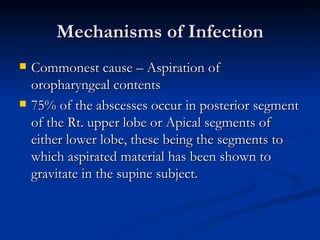
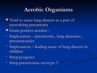
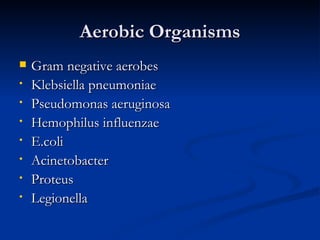
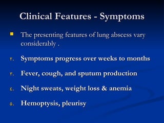
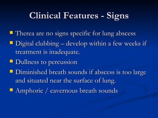
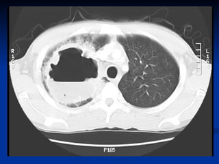
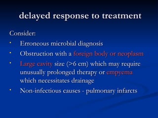
Lung Abscess
 Dr.Vemuri Chaitanya
definition
   A localized area of destruction of lung
    parenchyma in which infection by pyogenic
    organisms results in tissue necrosis &
    suppuration .
   It manifests radiographically as a cavity with an
    air – fluid levels.
Necrotizing Pneumonia

   necrosis with multiple micro abscesses
    form a larger cavitary lesion; actually
    represents a continuum of the same
    process(less than 2cm in diam)
Lung Abscess - Classification

   May be primary or secondary
   Primary = abscess in previously healthy
    patient or in a patient at risk for aspiration
   Secondary = associated bronchogenic
    neoplasm or immunocompromised patient.
Etiology
   Aspiration of Oropharyngeal flora
•   Dental / Periodontal sepsis
•   Paranasal sinus infection
•   Depressed conscoius level
•   Impaired laryngeal closure ( cuffed endotracheal tube,
    tracheostomy tube, recurrent laryngeal nerve palsy )
•   Disturbances of swallowing
•   Dealayed gastric emptying. / gerd / vomiting
Etiology
   Necrotizing pneumonia
•   Staph aureus
•   Strep milleri / intermedius
•   Klebsiella pneumoniae
•   Pseudomonas aeruginosa
Etiology
   Hematogenous spread from a distal site
•   UTI
•   Abdominal sepsis
•   Pelvic sepsis
•   Infective endocarditis
•   IV drug abuse
•   Infected IV cannulae
•   Septic thrombophlebitis
Etiology
 Pre existing lung disease
• Bronchiectasis
• Cystic fibrosis
• Bronchial obstruction : tumour, foreign body,
  cong.abn
 Infected pulmonary infarct
 Trauma
 Immunodeficiency
Mechanisms of Infection
   Commonest cause – Aspiration of
    oropharyngeal contents
   75% of the abscesses occur in posterior segment
    of the Rt. upper lobe or Apical segments of
    either lower lobe, these being the segments to
    which aspirated material has been shown to
    gravitate in the supine subject.
Other Mechanisms
   The development of lung abscess favoured by
    conditions that prevent normal clearance of
    pulmonary secretions – lung tumours,
    bronchiectasis , inhaled foreign bodies.
   Secondary infection – in cong.abn like
    bronchopulmonary sequestration & lung cysts
Microbiological characteristics
   Caused by a wide variety of different organisms
    & its common to obtain a mixed bacterial
    growth from single abscess when pus is cultured
   Anaerobes – 69% of community acquired cases
   Anaerobes – 7% hosp acquired cases
   Staph aureus, Klebsiella pneumoniae,
    Pseudomonas aeruginosa – imp role
Anaerobic organisms
   Most frequently implicated
   Main groups
•   Gram negative bacilli – Bacteroides- Bacteroides
    fragilis
•   Gram positive cocci mainly Peptostreptococcus
•   Long & thin gram negative rods –
    Fusobacterium – Fusobacterium nucleatum,
    Fusobacterium necrophorum
Aerobic Organisms
   Tend to cause lung abscess as a part of
    necrotizing pneumonia
   Gram positive aerobes :
•   Staph.aureus – pneumonia , lung abscesses ,
    pneumatoceles
•   Staph.aureus – leading cause of lung abscess in
    children
•   Strep.pyogenes
•   Strep.pneumoniae serotype 3
Aerobic Organisms
   Gram negative aerobes
•   Klebsiella pneumoniae
•   Pseudomonas aeruginosa
•   Hemophilus influenzae
•   E.coli
•   Acinetobacter
•   Proteus
•   Legionella
Other causes
   Tuberculosis & non tuberculous mycobacterial
    infection – fluid filled cavities – upper lobes / apical
    segments of lower lobes
   Fungal infection – Histoplasma capsulatum
                         Blastomyces dermatitidis
                         Coccidiodes immitis
                         Aspergillus
                         Cryptococcus neoformans
                         Candida
Other causes
   Major risk factors for all opportunistic fungal
    infections are neutropenia, coticosteroid use,
    HIV infection
   Single large lung abscess – Actinomyces israeli.
    This infection – lung infiltrate with honey comb
    of small abscess cavities that may communicate
    with chest wall with bony destruction and sinus
    formation
Pathology
   Most often -as a complication of aspiration pneumonia
   Oral anaerobes
   “Typical patient” is predisposed to aspiration due to
    compromised consciousness
    (ie, alcoholism, drug abuse, general anesthesia) or
     dysphagia
   Periodontal disease, especially gingivitis, with
    concentrations of bacteria in the gingival crevice as high as
    1011/mL
pathology
1.   Inoculum from gingival crevice reach lower airways -
     while the patient is in the recumbent position.
2.   Pneumonitis arises first but progresses to tissue necrosis
     after 7-14 days.
3.   Necrosis results in lung abscess and/or an empyema; the
     latter can be due to a bronchopleural fistula or direct
     extension of infection into the pleural space
pathology
   Lung abscesses begin as areas of pneumonia on which
    small zones of necrosis ( microabscesses ) develop
    within consolidated lung. Some of these areas coalesce
    to form single / sometimes multiple areas of
    suppuration and when they reach a size of 1 -2 cm dia –
    abscess.
   If the natural history of this pathological process is
    interupted at an early stage by an appropriate
    antimicrobial , then healing may be complete with no
    residual radiographic evidence of damage.
pathology
   If treatment is delayed / inadequate , the inflammatory
    process may progress , entering a chronic phase.
   Abscesses arising as a result of aspiration usually occur
    close to visceral pleural surface in dependent parts of
    lungs.
   ¾ ths of lung abscesses occur in posterior segement of
    right upper lobe or apical segement of either lower
    lobes, the anatomical disposition of these segmental
    bronchi accepting the passage of aspirated liquid in
    supine position most readily.
   Those d/t haematogenous spread can occur in any part
    of lungs
Clinical Features - Symptoms
    The presenting features of lung abscess vary
     considerably .

2.   Symptoms progress over weeks to months

3.   Fever, cough, and sputum production

4.   Night sweats, weight loss & anemia

5.   Hemoptysis, pleurisy
Clinical Features - Signs
   Therea are no signs specific for lung abscess
   Digital clubbing – develop within a few weeks if
    treatment is inadequate.
   Dullness to percussion
   Diminished breath sounds if abscess is too large
    and situated near the surface of lung.
   Amphoric / cavernous breath sounds
diagnosis
1.   CXR, CT CHEST
2.   Difficult to isolate anaerobic bacteria
3.   Generally, if symptoms and clinical setting right
     for anaerobic infection, generally treat
     empirically
4.   Gram stain:both +ve &-ve,mixed
5.   AFB & Anaerobic culture
diagnosis
   Transtracheal aspirates (TTA), transthoracic
    needle aspirates (TTNA), BAL, pleural fluid, or
    blood cultures allow uncontaminated specimens
   Bronchoscopy with quantitative cultures
    experience with anaerobic lung infections is
    limited
   Further, none of these specimens likely to yield
    anaerobes after antibiotic therapy initiated
diagnosis
•   For patients presenting less typically,
    differential diagnosis is broader and
    evaluation should include r/o TB with
    AFB sputum smear x 3, possible
    bronchoscopy for cx and biopsy
•   Blood culture
Differential diagnosis
   Cavitating lung cancer
   Localized empyema
   Infected bulla containing a fluid level
   Infected congenital pulmonary lesions
   Pulmonary haematoma
   Cavitated pneumoconiotic lesions
   Hiatus hernia
   Hydatid cysts
   Infection with paragonimus westermani
   Cavitating pulmonary infarcts
   Wegeners granulomatosis
Treatment – antibiotic therapy

1.   Ampi / Amoxicillin x orally
2.   Metronidazole 400mg TDS –Anaerobes
3.   Cry.penicillin & clindamycin +/-
     metronidazole IV – in hospitalised pts.
4.   Can change – according to sensitivity
Duration of treatment
   Debated
   Some advocate 4-6 weeks
   Most treat until radiographic abnormalities resolve ,
    generally requiring months of treatment
Surgical intervention
•   Surgery rarely required
•   Indications: failure of medical management, suspected
    neoplasm, or hemorrhage.
•   Predictors of poor response to antibiotic therapy alone:
    abscesses associated-
•   with an obstructed bronchus, large abscess (>6 cm in
    diameter), relatively resistant organisms, such as P. aeruginosa.
•   The usual procedure in such cases is a lobectomy or
    pneumonectomy
Treatment contd…
•   Alternative for patients who are considered
    poor operative risks is percutaneous
    drainage.
•   Bronchoscopy- may be done as a
    diagnostic procedure, especially to detect
    an underlying lesion, but is of relatively
    little use to facilitate drainage
Response to treatment
•   Usually show clinical improvement with ↓ fever within
    3-4 days after beginning antibiotics
   Should deffervesce in 7-10 days
   Persistent fevers beyond this time indicate delayed
    response, and such patients should undergo further
    diagnostic tests to define the underlying anatomy and
    microbiology of the infection
delayed response to treatment
Consider:
•  Erroneous microbial diagnosis
•  Obstruction with a foreign body or neoplasm
•  Large cavity size (>6 cm) which may require
   unusually prolonged therapy or empyema
   which necessitates drainage
•  Non-infectious causes - pulmonary infarcts
complications
1.   Empyema
2.   Bronchopleural –fistula
3.   Pneumothorax , pyoneumothorax
4.   Metastatic cerebral abscess
5.   Sepsis
6.   Fibrosis,bronchiectasis,amyloidosis
Thank You

Lung abscess

  • 1.
  • 2.
    definition  A localized area of destruction of lung parenchyma in which infection by pyogenic organisms results in tissue necrosis & suppuration .  It manifests radiographically as a cavity with an air – fluid levels.
  • 3.
    Necrotizing Pneumonia  necrosis with multiple micro abscesses form a larger cavitary lesion; actually represents a continuum of the same process(less than 2cm in diam)
  • 4.
    Lung Abscess -Classification  May be primary or secondary  Primary = abscess in previously healthy patient or in a patient at risk for aspiration  Secondary = associated bronchogenic neoplasm or immunocompromised patient.
  • 5.
    Etiology  Aspiration of Oropharyngeal flora • Dental / Periodontal sepsis • Paranasal sinus infection • Depressed conscoius level • Impaired laryngeal closure ( cuffed endotracheal tube, tracheostomy tube, recurrent laryngeal nerve palsy ) • Disturbances of swallowing • Dealayed gastric emptying. / gerd / vomiting
  • 6.
    Etiology  Necrotizing pneumonia • Staph aureus • Strep milleri / intermedius • Klebsiella pneumoniae • Pseudomonas aeruginosa
  • 7.
    Etiology  Hematogenous spread from a distal site • UTI • Abdominal sepsis • Pelvic sepsis • Infective endocarditis • IV drug abuse • Infected IV cannulae • Septic thrombophlebitis
  • 8.
    Etiology  Pre existinglung disease • Bronchiectasis • Cystic fibrosis • Bronchial obstruction : tumour, foreign body, cong.abn  Infected pulmonary infarct  Trauma  Immunodeficiency
  • 9.
    Mechanisms of Infection  Commonest cause – Aspiration of oropharyngeal contents  75% of the abscesses occur in posterior segment of the Rt. upper lobe or Apical segments of either lower lobe, these being the segments to which aspirated material has been shown to gravitate in the supine subject.
  • 10.
    Other Mechanisms  The development of lung abscess favoured by conditions that prevent normal clearance of pulmonary secretions – lung tumours, bronchiectasis , inhaled foreign bodies.  Secondary infection – in cong.abn like bronchopulmonary sequestration & lung cysts
  • 11.
    Microbiological characteristics  Caused by a wide variety of different organisms & its common to obtain a mixed bacterial growth from single abscess when pus is cultured  Anaerobes – 69% of community acquired cases  Anaerobes – 7% hosp acquired cases  Staph aureus, Klebsiella pneumoniae, Pseudomonas aeruginosa – imp role
  • 12.
    Anaerobic organisms  Most frequently implicated  Main groups • Gram negative bacilli – Bacteroides- Bacteroides fragilis • Gram positive cocci mainly Peptostreptococcus • Long & thin gram negative rods – Fusobacterium – Fusobacterium nucleatum, Fusobacterium necrophorum
  • 13.
    Aerobic Organisms  Tend to cause lung abscess as a part of necrotizing pneumonia  Gram positive aerobes : • Staph.aureus – pneumonia , lung abscesses , pneumatoceles • Staph.aureus – leading cause of lung abscess in children • Strep.pyogenes • Strep.pneumoniae serotype 3
  • 14.
    Aerobic Organisms  Gram negative aerobes • Klebsiella pneumoniae • Pseudomonas aeruginosa • Hemophilus influenzae • E.coli • Acinetobacter • Proteus • Legionella
  • 15.
    Other causes  Tuberculosis & non tuberculous mycobacterial infection – fluid filled cavities – upper lobes / apical segments of lower lobes  Fungal infection – Histoplasma capsulatum Blastomyces dermatitidis Coccidiodes immitis Aspergillus Cryptococcus neoformans Candida
  • 16.
    Other causes  Major risk factors for all opportunistic fungal infections are neutropenia, coticosteroid use, HIV infection  Single large lung abscess – Actinomyces israeli. This infection – lung infiltrate with honey comb of small abscess cavities that may communicate with chest wall with bony destruction and sinus formation
  • 17.
    Pathology  Most often -as a complication of aspiration pneumonia  Oral anaerobes  “Typical patient” is predisposed to aspiration due to compromised consciousness (ie, alcoholism, drug abuse, general anesthesia) or dysphagia  Periodontal disease, especially gingivitis, with concentrations of bacteria in the gingival crevice as high as 1011/mL
  • 18.
    pathology 1. Inoculum from gingival crevice reach lower airways - while the patient is in the recumbent position. 2. Pneumonitis arises first but progresses to tissue necrosis after 7-14 days. 3. Necrosis results in lung abscess and/or an empyema; the latter can be due to a bronchopleural fistula or direct extension of infection into the pleural space
  • 19.
    pathology  Lung abscesses begin as areas of pneumonia on which small zones of necrosis ( microabscesses ) develop within consolidated lung. Some of these areas coalesce to form single / sometimes multiple areas of suppuration and when they reach a size of 1 -2 cm dia – abscess.  If the natural history of this pathological process is interupted at an early stage by an appropriate antimicrobial , then healing may be complete with no residual radiographic evidence of damage.
  • 20.
    pathology  If treatment is delayed / inadequate , the inflammatory process may progress , entering a chronic phase.  Abscesses arising as a result of aspiration usually occur close to visceral pleural surface in dependent parts of lungs.  ¾ ths of lung abscesses occur in posterior segement of right upper lobe or apical segement of either lower lobes, the anatomical disposition of these segmental bronchi accepting the passage of aspirated liquid in supine position most readily.  Those d/t haematogenous spread can occur in any part of lungs
  • 21.
    Clinical Features -Symptoms  The presenting features of lung abscess vary considerably . 2. Symptoms progress over weeks to months 3. Fever, cough, and sputum production 4. Night sweats, weight loss & anemia 5. Hemoptysis, pleurisy
  • 22.
    Clinical Features -Signs  Therea are no signs specific for lung abscess  Digital clubbing – develop within a few weeks if treatment is inadequate.  Dullness to percussion  Diminished breath sounds if abscess is too large and situated near the surface of lung.  Amphoric / cavernous breath sounds
  • 23.
    diagnosis 1. CXR, CT CHEST 2. Difficult to isolate anaerobic bacteria 3. Generally, if symptoms and clinical setting right for anaerobic infection, generally treat empirically 4. Gram stain:both +ve &-ve,mixed 5. AFB & Anaerobic culture
  • 24.
    diagnosis  Transtracheal aspirates (TTA), transthoracic needle aspirates (TTNA), BAL, pleural fluid, or blood cultures allow uncontaminated specimens  Bronchoscopy with quantitative cultures experience with anaerobic lung infections is limited  Further, none of these specimens likely to yield anaerobes after antibiotic therapy initiated
  • 25.
    diagnosis • For patients presenting less typically, differential diagnosis is broader and evaluation should include r/o TB with AFB sputum smear x 3, possible bronchoscopy for cx and biopsy • Blood culture
  • 26.
    Differential diagnosis  Cavitating lung cancer  Localized empyema  Infected bulla containing a fluid level  Infected congenital pulmonary lesions  Pulmonary haematoma  Cavitated pneumoconiotic lesions  Hiatus hernia  Hydatid cysts  Infection with paragonimus westermani  Cavitating pulmonary infarcts  Wegeners granulomatosis
  • 31.
    Treatment – antibiotictherapy 1. Ampi / Amoxicillin x orally 2. Metronidazole 400mg TDS –Anaerobes 3. Cry.penicillin & clindamycin +/- metronidazole IV – in hospitalised pts. 4. Can change – according to sensitivity
  • 32.
    Duration of treatment  Debated  Some advocate 4-6 weeks  Most treat until radiographic abnormalities resolve , generally requiring months of treatment
  • 33.
    Surgical intervention • Surgery rarely required • Indications: failure of medical management, suspected neoplasm, or hemorrhage. • Predictors of poor response to antibiotic therapy alone: abscesses associated- • with an obstructed bronchus, large abscess (>6 cm in diameter), relatively resistant organisms, such as P. aeruginosa. • The usual procedure in such cases is a lobectomy or pneumonectomy
  • 34.
    Treatment contd… • Alternative for patients who are considered poor operative risks is percutaneous drainage. • Bronchoscopy- may be done as a diagnostic procedure, especially to detect an underlying lesion, but is of relatively little use to facilitate drainage
  • 35.
    Response to treatment • Usually show clinical improvement with ↓ fever within 3-4 days after beginning antibiotics  Should deffervesce in 7-10 days  Persistent fevers beyond this time indicate delayed response, and such patients should undergo further diagnostic tests to define the underlying anatomy and microbiology of the infection
  • 36.
    delayed response totreatment Consider: • Erroneous microbial diagnosis • Obstruction with a foreign body or neoplasm • Large cavity size (>6 cm) which may require unusually prolonged therapy or empyema which necessitates drainage • Non-infectious causes - pulmonary infarcts
  • 37.
    complications 1. Empyema 2. Bronchopleural –fistula 3. Pneumothorax , pyoneumothorax 4. Metastatic cerebral abscess 5. Sepsis 6. Fibrosis,bronchiectasis,amyloidosis
  • 38.