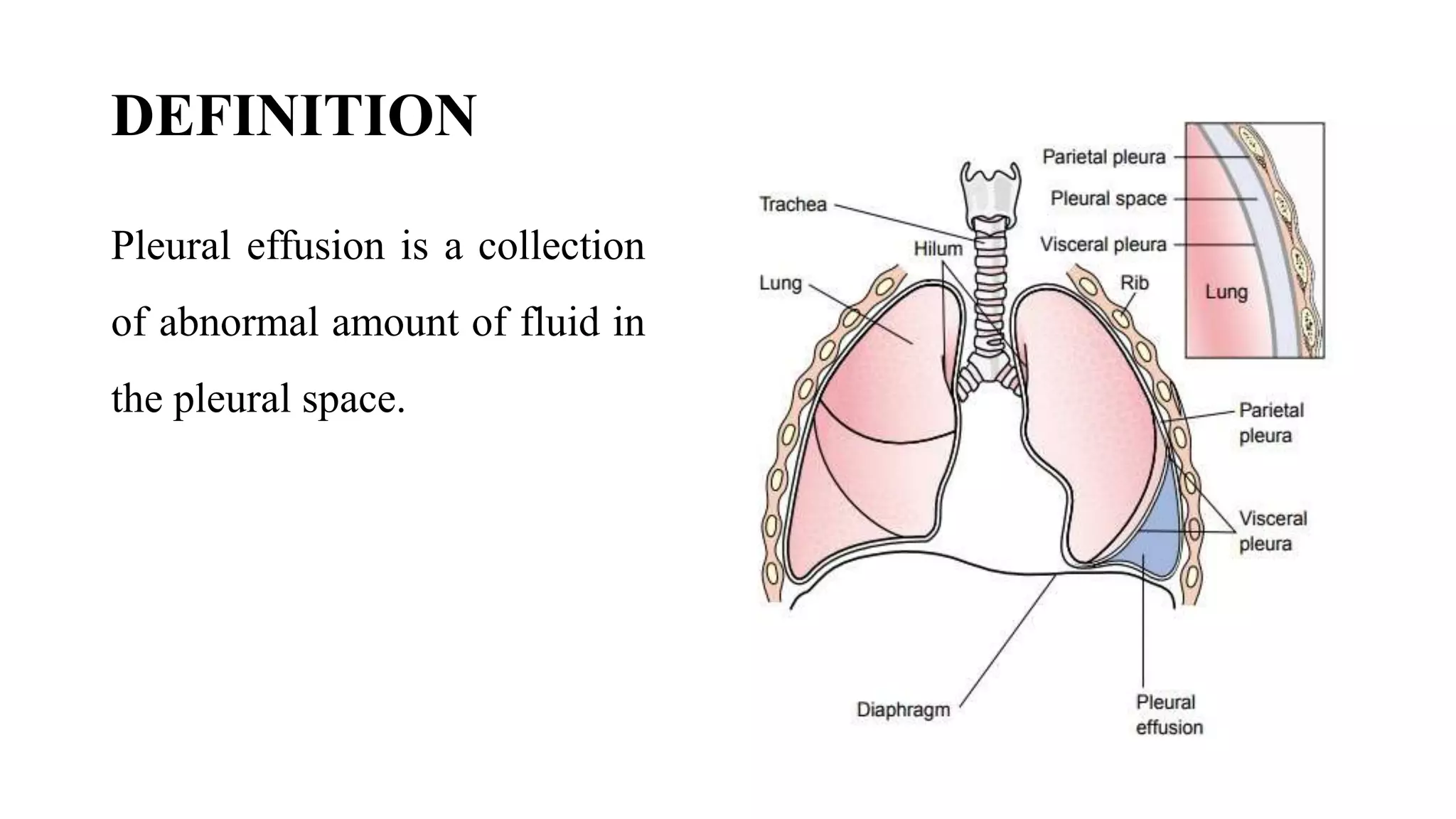
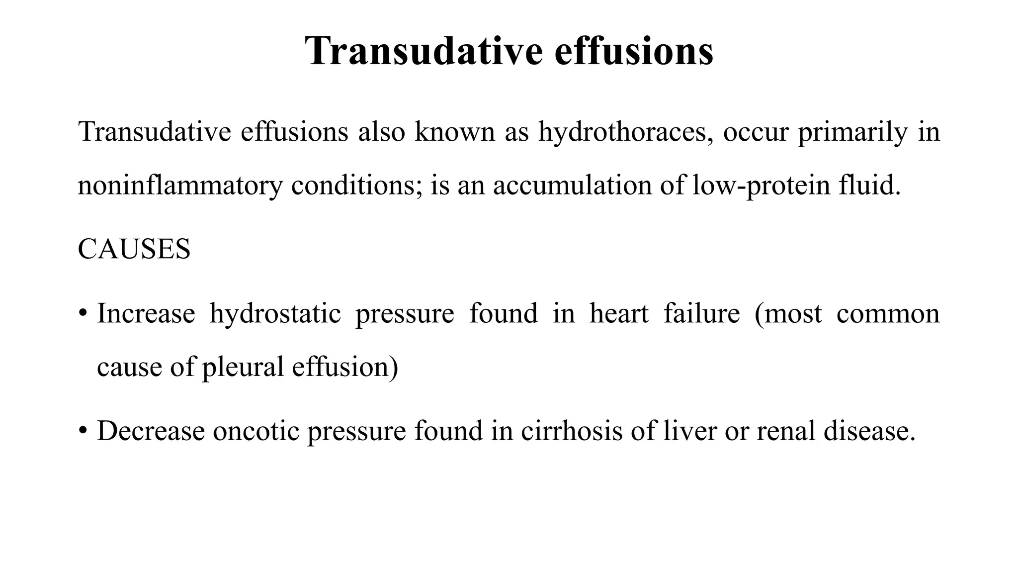
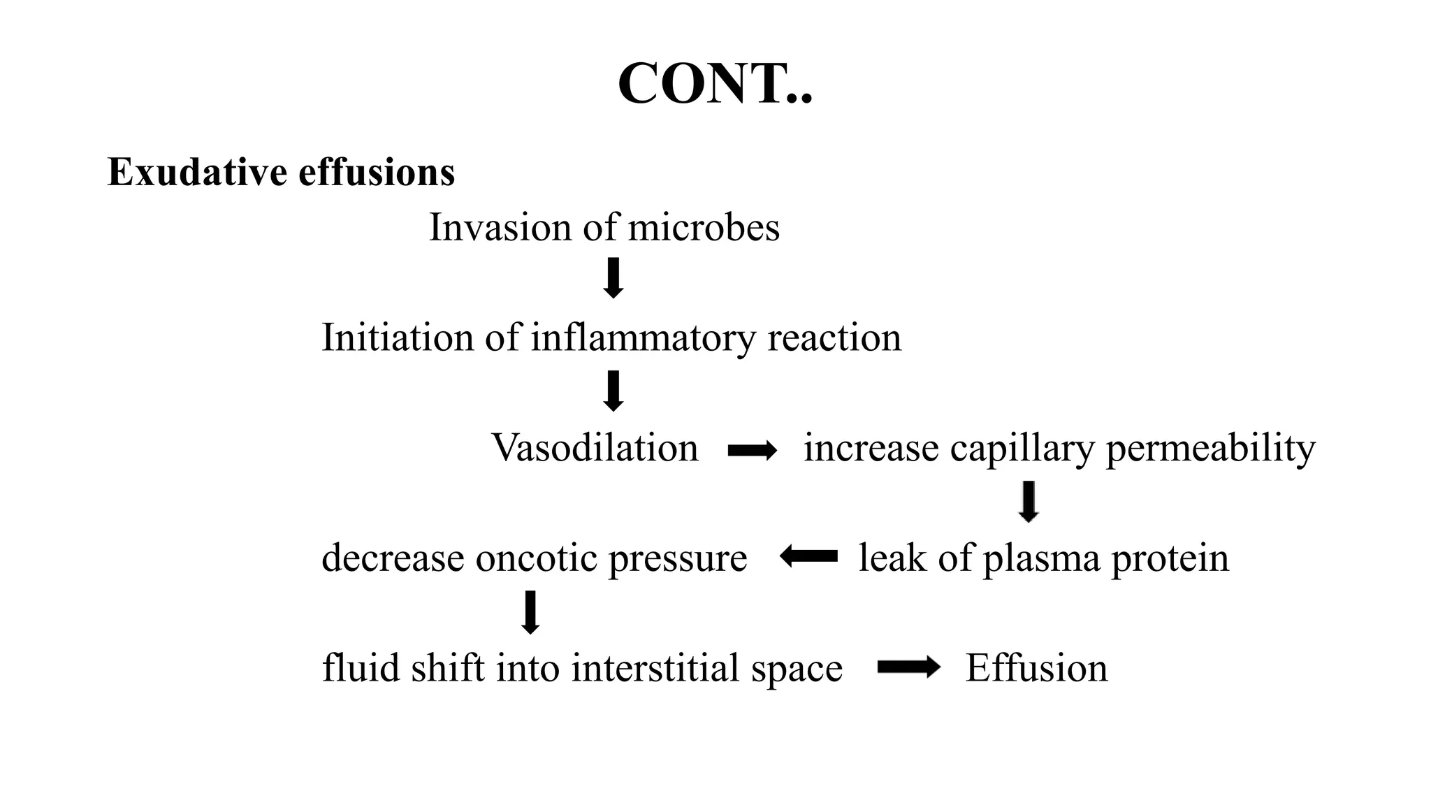
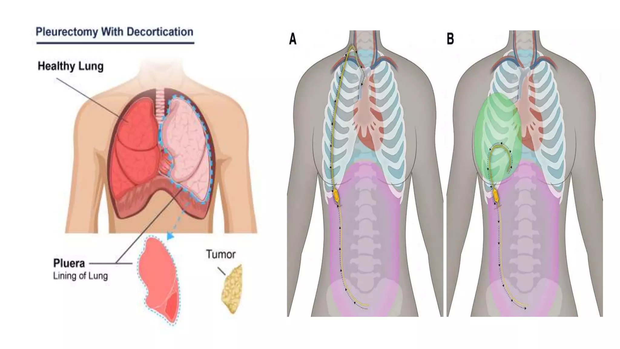
INTRODUCTION
• Pleural effusion, a collection of fluid in the pleural space, is rarely a primary
disease process but is usually secondary to other diseases
• The pleural space normally contains only about 10-20 ml of serous fluid.
• Pleural fluid normally seeps continually into the pleural space from the
capillaries lining the parietal pleura and is reabsorbed by the visceral pleural
capillaries and lymphatic system
• Any condition that interferes with either secretion or drainage of this fluid
leads to pleural effusion
DEFINITION
Pleural effusion is a collection
of abnormal amount of fluid in
the pleural space.
CLASSIFICATION
• Transudative effusions
• Exsudative effusions
Transudative effusions
Transudative effusions also known as hydrothoraces, occur primarily in
noninflammatory conditions; is an accumulation of low-protein fluid.
CAUSES
• Increase hydrostatic pressure found in heart failure (most common
cause of pleural effusion)
• Decrease oncotic pressure found in cirrhosis of liver or renal disease.
Exudative effusions
• Exudative effusions occur in an area of inflammation; is an
accumulation of high-protein fluid.
• An exudative effusion results from increased capillary permeability
characteristic of inflammatory reaction.
• This types of effusion occurs secondary to conditions such as
pulmonary malignancies, pulmonary infections and pulmonary
embolization.
PATHOPHYSIOLOGY
Transudative pleural effusions:
hydrostatic pressure,  oncotic pressure
Unable to remain the fluid with in a intravascular space
Fluid shift interstitial space
Effusion
CONT..
Exudative effusions
Invasion of microbes
Initiation of inflammatory reaction
Vasodilation increase capillary permeability
decrease oncotic pressure leak of plasma protein
fluid shift into interstitial space Effusion
CLINICAL MANIFESTATIONS
• When a small to moderate pleural effusion is present, dyspnea may be
absent or only minimal.
• Malignant effusion may result in dyspnea and coughing
• Pleuritic chest pain, fever, chills
• Dullness or flatness to percussion
• Decreased or absent breath sounds
DIAGNOSTIC EVALUATION
• Chest X-ray or ultrasound detects presence of fluid.
• Thoracentesis, biochemical, bacteriologic, and cytologic studies of
pleural fluid indicates cause.
MANAGEMENT
The objectives of treatment are
• to discover the underlying cause,
• to prevent reaccumulation of fluid and
• to relieve discomfort, dyspnea, and respiratory compromise
CONT..
General effusion
• Treatment is aimed at underlying cause (heart disease, infection).
• Thoracentesis is done to remove fluid, collect a specimen, and relieve
dyspnea
For Malignant Effusions
• Chest tube drainage or pleurodesis
• Radiation
• Chemotherapy
• Surgical pleurectomy
• Pleuroperitoneal shunt
NURSING MANAGEMENT
Nursing Assessment
• Obtain history of previous pulmonary condition
• Assess patient for dyspnea and tachypnea
• Auscultate and percuss lungs for abnormalities
Nursing Diagnosis
Ineffective Breathing Pattern related to collection of fluid in pleural
space as evidenced by dyspnea.
CONT..
Nursing Interventions
• Observe patient's breathing pattern, oxygen saturation
• Administer oxygen as indicated by dyspnea and hypoxemia.
• Assist with thoracentesis if indicated
• Maintain chest drainage as needed
• Provide care after pleurodesis.
• Administer prescribed analgesic.
COMPLICATION
Large effusion could lead to respiratory failure.
THANK YOU

Plural effusion_092054.pptx

  • 2.
    INTRODUCTION • Pleural effusion,a collection of fluid in the pleural space, is rarely a primary disease process but is usually secondary to other diseases • The pleural space normally contains only about 10-20 ml of serous fluid. • Pleural fluid normally seeps continually into the pleural space from the capillaries lining the parietal pleura and is reabsorbed by the visceral pleural capillaries and lymphatic system • Any condition that interferes with either secretion or drainage of this fluid leads to pleural effusion
  • 3.
    DEFINITION Pleural effusion isa collection of abnormal amount of fluid in the pleural space.
  • 4.
  • 5.
    Transudative effusions Transudative effusionsalso known as hydrothoraces, occur primarily in noninflammatory conditions; is an accumulation of low-protein fluid. CAUSES • Increase hydrostatic pressure found in heart failure (most common cause of pleural effusion) • Decrease oncotic pressure found in cirrhosis of liver or renal disease.
  • 6.
    Exudative effusions • Exudativeeffusions occur in an area of inflammation; is an accumulation of high-protein fluid. • An exudative effusion results from increased capillary permeability characteristic of inflammatory reaction. • This types of effusion occurs secondary to conditions such as pulmonary malignancies, pulmonary infections and pulmonary embolization.
  • 7.
    PATHOPHYSIOLOGY Transudative pleural effusions: hydrostaticpressure,  oncotic pressure Unable to remain the fluid with in a intravascular space Fluid shift interstitial space Effusion
  • 8.
    CONT.. Exudative effusions Invasion ofmicrobes Initiation of inflammatory reaction Vasodilation increase capillary permeability decrease oncotic pressure leak of plasma protein fluid shift into interstitial space Effusion
  • 9.
    CLINICAL MANIFESTATIONS • Whena small to moderate pleural effusion is present, dyspnea may be absent or only minimal. • Malignant effusion may result in dyspnea and coughing • Pleuritic chest pain, fever, chills • Dullness or flatness to percussion • Decreased or absent breath sounds
  • 10.
    DIAGNOSTIC EVALUATION • ChestX-ray or ultrasound detects presence of fluid. • Thoracentesis, biochemical, bacteriologic, and cytologic studies of pleural fluid indicates cause.
  • 11.
    MANAGEMENT The objectives oftreatment are • to discover the underlying cause, • to prevent reaccumulation of fluid and • to relieve discomfort, dyspnea, and respiratory compromise
  • 12.
    CONT.. General effusion • Treatmentis aimed at underlying cause (heart disease, infection). • Thoracentesis is done to remove fluid, collect a specimen, and relieve dyspnea For Malignant Effusions • Chest tube drainage or pleurodesis • Radiation • Chemotherapy • Surgical pleurectomy • Pleuroperitoneal shunt
  • 14.
    NURSING MANAGEMENT Nursing Assessment •Obtain history of previous pulmonary condition • Assess patient for dyspnea and tachypnea • Auscultate and percuss lungs for abnormalities Nursing Diagnosis Ineffective Breathing Pattern related to collection of fluid in pleural space as evidenced by dyspnea.
  • 15.
    CONT.. Nursing Interventions • Observepatient's breathing pattern, oxygen saturation • Administer oxygen as indicated by dyspnea and hypoxemia. • Assist with thoracentesis if indicated • Maintain chest drainage as needed • Provide care after pleurodesis. • Administer prescribed analgesic.
  • 16.
    COMPLICATION Large effusion couldlead to respiratory failure.
  • 17.