Lung abscess
•Download as PPT, PDF•
30 likes•15,970 views

om verma msc lecturer medical surgical nursing

Recommended
More Related Content
What's hot
Similar to Lung abscess
Similar to Lung abscess (20)
More from OM VERMA
More from OM VERMA (20)
Recently uploaded
Recently uploaded (20)
Lung abscess
- 1. MR .OM VERMA
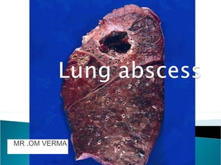
- 2. Localized suppurative inflammation of the lung. Necrosis of the pulmonary tissue & formation of cavities containing necrotic debris or fluid caused by microbial infection. A localized area of destruction of lung parenchyma in which infection by pyogenic organisms results in tissue necrosis & suppuration . It manifests radio graphically as a cavity with an air – fluid levels.
- 3. Lung Abscess is a localized necrotic lesion of the lung parenchyma containing purulent material that collapses and forms a cavity. It is generally caused by aspiration of anaerobic bacteria.
- 5. Location: Dependent areas (posterior segments of upper & lower lobes). Stages: Pneumonic stage: patch of consolidation. Rupture: central necrosis, communication with a bronchus & expec- toration of the liquid centre. Acute cavitations: irregular thick wall, shaggy ( khurdura) inner margin & surrounding consolidation (collection). Chronic cavitation: thin wall, smooth inner margin & subsidence of surrounding inflammatory reaction.
- 6. Complication of pneumonia: Staph pneumonia. Klebsiella pneumonia. T.B.
- 7. Lung abscess with bulging fissure ( a long narrow opening or line of breakage ) sign.
- 8. EMBOLIC ABSCESSES an abscess arising distal to the point of arrest of a septic embolus. A septic embolism is a type of embolism that is infected with bacteria, resulting in the formation of pus. a clot or other plug, usually part or all of a THROMBUS
- 10. Infected catheters. Infected pacemakers. Tricuspid endocarditic ( IV drug abusers).
- 11. ETOLOGY
- 12. Hematogenous ( originating in the blood. 2. producing blood or components of blood. 3. distributed orspread by way of the bloodstream, as in metastases of tumors or in infections; blood-borne. spread from a distal site) • UTI • Abdominal sepsis • Pelvic sepsis • Infective endocarditic • IV drug abuse • Infected IV cannulae • Septic thrombophlebitis = venous thrombosis, inflammation, and bacteremia.
- 13. • Bronchiectasis • Cystic fibrosis • Bronchial obstruction : tumour, foreign body, cong.abn Infected pulmonary infarct Trauma Immunodeficiency = immunodeficiency disease characterized by eczema, recurrent staphylococcal skin abscesses, recurrent lung infections, eosinophilia (a high number of eosinophils in the blood) and high serum levels of IgE.
- 14. Lung Abscess pyogenic lung infection/pneumonia, necrotizing pneumonia. ... The most frequent cause is aspiration of anaerobic organisms from the mouth in those predisposed to ... Penetrating pulmonary trauma - eg, a stab wound.
- 15. Tuberculosis & non tuberculous mycobacterial infection – fluid filled cavities – upper lobes / apical segments of lower lobes Fungal infection – Histoplasma capsulatum Blastomyces dermatitidis Coccidiodes immitis Aspergillus Cryptococcus neoformans Candida
- 16. Aspiration of oropharyngeal or gastric secretion. 2) Septic emboli. Necrotizing pneumonia Necrotizing tumors Gram negative organisms. (klebsiella) Anaerobic bacilli (Bacterorides Carcinoma of the lung Parasitic and fungal diseases of the lung. TB
- 17. Lung abscess starts as an area of pneumonia Small zones of necrosis Coalesce together to from one or more large cavities of 1-2 c.m Progressive and enlargement to from the abscess cavity The abscess cavity well erode( increase) a bronchus
- 18. Expec-toration of purulent sputum with air fluid formation in the cavity Fate 1. infection of the other lung 2. Open into pleura –pyopneumothorax 3. Hematogenous spread
- 19. The presenting features of lung abscess vary considerably . 1. Symptoms progress over weeks to months 2. Fever, cough, and sputum production 3. Night sweats, weight loss & anemia 4. Hemoptysis, is the coughing up of blood or blood-stained mucus from the bronchi, larynx, trachea, or lungs.
- 20. Digital clubbing – develop within a few weeks if treatment is inadequate. Dullness to percussion Diminished breath sounds if abscess is too large and situated near the surface of lung. Amphoric / cavernous breath sounds Cough with foul smelling purulent sputum. Fever with shivering Night sweats Chest pain Shortness of breath Lethargy ) Finger clubbing
- 21. Leukocytosis refers to an increase in the total number of WBCs Anorexia Weight loss Weakness Dyspnea
- 22. Lung abscess Acute < 6 weeks. Chronic > 6 weeks
- 23. CT SCAN = thick-walled, usually round cavity with irregular margins forming an acute angle with chest wall, no signs of compression of surrounding lung . Bronchoscopy proximal airway obstruction by a tumour or foreign body LUNGS XRY Chest x-rays nearly Identifying the lung abscess as a cavity filled with fluid and air.
- 24. Sputum Gram Stain: May occasionally be helpful if there is a large number of white blood cells and bacteria consistent with oropharyngeal flora. - X RY Irregularly sharp cavity with an air-fluid level inside Chest physiotherapy and postural drainage and postural drainage may improve clearance of the purulent and necrotic abscess contents
- 25. Arterial Blood Gas Test The arterial blood gas test is a test used to check the level of oxygen and carbon dioxide in your blood. A doctor or nurse will take blood from the artery in your wrist. Then, they will send the blood to a lab for testing. The results of this test indicate the amount of oxygen and carbon dioxide in your blood.
- 26. Pulse Oximetry Test The doctor will measure your oxygen level using a small sensor that’s placed on the tip of your finger to see if you are getting enough oxygen. This is called the pulse oximetry test Physical Examination During a physical exam, your doctor will listen for abnormal sounds in your lungs and heart using a stethoscope.
- 27. 1 Amoxicillin x orally 1. Metronidazole 400mg TDS –Anaerobes 2. Cry.penicillin & clindamycin +/- metronidazole IV – in hospitalised pts. 3. Can change – according to sensitivity
- 28. COMPLICATION
- 29. Caused by spread of infection into the pleural space or by contamination of the pleural cavity after percutaneous drainage. Amyloidosis is a group of diseases in which abnormal protein, known as amyloid fibrils, builds up in tissue. Hemoptysis is the coughing up of blood or blood-stained mucus from the bronchi, larynx, trachea, or lungs.