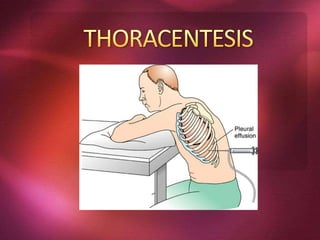
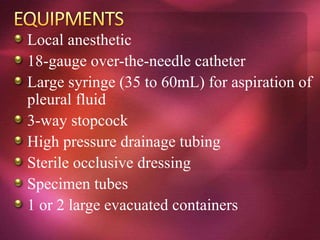
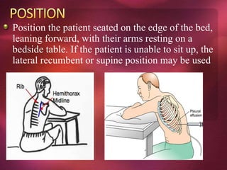
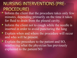
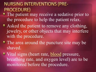
Define the Thoracentesis
Describe the indications for thoracentesis
Describe the contraindications for
thoracentesis
Describe the procedure for thoracentesis
Describe the complications of thoracentesis
and relevant management strategies
Thoracentesis (thor-a-sen-tee-sis) is a procedure
that is done to remove a sample of fluid from
around the lung.
The lung is covered with a tissue called the pleura.
The inside of the chest is also lined with pleura.
The space between these two areas is called the
pleural space.
This space normally contains just a thin layer of
fluid, however, some conditions such as
pneumonia, some types of cancer, or congestive
heart failure may cause excessive fluid to develop
(pleural effusion).
Thoracentesis, also known as pleural fluid
analysis, is a procedure in which a needle
is inserted through the back of the chest
wall into the pleural space (a space that
exists between the two lungs and the
anterior chest wall) to remove fluid or air.
Pleural fluid analysis is the microscopic
and chemical lab analysis of the fluid
obtained during thoracentesis.
Thoracentesis is a procedure in which the
chest wall is punctured for aspiration of
pleural fluid.
It is used to determine the etiology of
pleural fluid (diagnostic thoracentesis), to
relieve dyspnea caused by pleural fluid
(therapeutic thoracentesis), and,
occasionally, to perform pleurodesis.
Diagnostic: determination of pleural effusion
etiology (e.g. transudative versus exudative)
usually requires the removal of 50 to 100mL of
pleural fluid for laboratory studies. Most new
effusions require diagnostic thoracentesis, an
exception being a new effusion with a clear clinical
diagnosis (e.g. CHF) with no evidence for
superimposed pleural space infection
Therapeutic: reduce dyspnea and respiratory
compromise in patients with large pleural
effusions. This is typically achieved by removing a
much larger volume of fluid compared to the
diagnostic thoracentesis
Local infection over proposed site of thoracentesis
(e.g. cellulitis, herpes zoster). Select another entry
site
Uncontrolled bleeding
Coagulopathy is a relative contraindication;
some data suggest it is safe to perform
thoracentesis in patients with mild PTT elevations
(<1.5 times the upper limit of normal).
Caution must be exerted when performing
thoracentesis in mechanically ventilated
patients. The positive pressure of the
ventilator may expand the lung to greater
than normal volumes, increasing the
potential risk of pneumothorax.
Ultrasound-guided thoracentesis is
recommended in this situation
Defer thoracentesis in patients with severe
hemodynamic or respiratory compromise
until the underlying condition is stabilized
Thoracentesis may be performed for
diagnostic and/or therapeutic reasons.
The diagnostic use of a thoracentesis
involves pleural fluid analysis to
distinguish between exudate, which may
result from inflammatory or malignant
conditions, and transudate, which may
result from failure of organ systems that
affect fluid balance in the body. This
analysis aids in determining the cause of
the abnormality.
PLEURAL EFFUSION
INFECTION
TUMOR (Some types of tumors spread to the
lung or the pleura (the lining of the lung and
chest wall). This can cause fluid to build up in
the chest. A thoracentesis may be done to help
make a diagnosis. Fluid that is drained can be
examined in the laboratory to see if cancer cells
are present. This type of pleural effusion is
called a Malignant Pleural Effusion (MPE).
The most notable potential complication
after thoracentesis is the developmnent of a
pneumothorax.
1. Pain during placement
2. Bleeding—(During insertion of the needle,
a blood vessel in the skin or chest wall
may be accidentally nicked. Bleeding is
usually minor and stops on its own)
Air was aspirated from the pleural space
during the procedure
The patient develops chest pain,dyspnea,
or hypoxemia during or after the procedure
multiple needle insertions were required
Coughing
localized infection
Hemothorax and intraabdominal-organ injury.
Air embolism, and post-expansion pulmonary
edema. (Post-expansion pulmonary
edema is rare and can most likely be
avoided by limiting therapeutic
aspirations to less than 1500mL)
Pre-packaged kits are typically available for the
procedure. You should be familiar with the type
used at your particular institution.
Antiseptic solution
Sterile gauze
Sterile drape
Sterile gloves
Syringe and 22 and 25-gauge needles for
local anesthetic injection
Local anesthetic
18-gauge over-the-needle catheter
Large syringe (35 to 60mL) for aspiration of
pleural fluid
3-way stopcock
High pressure drainage tubing
Sterile occlusive dressing
Specimen tubes
1 or 2 large evacuated containers
Position the patient seated on the edge of the bed,
leaning forward, with their arms resting on a
bedside table. If the patient is unable to sit up, the
lateral recumbent or supine position may be used
IV access should be
established before the
procedure in most cases.
Atropine should be on
hand in case of profound
vasovagal response, and
supplemental oxygen
should be administered
throughout the procedure
as necessary.
Check the doctor’s order.
Identify the client.
Asked patient to sign a consent form that gives
your permission to do the test. Read the form
carefully and ask questions if something is not
clear.
Explain and emphasize the importance of the
procedure.
Inform that she will be experiencing mild pain
on the site where the needle was pricked
Inform the client that the procedure takes only few
minutes, depending primarily on the time it takes
for fluid to drain from the pleural cavity.
Inform the client not to cough while the needle is
inserted in order to avoid puncturing the lung
Explain when and where the procedure will occur
and who will be present.
Explain the procedure to the patient and SO,
reinforcing what the physician has previously
explained to the patient/SO
The patient may receive a sedative prior to
the procedure to help the patient relax.
Asked the patient to remove any clothing,
jewelry, or other objects that may interfere
with the procedure.
The area around the puncture site may be
shaved.
Vital signs (heart rate, blood pressure,
breathing rate, and oxygen level) are to be
monitored before the procedure.
Support the client verbally and describe the steps
of the procedure as needed.
Vital signs (heart rate, blood pressure, breathing
rate, and oxygen level) are to be monitored during
the procedure.
The patient may receive supplemental oxygen as
needed, through a face mask or nasal cannula
(tube).
Observe the client for signs of distress, such as
dyspnea, pallor, and coughing
Place the patient in a sitting position with arms
raised and resting on an overbed table.
The skin at the puncture site will be cleansed
with an antiseptic solution.
The patient will receive a local anesthetic at
the site where the thoracentesis is to be
performed.
Don’t remove more than 1000 ml of fluid
from the pleural cavity within first 30
minutes.
Place a small sterile dressing over the site of
the puncture.
Observe changes in the client’s cough, sputum,
respiratory depth, and breath sounds, and note
complaints of chest pain.
Position the client appropriately
Some agency protocols recommend that the client
lie on the unaffected side with the head of the bed
elevated 30 degrees for at least 30 minutes
because this position facilitates expansion of the
affected lung and eases respirations
Position the patient in a side-lying position with
the unaffected side down for an hour or longer.
Include date and time performed; the primary
care provider’s name; the amount, color, and
clarity of fluid drained; and nursing assessments
and interventions provided.
Transport the specimens to the laboratory.
The dressing over the puncture site will be
monitored for bleeding or other drainage.
Monitor patient’s blood pressure, pulse, and
breathing until are stable.
Document all relevant information.
The pleural fluid should be sent for the
following investigations if its etiology is
uncertain:
Cell count and differential
Protein level
Lactate Dehydrogenase level
pH
Cytology
Culture and sensitivities

Thoracentesis

  • 3.
    Define the Thoracentesis Describethe indications for thoracentesis Describe the contraindications for thoracentesis Describe the procedure for thoracentesis Describe the complications of thoracentesis and relevant management strategies
  • 4.
    Thoracentesis (thor-a-sen-tee-sis) isa procedure that is done to remove a sample of fluid from around the lung. The lung is covered with a tissue called the pleura. The inside of the chest is also lined with pleura. The space between these two areas is called the pleural space. This space normally contains just a thin layer of fluid, however, some conditions such as pneumonia, some types of cancer, or congestive heart failure may cause excessive fluid to develop (pleural effusion).
  • 5.
    Thoracentesis, also knownas pleural fluid analysis, is a procedure in which a needle is inserted through the back of the chest wall into the pleural space (a space that exists between the two lungs and the anterior chest wall) to remove fluid or air. Pleural fluid analysis is the microscopic and chemical lab analysis of the fluid obtained during thoracentesis.
  • 6.
    Thoracentesis is aprocedure in which the chest wall is punctured for aspiration of pleural fluid. It is used to determine the etiology of pleural fluid (diagnostic thoracentesis), to relieve dyspnea caused by pleural fluid (therapeutic thoracentesis), and, occasionally, to perform pleurodesis.
  • 7.
    Diagnostic: determination ofpleural effusion etiology (e.g. transudative versus exudative) usually requires the removal of 50 to 100mL of pleural fluid for laboratory studies. Most new effusions require diagnostic thoracentesis, an exception being a new effusion with a clear clinical diagnosis (e.g. CHF) with no evidence for superimposed pleural space infection Therapeutic: reduce dyspnea and respiratory compromise in patients with large pleural effusions. This is typically achieved by removing a much larger volume of fluid compared to the diagnostic thoracentesis
  • 8.
    Local infection overproposed site of thoracentesis (e.g. cellulitis, herpes zoster). Select another entry site Uncontrolled bleeding Coagulopathy is a relative contraindication; some data suggest it is safe to perform thoracentesis in patients with mild PTT elevations (<1.5 times the upper limit of normal).
  • 9.
    Caution must beexerted when performing thoracentesis in mechanically ventilated patients. The positive pressure of the ventilator may expand the lung to greater than normal volumes, increasing the potential risk of pneumothorax. Ultrasound-guided thoracentesis is recommended in this situation Defer thoracentesis in patients with severe hemodynamic or respiratory compromise until the underlying condition is stabilized
  • 10.
    Thoracentesis may beperformed for diagnostic and/or therapeutic reasons. The diagnostic use of a thoracentesis involves pleural fluid analysis to distinguish between exudate, which may result from inflammatory or malignant conditions, and transudate, which may result from failure of organ systems that affect fluid balance in the body. This analysis aids in determining the cause of the abnormality.
  • 11.
    PLEURAL EFFUSION INFECTION TUMOR (Sometypes of tumors spread to the lung or the pleura (the lining of the lung and chest wall). This can cause fluid to build up in the chest. A thoracentesis may be done to help make a diagnosis. Fluid that is drained can be examined in the laboratory to see if cancer cells are present. This type of pleural effusion is called a Malignant Pleural Effusion (MPE).
  • 12.
    The most notablepotential complication after thoracentesis is the developmnent of a pneumothorax. 1. Pain during placement 2. Bleeding—(During insertion of the needle, a blood vessel in the skin or chest wall may be accidentally nicked. Bleeding is usually minor and stops on its own)
  • 13.
    Air was aspiratedfrom the pleural space during the procedure The patient develops chest pain,dyspnea, or hypoxemia during or after the procedure multiple needle insertions were required
  • 14.
    Coughing localized infection Hemothorax andintraabdominal-organ injury. Air embolism, and post-expansion pulmonary edema. (Post-expansion pulmonary edema is rare and can most likely be avoided by limiting therapeutic aspirations to less than 1500mL)
  • 17.
    Pre-packaged kits aretypically available for the procedure. You should be familiar with the type used at your particular institution. Antiseptic solution Sterile gauze Sterile drape Sterile gloves Syringe and 22 and 25-gauge needles for local anesthetic injection
  • 18.
    Local anesthetic 18-gauge over-the-needlecatheter Large syringe (35 to 60mL) for aspiration of pleural fluid 3-way stopcock High pressure drainage tubing Sterile occlusive dressing Specimen tubes 1 or 2 large evacuated containers
  • 19.
    Position the patientseated on the edge of the bed, leaning forward, with their arms resting on a bedside table. If the patient is unable to sit up, the lateral recumbent or supine position may be used
  • 20.
    IV access shouldbe established before the procedure in most cases. Atropine should be on hand in case of profound vasovagal response, and supplemental oxygen should be administered throughout the procedure as necessary.
  • 21.
    Check the doctor’sorder. Identify the client. Asked patient to sign a consent form that gives your permission to do the test. Read the form carefully and ask questions if something is not clear. Explain and emphasize the importance of the procedure. Inform that she will be experiencing mild pain on the site where the needle was pricked
  • 22.
    Inform the clientthat the procedure takes only few minutes, depending primarily on the time it takes for fluid to drain from the pleural cavity. Inform the client not to cough while the needle is inserted in order to avoid puncturing the lung Explain when and where the procedure will occur and who will be present. Explain the procedure to the patient and SO, reinforcing what the physician has previously explained to the patient/SO
  • 23.
    The patient mayreceive a sedative prior to the procedure to help the patient relax. Asked the patient to remove any clothing, jewelry, or other objects that may interfere with the procedure. The area around the puncture site may be shaved. Vital signs (heart rate, blood pressure, breathing rate, and oxygen level) are to be monitored before the procedure.
  • 24.
    Support the clientverbally and describe the steps of the procedure as needed. Vital signs (heart rate, blood pressure, breathing rate, and oxygen level) are to be monitored during the procedure. The patient may receive supplemental oxygen as needed, through a face mask or nasal cannula (tube). Observe the client for signs of distress, such as dyspnea, pallor, and coughing Place the patient in a sitting position with arms raised and resting on an overbed table.
  • 25.
    The skin atthe puncture site will be cleansed with an antiseptic solution. The patient will receive a local anesthetic at the site where the thoracentesis is to be performed. Don’t remove more than 1000 ml of fluid from the pleural cavity within first 30 minutes. Place a small sterile dressing over the site of the puncture.
  • 26.
    Observe changes inthe client’s cough, sputum, respiratory depth, and breath sounds, and note complaints of chest pain. Position the client appropriately Some agency protocols recommend that the client lie on the unaffected side with the head of the bed elevated 30 degrees for at least 30 minutes because this position facilitates expansion of the affected lung and eases respirations Position the patient in a side-lying position with the unaffected side down for an hour or longer.
  • 27.
    Include date andtime performed; the primary care provider’s name; the amount, color, and clarity of fluid drained; and nursing assessments and interventions provided. Transport the specimens to the laboratory. The dressing over the puncture site will be monitored for bleeding or other drainage. Monitor patient’s blood pressure, pulse, and breathing until are stable. Document all relevant information.
  • 28.
    The pleural fluidshould be sent for the following investigations if its etiology is uncertain: Cell count and differential Protein level Lactate Dehydrogenase level pH Cytology Culture and sensitivities