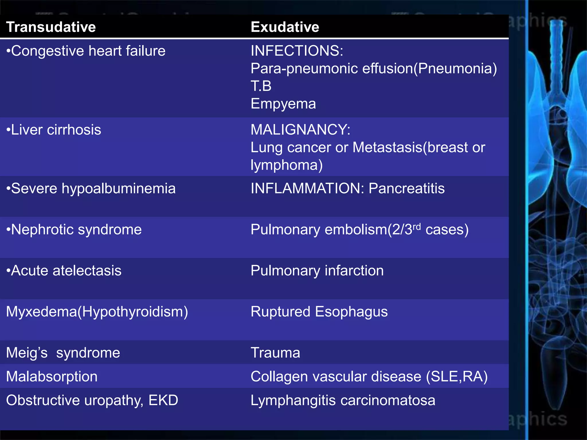
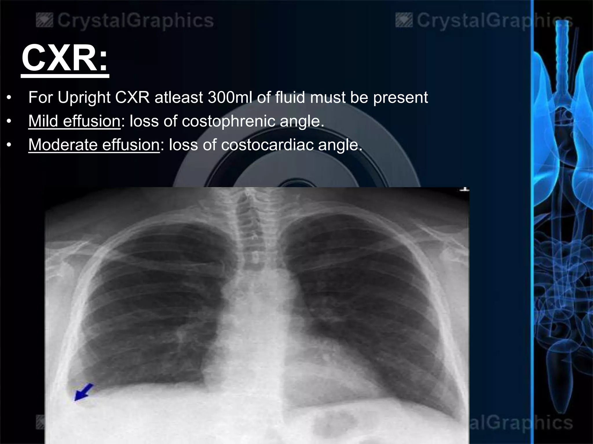
PLEURAL
EFFUSION
By
Ayman Bint Sultan(3rd yr)
Jinnah Sindh Medical Uni, Karachi
• Build-up of excess fluid between the
layers of the pleura outside the lungs.
NORMALLY,
• Lung are enclosed by pleura consisting
of two layers, the one enclosing lungs is
visceral pleura and other covering up
chest wall, is the parietal pleura.
• Pleural fluid is secreted by the parietal
layer of the pleura and reabsorbed by
the lymphatics in the most dependent
parts of the parietal pleura, primarily the
diaphragmatic and mediastinal regions.
• There is normally 5-15ml of fluid between the
pleura and more then 25 ml is termed as
pleural effusion, symptoms may not occur
untill there is accumlation of 300ml or
more.Normally this fluid serve for lubrication
while breathing in and out. Slippery due
to proteins-Albumin
• Types of Pleural Effusion:
• Exudative pleural effuisions occur when the pleura
is damaged,( Inflammed capillaries are leaky)(large
proteins-LDH, immune cells and fluid leaked) e.g., by
trauma, infection or malignancy which increases the
capillary permeability resulting in effusion, however,
Transudative pleural effusions develop when there
is either excessive production of pleural fluid
(inc.hydrostatic pressure) or the resorption capacity is
reduced (dec.oncotic pressure).
• Too little fluid drained by lymphatics may result in
lymphatic effusion. (Chylothorax.. Thoracic duct
disrupted)
TRANSUDATE EXUDATE
Main causes
↑ hydrostatic
pressure,
↓ colloid
osmotic pressure
Inflammation-
Increased vascular
permeability
Appearance Clear Cloudy
Specific gravity < 1.012 >1.020
Protein content < 2.5 g/dL > 2.9 g/dL
Fluid protein/
serum protein(Ratio)
< 0.5 > 0.5
SAAG = Serum
[albumin] - Effusion
[albumin]
> 1.2 g/dL < 1.2 g/dL
Fluid LDH
upper limit for serum
< 0.6 or < 2⁄3
0.6 or > 2⁄3
Cholesterol content < 45 mg/dL > 45 mg/dL
TO DIFFER B/W TRAN. & EXUDATIVE:
An exudate meets one or more of the following, while a transudate meets
none.
Transudative Exudative
•Congestive heart failure INFECTIONS:
Para-pneumonic effusion(Pneumonia)
T.B
Empyema
•Liver cirrhosis MALIGNANCY:
Lung cancer or Metastasis(breast or
lymphoma)
•Severe hypoalbuminemia INFLAMMATION: Pancreatitis
•Nephrotic syndrome Pulmonary embolism(2/3rd cases)
•Acute atelectasis Pulmonary infarction
Myxedema(Hypothyroidism) Ruptured Esophagus
Meig’s syndrome Trauma
Malabsorption Collagen vascular disease (SLE,RA)
Obstructive uropathy, EKD Lymphangitis carcinomatosa
Other fluids in pleural cavity:
• Other fluids can be classified as:
• Serous fluid (hydrothorax)
• Blood (Heamothorax)
• Chyle (chylothorax)
• Pus (pyothorax or empyema)
• Urine (urinothorax)
SYMPTOMS..
• Commom symptoms include:
• Pain on breathing=Dyspnea =Pleuritic
pain(When the pleura becomes inflamed,
the layers rub together, causing chest
pain. This is known as pleuritic pain.)
• Shortness of breath
• Cough
COMPLICATIONS:
• The potential complications associated with pleural
effusion are:
• Lung scarring,
• Pneumothorax (collapse of the lung) as a complication
of thoracentesis,
• Empyema (a collection of pus within the pleural
space), and
• Sepsis (blood infection) sometimes leading to death.
Diagnosis:
• History+examination+Labs
On Examination…
• Inspection; Dec. lung expansion and movement
• Palpitation: Shift of trachea or Mediastinum (apex beat) towards
healthy side.
• Decreased vocal fremitus and tactile fremitus.
• Percussion: ‘Stony Dull’ note
• Auscultation: In some cases , faint bronchial breathig may be
heard or findings associated with other lung infections i.e pleuritic
rub , crackles etc.
• Look for aspiration marks and signs of associated disease:
malignancy ( cachexia, clubbing, lymphadenopathy, radiation
marks, mastectomy scar); stigmata of chronic liver disease;
cardiac failure; hypothyroidism; rheumatoid arthritis ;butterfly rash
of SLE.
CXR:
• For Upright CXR atleast 300ml of fluid must be present
• Mild effusion: loss of costophrenic angle.
• Moderate effusion: loss of costocardiac angle.
• Severe effusion: seen as water-dense shadows with
concave upper borders.
• ‘Meniscus sign’
• Lateral decubitus will show shift of fluid, and are more
sensitive and can detect as little as 50ml of fluid.
• CT SCAN accurate for diagnosis,characterize presence,size and
characterstics of effusion.
• Ultrasound is useful in identifying the presence of pleural fluid and
in guiding diagnostic or therapeutic aspiration.
• Diagnostic aspiration; Thoracocentesis: Percuss the
upper border of the pleural effusion and choose a site 1 or 2 intercostal
spaces below it .
• Infiltrate down to the pleura with 5–10mL of 1% lidocaine. Attach a 21G
needle to a syringe and insert it just above the upper border of an
appropriate rib (avoids neurovascular bundle). Draw off 10–30mL of
pleural fluid and send it to the lab for clinical chemistry (protein, glucose,
pH, LDH, amylase), bacteriology (microscopy and culture, auramine
stain, TB culture), cytology, and, if indicated, immunology (rheumatoid
factor, ANA, complement).
• If pleural fluid analysis is inconclusive, consider parietal pleural biopsy.
• Should be done in all patients who have pleural fluid at 10 mm in
thickness on CT.
• Thoracoscopic or CT-guided pleural biopsy increases
diagnostic yield (by enabling direct visualization of the pleural cavity and
biopsy of suspicious areas)
Pleural Fluid Analysis;
MANAGEMENT
• Treat the underlying cause.
• • Drainage: If the effusion is symptomatic, drain it, repeatedly if
necessary . Fluid is best removed slowly (0.5–1.5L/24h). It may be
aspirated in the same way as a diagnostic tap, or using an intercoastal
drain.
• • Pleurodesis with (talc, bleomycin or tetracyclin) may be helpful for
recurrent effusions. It’s a process in which two pleural surfaces are
scarred with each other so no fluid can accumulate in
between.Thorascopic mechanical pleurodesis is most effective for
malignant effusions.
• MEDICATION: The use of medications for pleural effusions depends on
the underlying cause. Antibiotics are used when there is an infectious
cause, whereby diuretics such as furosemide (Lasix) may be used to
slowly help reduce the size of the pleural effusion.
• • Surgery: Persistent collections and increasing pleural thickness (on
ultrasound) requires surgery.
Pleural effusion
Pleural effusion

Pleural effusion

  • 1.
    PLEURAL EFFUSION By Ayman Bint Sultan(3rdyr) Jinnah Sindh Medical Uni, Karachi
  • 2.
    • Build-up ofexcess fluid between the layers of the pleura outside the lungs.
  • 3.
    NORMALLY, • Lung areenclosed by pleura consisting of two layers, the one enclosing lungs is visceral pleura and other covering up chest wall, is the parietal pleura. • Pleural fluid is secreted by the parietal layer of the pleura and reabsorbed by the lymphatics in the most dependent parts of the parietal pleura, primarily the diaphragmatic and mediastinal regions.
  • 4.
    • There isnormally 5-15ml of fluid between the pleura and more then 25 ml is termed as pleural effusion, symptoms may not occur untill there is accumlation of 300ml or more.Normally this fluid serve for lubrication while breathing in and out. Slippery due to proteins-Albumin
  • 5.
    • Types ofPleural Effusion: • Exudative pleural effuisions occur when the pleura is damaged,( Inflammed capillaries are leaky)(large proteins-LDH, immune cells and fluid leaked) e.g., by trauma, infection or malignancy which increases the capillary permeability resulting in effusion, however, Transudative pleural effusions develop when there is either excessive production of pleural fluid (inc.hydrostatic pressure) or the resorption capacity is reduced (dec.oncotic pressure). • Too little fluid drained by lymphatics may result in lymphatic effusion. (Chylothorax.. Thoracic duct disrupted)
  • 6.
    TRANSUDATE EXUDATE Main causes ↑hydrostatic pressure, ↓ colloid osmotic pressure Inflammation- Increased vascular permeability Appearance Clear Cloudy Specific gravity < 1.012 >1.020 Protein content < 2.5 g/dL > 2.9 g/dL Fluid protein/ serum protein(Ratio) < 0.5 > 0.5 SAAG = Serum [albumin] - Effusion [albumin] > 1.2 g/dL < 1.2 g/dL Fluid LDH upper limit for serum < 0.6 or < 2⁄3 0.6 or > 2⁄3 Cholesterol content < 45 mg/dL > 45 mg/dL
  • 7.
    TO DIFFER B/WTRAN. & EXUDATIVE: An exudate meets one or more of the following, while a transudate meets none.
  • 8.
    Transudative Exudative •Congestive heartfailure INFECTIONS: Para-pneumonic effusion(Pneumonia) T.B Empyema •Liver cirrhosis MALIGNANCY: Lung cancer or Metastasis(breast or lymphoma) •Severe hypoalbuminemia INFLAMMATION: Pancreatitis •Nephrotic syndrome Pulmonary embolism(2/3rd cases) •Acute atelectasis Pulmonary infarction Myxedema(Hypothyroidism) Ruptured Esophagus Meig’s syndrome Trauma Malabsorption Collagen vascular disease (SLE,RA) Obstructive uropathy, EKD Lymphangitis carcinomatosa
  • 10.
    Other fluids inpleural cavity: • Other fluids can be classified as: • Serous fluid (hydrothorax) • Blood (Heamothorax) • Chyle (chylothorax) • Pus (pyothorax or empyema) • Urine (urinothorax)
  • 11.
    SYMPTOMS.. • Commom symptomsinclude: • Pain on breathing=Dyspnea =Pleuritic pain(When the pleura becomes inflamed, the layers rub together, causing chest pain. This is known as pleuritic pain.) • Shortness of breath • Cough
  • 12.
    COMPLICATIONS: • The potentialcomplications associated with pleural effusion are: • Lung scarring, • Pneumothorax (collapse of the lung) as a complication of thoracentesis, • Empyema (a collection of pus within the pleural space), and • Sepsis (blood infection) sometimes leading to death.
  • 13.
  • 15.
    On Examination… • Inspection;Dec. lung expansion and movement • Palpitation: Shift of trachea or Mediastinum (apex beat) towards healthy side. • Decreased vocal fremitus and tactile fremitus. • Percussion: ‘Stony Dull’ note • Auscultation: In some cases , faint bronchial breathig may be heard or findings associated with other lung infections i.e pleuritic rub , crackles etc. • Look for aspiration marks and signs of associated disease: malignancy ( cachexia, clubbing, lymphadenopathy, radiation marks, mastectomy scar); stigmata of chronic liver disease; cardiac failure; hypothyroidism; rheumatoid arthritis ;butterfly rash of SLE.
  • 16.
    CXR: • For UprightCXR atleast 300ml of fluid must be present • Mild effusion: loss of costophrenic angle. • Moderate effusion: loss of costocardiac angle.
  • 17.
    • Severe effusion:seen as water-dense shadows with concave upper borders. • ‘Meniscus sign’ • Lateral decubitus will show shift of fluid, and are more sensitive and can detect as little as 50ml of fluid.
  • 18.
    • CT SCANaccurate for diagnosis,characterize presence,size and characterstics of effusion. • Ultrasound is useful in identifying the presence of pleural fluid and in guiding diagnostic or therapeutic aspiration. • Diagnostic aspiration; Thoracocentesis: Percuss the upper border of the pleural effusion and choose a site 1 or 2 intercostal spaces below it . • Infiltrate down to the pleura with 5–10mL of 1% lidocaine. Attach a 21G needle to a syringe and insert it just above the upper border of an appropriate rib (avoids neurovascular bundle). Draw off 10–30mL of pleural fluid and send it to the lab for clinical chemistry (protein, glucose, pH, LDH, amylase), bacteriology (microscopy and culture, auramine stain, TB culture), cytology, and, if indicated, immunology (rheumatoid factor, ANA, complement). • If pleural fluid analysis is inconclusive, consider parietal pleural biopsy. • Should be done in all patients who have pleural fluid at 10 mm in thickness on CT. • Thoracoscopic or CT-guided pleural biopsy increases diagnostic yield (by enabling direct visualization of the pleural cavity and biopsy of suspicious areas)
  • 19.
  • 21.
    MANAGEMENT • Treat theunderlying cause. • • Drainage: If the effusion is symptomatic, drain it, repeatedly if necessary . Fluid is best removed slowly (0.5–1.5L/24h). It may be aspirated in the same way as a diagnostic tap, or using an intercoastal drain. • • Pleurodesis with (talc, bleomycin or tetracyclin) may be helpful for recurrent effusions. It’s a process in which two pleural surfaces are scarred with each other so no fluid can accumulate in between.Thorascopic mechanical pleurodesis is most effective for malignant effusions. • MEDICATION: The use of medications for pleural effusions depends on the underlying cause. Antibiotics are used when there is an infectious cause, whereby diuretics such as furosemide (Lasix) may be used to slowly help reduce the size of the pleural effusion. • • Surgery: Persistent collections and increasing pleural thickness (on ultrasound) requires surgery.