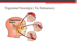
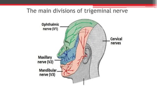
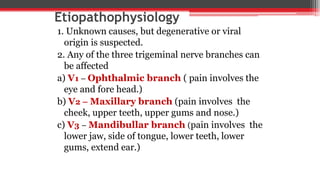
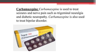
CRANIAL NERVE DISORDERS & ITS
MANAGEMNET
Types of cranial nerve disorders
1. Bell’s palsy
2. Trigeminal Neuralgia ( Tic Douloreux)
3. Cranial & spinal neuropathies
BELL’S PALSY ( Facial plasy)
• Bell’s palsy (facial paralysis) is due to unilateral inflammation of
the ( CN VII Facial nerve) seventh cranial nerve, which results in
weakness or paralysis of the facial muscles on the affected side.
• That most often occurs unilaterally.
• Generally self-limiting. With or without treatment, most clients
improve significantly within 2 weeks and about 80% recover
completely within 3 months.
• in very rare cases the symptoms may never completely resolve or
may recur.
Etiological factors
1. Causes is unknown.
2. Although possible causes may include vascular ischemia, viral
disease (herpes simplex, herpes zoster), autoimmune disease, or a
combination of all of these factors.
3. HIV infection
4. Lyme disease
5. Middle ear infection and Sarcoidosis
Clinical manifestations
1. Acute onset of unilateral upper and lower facial
paralysis ( over a 48 hours period).
2. Paralysis of ipsilateral side of face from vertex
of scalp to chin.
3. Facial muscle weak throughout forehead,
check, and chin, can affect speech and taste,
distort face, decreasing tearing and cause
posterior auricular pain.
4. Inability to close eye and painful eye sensation.
5. Photophobia.
6. Hyperacusis on the affected side.
Clinical manifestations
Diagnostic evaluations
1. Bells palsy can be diagnosed just by taking a
health history and doing a complete physical
examination.
2. History to determine previous illness, onset of
paralysis and associated symptoms.
3. Exclusion of lesions that mimic Bell’s palsy, such
as tumor, infection ( Lyme disease, Meningitis)
trauma,, stroke or other conditions.
4. Neurological examination
5. CT-scan and EMG
Medical management
• The objectives of treatment are to maintain the
muscle tone of the face and to prevent or
minimize denervation.
• The patient should be reassured that no stroke
has occurred and that spontaneous recovery
occurs within 3 to 5 weeks in most patients.
Medical management
1. Corticosteroids therapy may be started
early to decrease inflammation ( e.g.
Prednisone 1 mg/kg/day for 10 to 14 days.
2. when using Corticosteroids therapy for
the treatment of bell’s palsy, take cautions
should be used client with TB, peptic ulcer, DM,
renal & hepatic dysfunction or malignant
hypertension.
3. Eye care is essential maintain lubrication and moisture if unable
to close the eye. May be need to be patched during sleeping.
4. Physical therapy , electrical stimulation to maintain muscle tone.
5. Biofeed back as adjunct therapy
6. Mild analgesics to relieve pain
Complications
1. Corneal ulcerations
2. Impairment of vision
3. Psychosocial adjustment to prolonged paralysis
Trigeminal Neuralgia ( Tic Duloureux)
Introduction
• The trigeminal nerve which is divided into three
branches, is responsible for chewing, for
producing saliva and tears, and for sending
facial sensations to the brain.
• When this nerve breaks down for some reason,
it can trigger brief but agonizing sizzles of pain
on one side of the face.
• This condition is unusual in those under age 50
and more often occurs after 70.
Trigeminal Neuralgia ( Tic Duloureux)
• Trigeminal Neuralgia is a disorder of the
trigeminal nerve (the fifth cranial nerve) that
causes episodes of sharp, stabbing pain in the
cheek, lips, gums, or chin on one side of the face.
Or
• Trigeminal Neuralgia ( Tic Duloureux) is
an intensely painful neurologic condition
that affects one or more branches of the
fifth cranial nerve.
The main divisions of trigeminal nerve
Etiopathophysiology
1. Unknown causes, but degenerative or viral
origin is suspected.
2. Any of the three trigeminal nerve branches can
be affected
a) V1 – Ophthalmic branch ( pain involves the
eye and fore head.)
b) V2 – Maxillary branch (pain involves the
cheek, upper teeth, upper gums and nose.)
c) V3 – Mandibullar branch (pain involves the
lower jaw, side of tongue, lower teeth, lower
gums, extend ear.)
• The main cause is damage to nerve leading to
demyelination of nerve leading to stabbing,
severe, shock like pain of neuralgia results.
FACTORS CAUSING DAMAGE ARE-
1. Old age
2.Infection
3.Multiple sclerosis
4. Pressure on nerves
5. Diabetes
Clinical manifestations
1. Sudden severe episodes of intense facial
pain localized to one or more branches of the
TN nerve lasting less than 30 to 6o seconds.
2. pain may occur spontaneously or be
precipitated by activation of trigger points, such
as touching the face, talking, chewing, and
brushing of the teeth.
3. Pain always unilateral & does not cross
midline.
4. Some clients will experience numbness esp.
around the mouth.
Diagnostic evaluations
1. History of characteristic symptoms
and pattern.
2. Neurologic & cranial nerve
examination.
3. CT –scan & MRI
Medical Management
PHARMACOLOGIC
1. Use of carbamazepine is first and most
effective medication used to treat the condition.
2. Other drugs such as:
a) Imipramine
b) Phenytoin
c) Diavlproex
d) Gabapentin and othe AED may be used
Carbamazepine Carbamazepine is used to treat
seizures and nerve pain such as trigeminal neuralgia
and diabetic neuropathy. Carbamazepine is also used
to treat bipolar disorder.
• Side effects include nausea, dizziness
drowsiness, and aplastic anemia. The patient is
monitored for bone marrow depression during
long-term therapy.
SURGICAL MANAGEMENT
• When these methods fail to relieve pain, a number
of surgical options are available. The choice of
procedure depends on the patient’s preference and
health status.
Microvascular Decompression of
the Trigeminal Nerve
• An intracranial approach can be used to
decompress the trigeminal nerve.
• This procedure relieves facial pain while
preserving normal sensation, but it is a major
procedure, involving a craniotomy.
• The postoperative management is the same as
for other intracranial surgeries.
Percutaneous Radiofrequency Trigeminal
Gangliolysis.
• It directs low- voltage stimulation of nerve by electrode inserted
through foramen ovale.
• Under local anesthesia, the needle is introduced through the
cheek on the affected side. Under fluoroscopic guidance, the
needle electrode is guided through the foramen magnum into the
gasserian ganglion.
Complications
1. Anorexia and weight loss
2. Dehydration
3. Anxiety and fear
4. Depression, social isolation and suicidal
ideations in extreme cases.
Nursing Management
1. Take history of the pain, including duration,
severity, and aggravating factors.
2. Assess the nutritional status.
3. Assess for anxiety and depression, including
problems with sleep, social interaction etc.
Nursing Management
• Preventing pain
• Providing postoperative care : postoperative
neurologic assessments are conducted to
evaluate the patient for facial motor and sensory
deficits in each of the three branches of the
trigeminal nerve.
• If the surgery results in sensory deficits to the
affected side of the face, the patient is instructed
not to rub the eye, because pain will not be felt if
there is injury.

Cranial nerve disorders

  • 1.
    CRANIAL NERVE DISORDERS& ITS MANAGEMNET
  • 2.
    Types of cranialnerve disorders 1. Bell’s palsy 2. Trigeminal Neuralgia ( Tic Douloreux) 3. Cranial & spinal neuropathies
  • 3.
    BELL’S PALSY (Facial plasy) • Bell’s palsy (facial paralysis) is due to unilateral inflammation of the ( CN VII Facial nerve) seventh cranial nerve, which results in weakness or paralysis of the facial muscles on the affected side.
  • 4.
    • That mostoften occurs unilaterally. • Generally self-limiting. With or without treatment, most clients improve significantly within 2 weeks and about 80% recover completely within 3 months. • in very rare cases the symptoms may never completely resolve or may recur.
  • 5.
    Etiological factors 1. Causesis unknown. 2. Although possible causes may include vascular ischemia, viral disease (herpes simplex, herpes zoster), autoimmune disease, or a combination of all of these factors. 3. HIV infection 4. Lyme disease 5. Middle ear infection and Sarcoidosis
  • 6.
    Clinical manifestations 1. Acuteonset of unilateral upper and lower facial paralysis ( over a 48 hours period). 2. Paralysis of ipsilateral side of face from vertex of scalp to chin. 3. Facial muscle weak throughout forehead, check, and chin, can affect speech and taste, distort face, decreasing tearing and cause posterior auricular pain. 4. Inability to close eye and painful eye sensation. 5. Photophobia. 6. Hyperacusis on the affected side.
  • 7.
  • 8.
  • 9.
    Diagnostic evaluations 1. Bellspalsy can be diagnosed just by taking a health history and doing a complete physical examination. 2. History to determine previous illness, onset of paralysis and associated symptoms. 3. Exclusion of lesions that mimic Bell’s palsy, such as tumor, infection ( Lyme disease, Meningitis) trauma,, stroke or other conditions. 4. Neurological examination 5. CT-scan and EMG
  • 10.
    Medical management • Theobjectives of treatment are to maintain the muscle tone of the face and to prevent or minimize denervation. • The patient should be reassured that no stroke has occurred and that spontaneous recovery occurs within 3 to 5 weeks in most patients.
  • 11.
    Medical management 1. Corticosteroidstherapy may be started early to decrease inflammation ( e.g. Prednisone 1 mg/kg/day for 10 to 14 days. 2. when using Corticosteroids therapy for the treatment of bell’s palsy, take cautions should be used client with TB, peptic ulcer, DM, renal & hepatic dysfunction or malignant hypertension.
  • 12.
    3. Eye careis essential maintain lubrication and moisture if unable to close the eye. May be need to be patched during sleeping. 4. Physical therapy , electrical stimulation to maintain muscle tone. 5. Biofeed back as adjunct therapy 6. Mild analgesics to relieve pain
  • 13.
    Complications 1. Corneal ulcerations 2.Impairment of vision 3. Psychosocial adjustment to prolonged paralysis
  • 14.
    Trigeminal Neuralgia (Tic Duloureux)
  • 15.
    Introduction • The trigeminalnerve which is divided into three branches, is responsible for chewing, for producing saliva and tears, and for sending facial sensations to the brain. • When this nerve breaks down for some reason, it can trigger brief but agonizing sizzles of pain on one side of the face. • This condition is unusual in those under age 50 and more often occurs after 70.
  • 16.
    Trigeminal Neuralgia (Tic Duloureux) • Trigeminal Neuralgia is a disorder of the trigeminal nerve (the fifth cranial nerve) that causes episodes of sharp, stabbing pain in the cheek, lips, gums, or chin on one side of the face. Or • Trigeminal Neuralgia ( Tic Duloureux) is an intensely painful neurologic condition that affects one or more branches of the fifth cranial nerve.
  • 17.
    The main divisionsof trigeminal nerve
  • 18.
    Etiopathophysiology 1. Unknown causes,but degenerative or viral origin is suspected. 2. Any of the three trigeminal nerve branches can be affected a) V1 – Ophthalmic branch ( pain involves the eye and fore head.) b) V2 – Maxillary branch (pain involves the cheek, upper teeth, upper gums and nose.) c) V3 – Mandibullar branch (pain involves the lower jaw, side of tongue, lower teeth, lower gums, extend ear.)
  • 19.
    • The maincause is damage to nerve leading to demyelination of nerve leading to stabbing, severe, shock like pain of neuralgia results. FACTORS CAUSING DAMAGE ARE- 1. Old age 2.Infection 3.Multiple sclerosis 4. Pressure on nerves 5. Diabetes
  • 20.
    Clinical manifestations 1. Suddensevere episodes of intense facial pain localized to one or more branches of the TN nerve lasting less than 30 to 6o seconds. 2. pain may occur spontaneously or be precipitated by activation of trigger points, such as touching the face, talking, chewing, and brushing of the teeth. 3. Pain always unilateral & does not cross midline. 4. Some clients will experience numbness esp. around the mouth.
  • 21.
    Diagnostic evaluations 1. Historyof characteristic symptoms and pattern. 2. Neurologic & cranial nerve examination. 3. CT –scan & MRI
  • 22.
    Medical Management PHARMACOLOGIC 1. Useof carbamazepine is first and most effective medication used to treat the condition. 2. Other drugs such as: a) Imipramine b) Phenytoin c) Diavlproex d) Gabapentin and othe AED may be used
  • 23.
    Carbamazepine Carbamazepine isused to treat seizures and nerve pain such as trigeminal neuralgia and diabetic neuropathy. Carbamazepine is also used to treat bipolar disorder.
  • 24.
    • Side effectsinclude nausea, dizziness drowsiness, and aplastic anemia. The patient is monitored for bone marrow depression during long-term therapy.
  • 25.
    SURGICAL MANAGEMENT • Whenthese methods fail to relieve pain, a number of surgical options are available. The choice of procedure depends on the patient’s preference and health status.
  • 26.
    Microvascular Decompression of theTrigeminal Nerve • An intracranial approach can be used to decompress the trigeminal nerve. • This procedure relieves facial pain while preserving normal sensation, but it is a major procedure, involving a craniotomy. • The postoperative management is the same as for other intracranial surgeries.
  • 27.
    Percutaneous Radiofrequency Trigeminal Gangliolysis. •It directs low- voltage stimulation of nerve by electrode inserted through foramen ovale. • Under local anesthesia, the needle is introduced through the cheek on the affected side. Under fluoroscopic guidance, the needle electrode is guided through the foramen magnum into the gasserian ganglion.
  • 28.
    Complications 1. Anorexia andweight loss 2. Dehydration 3. Anxiety and fear 4. Depression, social isolation and suicidal ideations in extreme cases.
  • 29.
    Nursing Management 1. Takehistory of the pain, including duration, severity, and aggravating factors. 2. Assess the nutritional status. 3. Assess for anxiety and depression, including problems with sleep, social interaction etc.
  • 30.
    Nursing Management • Preventingpain • Providing postoperative care : postoperative neurologic assessments are conducted to evaluate the patient for facial motor and sensory deficits in each of the three branches of the trigeminal nerve. • If the surgery results in sensory deficits to the affected side of the face, the patient is instructed not to rub the eye, because pain will not be felt if there is injury.