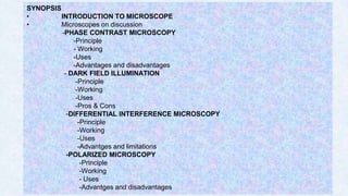
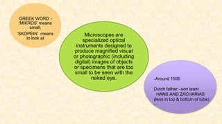
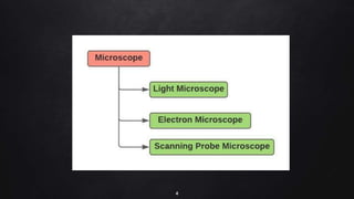
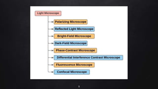
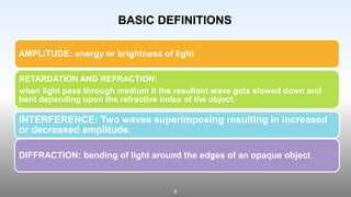
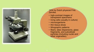
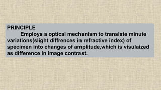
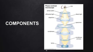
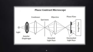
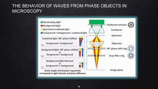
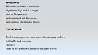
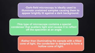
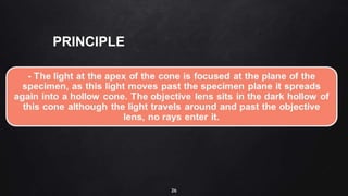
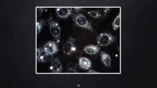
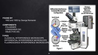
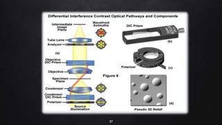
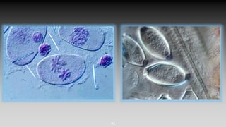
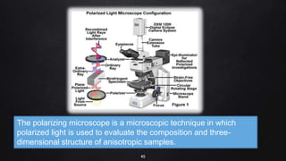
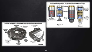
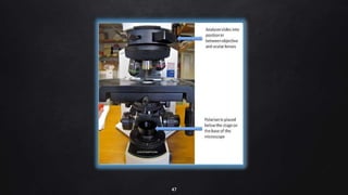
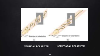
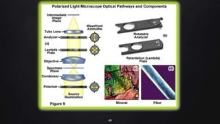
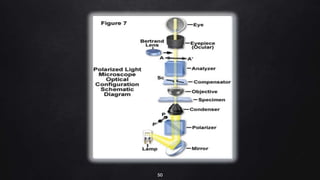
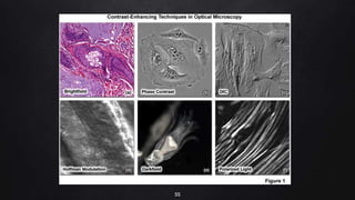
2. 2
SYNOPSIS
• INTRODUCTION TO MICROSCOPE
• Microscopes on discussion
-PHASE CONTRAST MICROSCOPY
-Principle
- Working
-Uses
-Advantages and disadvantages
- DARK FIELD ILLUMINATION
-Principle
-Working
-Uses
-Pros & Cons
-DIFFERENTIAL INTERFERENCE MICROSCOPY
-Principle
-Working
-Uses
-Advantges and limitations
-POLARIZED MICROSCOPY
-Principle
-Working
- Uses
-Advantges and disadvantages
3. 3
Microscopes are
specialized optical
instruments designed to
produce magnified visual
or photographic (including
digital) images of objects
or specimens that are too
small to be seen with the
naked eye.
GREEK WORD –
‘MIKROS’ means
small,
’SKOPEIN’ means
to look at
-Around 1595
Dutch father –son team
HANS AND ZACHARIAS
(lens in top & bottom of tube)
-
6. 6
AMPLITUDE: energy or brightness of light
RETARDATION AND REFRACTION:
when light pass through medium it the resultant wave gets slowed down and
bent depending upon the refractive index of the object.
INTERFERENCE: Two waves superimposing resulting in increased
or decreased amplitude.
DIFFRACTION: bending of light around the edges of an opaque object.
BASIC DEFINITIONS
8. .
8
1934 by Dutch physicist Frits
Zernike.
• high-contrast images of
transparent specimens
• living cells (usually in culture)
• microorganisms
• thin tissue slices
• lithographic patterns
• fibers, latex dispersions, glass
fragments, and subcellular
particles (including nuclei and
other organelles).
9. 9
PRINCIPLE
Employs a optical mechanism to translate minute
variations(slight diffrences in refractive index) of
specimen into changes of amplitude,which is visulaized
as difference in image contrast.
12. 12
THE ANNULAR DIAPHRAGM
- situated below the condenser.
-made up of a circular disc having a circular annular groove.
-The light rays are allowed to pass through the annular groove.
-Through the annular groove of the annular diaphragm, the light rays fall on the specimen or object
to be studied.
-At the back focal plane of the objective develops an image.
-The annular phase plate is placed at this back focal plane.
-The direct light rays pass through the annular groove whereas the diffracted light rays pass
through the region outside the groove.
THE PHASE PLATE
-The phase plate is a transparent disc.
-It is either a negative phase plate having a thick circular area or a positive phase plate
having a thin circular groove.
-This thick or thin area in the phase plate is called the conjugate area.
-Depending upon the different refractive indices of different cell components, the object to
be studied shows a different degree of contrast in this microscope
18. 18
To produce high-contrast images of
transparent specimens,
-Living cells (usually in culture),
-Microorganisms
-Thin tissue slices
-Lithographic patterns
-Fibers
-Latex dispersions
-Glass fragments
-Subcellular particles .
-Living cells can be observed in their
natural state without previous fixation.
-It makes a highly transparent object
more visible.
USES
20. 20
A quantitative phase-contrast microscopy image of cells in culture. The height
and color of an image point correspond to the optical thickness, which only
depends on the object's thickness and the relative refractive index. The
volume of an object can thus be determined when the difference in refractive
index between the object and the surrounding media is known.
24. Found by J J Lister
Standard bright field condenser is
replaced with a single or double-reflecting
dark field condenser.
25. -Dark-field microscopy is ideally used to
illuminate unstained samples causing them to
appear brightly lit against a dark background.
-This type of microscope contains a special
condenser that scatters light and causes it to reflect
off the specimen at an angle.
-Rather than illuminating the sample with a filled
cone of light, the condenser is designed to form a
hollow cone of light.
28. 28
WORKING
-A dark field microscope is arranged so that the light source is blocked off, causing light to
scatter as it hits the specimen.
-This is ideal for making objects with refractive values similar to the background appear bright
against a dark background.
-When light hits an object, rays are scattered in all azimuths or directions. The design of the
dark field microscope is such that it removes the dispersed light,so that only the scattered
beams hit the sample.
-The introduction of a condenser and/or stop below the stage ensures that these light rays will
hit the specimen at different angles, rather than as a direct light source above/below the object.
-The result is a “cone of light” where rays are diffracted, reflected and/or refracted off the object,
ultimately, allowing the individual to view a specimen in dark field.
31. 31
USES
-Dark field illumination is most readily set up at low magnifications (up to 100x), although it can be used with any dry objective
lens.
-Initial examination of suspensions of cells such as yeast, bacteria, small protists, or cell and tissue fractions including cheek
epithelial cells, chloroplasts, mitochondria.
-Initial survey and observation at low powers of pond water samples, hay or soil infusions, purchased protist or metazoan
cultures.
-Examination of lightly stained prepared slides.
-Initial location of any specimen of very small size for later viewing at higher power.
-Determination of motility in cultures.
LIMITATIONS
-The main limitation of dark-field microscopy is the low light levels seen in the final image.
-The sample must be very strongly illuminated, which can cause damage to the sample
35. 35
FOUND BY
1952 and 1955 by George Nomarski
COMPONENTS
POLARISER
CONDENSER DIC
OBJECTIVE DIC
TYPES
CLASSICAL INTERFERENCE MICROSCOPE
DIFFERENTIAL INTERFERENCE MICROSCOPE
FLUORESCENCE INTERFERENCE MICROSCOPE
36. 36
PRINCIPLE
- Derives the contrast from difference in refractive index of the
specimen.
-Does not rely on diffraction by the object for interference,generates
mutual interfering beams which produces the contrast.
-Single ray of light splits one traversing and other missing the
specimen but interacting with the background,then it is confined at the
image plane with wave interference.
41. 41
USES:
-Live unstained sections are visible
-Smear, individual cell, water-borne single celled organism
-High resolution and clarity
-Lacks halo effect
-Imaging the fibrous structures of nerve and muscle
-Imaging mitotic spindles.
-Imaging cellular nucleic structure
42. 42
LIMITATIONS
-Thick samples and non biological samples cannot be viewed
-Requires transparent samples of same refractive index.
ADVANTAGES:
- Adjoining regions does not obscure the images of the smaller specimen,so
that even the minute appendages are known .
-Moving organelles inside the cells can be viewed clearly.
45. 45
The polarizing microscope is a microscopic technique in which
polarized light is used to evaluate the composition and three-
dimensional structure of anisotropic samples.
51. 51
USES
-This technique is to highlight the features of various substances such as
crystals, fibers, and minerals.
-When both filters are aligned in the light path, they are at right angles to each other. When adjusted,
however, light is able to pass through the filters at different angles, allowing you to see different
aspects of the specimen.
-Light will either travel through an isotropic specimen, making it appear dark, or be reflected
by an anisotropic specimen, making it appear bright in contrast.
-Some birefringent materials that are commonly evaluated by polarising light microscopy include:
Bone
Teeth
Striated
muscle
Urine
crystals
Gout
crystals
Amyloid
52. 52
BIREFRINGENCE
-Birefringence is the optical property of a
material having a refractive index that depends
on the polarization and propagation direction of
light.
- These optically anisotropic materials are
said to be birefringent.
- Made of mica or layers of cellotape.
53. 53
TYPES
-Intrinsic
Type of anisotropy due to assymetrical alignment of chemical bonds,ions and
molecules (eg:collagen,muscle fibres)
-Strain B
when a dielectric substance is subjected to mechanical stress,their patterns
are distorted and gives rise to birefringence.
(eg:Electric tissue fibres under stress)
-Positive B
if the ordinary ray is parallel to the length of the crystal ,its positive
birefringence.
-Negative B
if the ordinary ray is perpendicular to the long axis ,its negative
birefringence.
54. 54
APPLICATIONS
-To know the crystalline stucture of enamel,dentin and cementum
-To study the remineralization and demineralization
-To know the cytoskeleton of organisms
-In urine analysis,to view the crystals with the presence of any
pathology.
-Used to study the elastic fibres,collagen fibres.
-Diagnosis of Amyloidosis-APPLE GREEN BIREFRINGENCE.