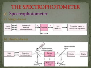
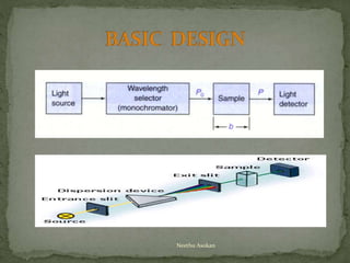
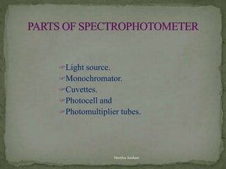
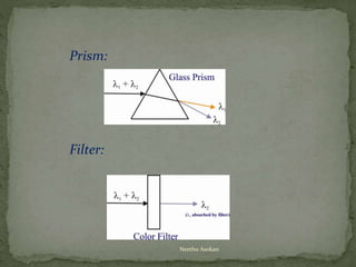
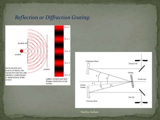
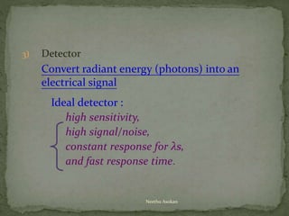
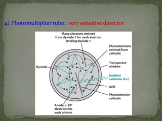
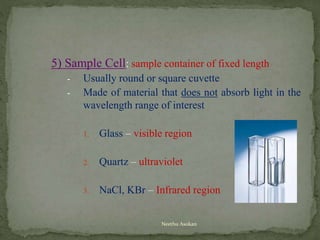
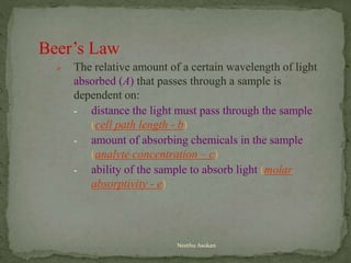
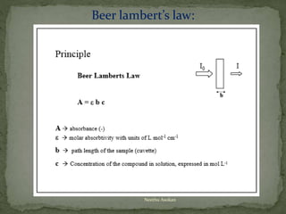
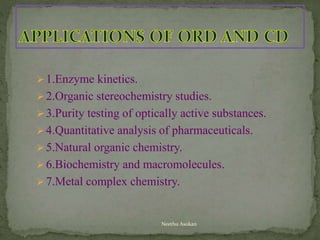
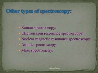
BY,
NEETHU ASOKAN
Neethu Asokan
FUNDAMENTALS OF SPECTROPHOTOMETRY
1.) Colorimetry
 An analytical technique in which the concentration of an analyte
is measured by its ability to produce or change the color of a
solution.
- Changes in the solution’s ability to absorb light.
2.) Spectrophotometry
 Any technique that uses light to measure chemical concentrations.
 A colorimetric method where an instrument is used to determine the
amount of analyte in a sample by the sample’s ability or inability to
absorb light at a certain wavelength.
Colorimetry
Instrumental Methods
(spectrophotometry)
Non-Instrumental Methods
Neethu Asokan
• History began with Issac Newton’s optic
experiments; later developed by Joseph von
Fraunhofer.
• Spectrophotometric techniques are used to
measure the concentration of solutes in solution by
measuring the amount of light that is absorbed by
the solution in a cuvette placed in the
spectrophotometer.
Neethu Asokan
 The spectrophotometer can measure the amount of light or
electromagnetic radiation (of certain frequency) transmitted or absorbed by
the solution.
 The study of how the chemical compound interacts with different
wavelengths in a given region of electromagnetic radiation is called
spectrochemical analysis.
 Spectroradiometers, which operate almost like the visible region
spectrophotometers, are designed to measure the spectral density of
illuminants. Applications may include evaluation and categorization of
lighting for sales by the manufacturer, or for the customers to confirm
the lamp they decided to purchase is within their specifications.
Components:
 The light source shines onto or through the sample.
 The sample transmits or reflects light.
 The detector detects how much light was reflected from or transmitted
through the sample.
 The detector then converts how much light the sample transmitted or
reflected into a number.
Neethu Asokan
Neethu Asokan
1) Spectrophotometer
a) Single-beam
b) Double-beam
Neethu Asokan
Light source.
Monochromator.
Cuvettes.
Photocell and
Photomultiplier tubes.
Neethu Asokan
Ultraviolet (UV) Spectrophotometers.
 Uses ultraviolet light of wave lengths from
200 nm to 350 nm.
Visible (VIS) Light Spectrophotometers.
 Uses visible light (white light) of wave
lengths from 350 nm to 700 nm.
Neethu Asokan
Neethu Asokan
Neethu Asokan
1) Light source
a. Tungsten lamp:
Vis. near IR (320 nm~2500 nm)
b. Deuterium are lamp: UV (200~400 nm)
c. Electric discharge lamp + Hg(g) or Xenon:
Vis & UV
d. Globar (silicon carbide rod):
IR (5000~200 cm-1)
e. Laser: intense monochromatic sources.
Tungsten lamp
Deuterium lamp
Neethu Asokan
2) Monochromator consists:
(1) lenses or mirrors: focus the radiation
(2) entrance and exit slits: restrict unwanted and control the
spectral purity of radiation.
(3) dispersing medium: separate the l of polychromatic
radiation from the source.
(a) prism and (b) diffraction grating
Prism:
Filter:
Neethu Asokan
Reflection or Diffraction Grating:
Neethu Asokan
3) Detector
Convert radiant energy (photons) into an
electrical signal
Ideal detector :
high sensitivity,
high signal/noise,
constant response for λs,
and fast response time.
Neethu Asokan
4) Photomultiplier tube: very sensitive detector
5) Sample Cell: sample container of fixed length
- Usually round or square cuvette
- Made of material that does not absorb light in the
wavelength range of interest
1. Glass – visible region
2. Quartz – ultraviolet
3. NaCl, KBr – Infrared region
Neethu Asokan
Beer’s Law
 The relative amount of a certain wavelength of light
absorbed (A) that passes through a sample is
dependent on:
- distance the light must pass through the sample
(cell path length - b)
- amount of absorbing chemicals in the sample
(analyte concentration – c)
- ability of the sample to absorb light (molar
absorptivity - e)
Neethu Asokan
Beer lambert’s law:
Neethu Asokan
HOW A
SPECTROPHOTOMETER
WORKS
Neethu Asokan
Neethu Asokan
Visible Spectrophotometer
 White light hits the prism or grating, it is split
into the colors of the rainbow (Visible
Spectrum).
 The wavelength knob rotates the
prism/grating, directing different color of
light toward the sample.
Neethu Asokan
 The wavelength of light produced by the
tungsten lamp range from about 350 nm
(Violet light) to 700 nm (red light).
 The molecules in the sample either
absorb or Transmit the light energy of
one wavelength or another.
Neethu Asokan
 The detector measures the amount of
light being transmitted by the sample
and reports that value directly (%
transmittance) or converts it to the
amount of light absorbed in absorbance
units (au) using Beers Law.
Neethu Asokan
 Turn the spectrophotometer on at least 10
minutes before using.
 Set the wavelength desired using the knob.
 With the chamber empty and closed, adjust
the machine to read 0%.
Neethu Asokan
 Insert a clean sample tube containing at
least 3 ml of distilled water into the
chamber. The outside of the sample tube
must be wiped clean using a Kleenex tissue
because fingerprints will be read by the
machine.
 Adjust the absorbance to 0.
 Insert a clean sample tube containing the
light absorbing sample (at least 3 ml.).
Read the absorbance.
Neethu Asokan
 Flame photometry is based on the
measurement of intensity of light emitted
when a metal is introduced into a flame.
 The wavelength of the colour indicates
the quantity of the element present.
 Also called as Atomic emission
spectrophotometry.
Neethu Asokan
 When a liquid sample containing a metallic salt.
 Solution is introduced into a flame ,the process
involved in flame photometry are complex, but a
simplified version of the events is listed below:
1.The solvent is vaporized, leaving particles of the
solid salt.
2.The salt vapourized or convert into the gaseous
state.
3.A part or all of the gaseous molecules are
progressively dissociates to give free neutral atoms or
radicals. These neutral atoms are exited by the
thermal energy of the flame.
Principle
Neethu Asokan
 The excited atoms which are unstable quickly emit
photons and return to lower energy state, eventually
reaching the unexcited state.
 The measurement of the emitted photons,i.e, radiation
forms the basis of flame photometry.
Instrumentation:
 The instrument possesses the same basic component as
that of a spectroscopic apparatus.
 The flame photometer also includes a burner, which is
utilized for burning the sample solution and exiting the
atoms produced in the flame after burning.
Neethu Asokan
Neethu Asokan
Burner.
Mirror.
Slits.
Monochromator.
Filters.
Detectors.
Neethu Asokan
 Flame photometry is to detect and quantify the elements
 Sodium,
 Potassium,
 Lithium,
 Magnesium,
 Calcium,
 Strontium and
 Barium.
Neethu Asokan
 Nepholometry and tubidimetry are techniques of
analysis that are closely allied to colorimetric.
 Both nepholometry and tubidimetry are based on the
scattering of light by non-transparent particle carbonates
such as BaCO3,
 cyanides such as silver cyanide, calcium as oxalate or
oleate and zinc as Ferro cyanide .
 Out of all these ,sulphate determination is of total
sulphurincoke, coal, coils ,rubbers, plastics, and other
organic substances.
Neethu Asokan
 Inorganic analysis the important use of nepholometry and
tubidimetry are the determination of sulphates such as BaSO4.
 Carbonates such as BaSO3,chloride such as Agcl, flurides such
as CaF4.
 Cyanide such as silvered cyanide, calcium as oxalate or oleate
and zinc as Ferro cyanide.
 Organic analysis in food and beverages ,turbid meter is used
for the analysis of turbidity in sugar products, and clarity of
citrus juices.
 Another interesting application is in the determination of
benzene in alcohol by dilution with water to make an
immiscible suspension.
Neethu Asokan
 Another important application of nepholometry and
tubidimetry is in the determination of carbon dioxide.
The method involves bubbling of the gas through an
alkaline solution of a barium salt and then analysis the
barium carbonate suspension with nepholometry or
tubidimetry.
 Other uses of tubidimetry are Organic analysis.
Biochemical analysis, Air and water pollution,
Tubidimetry titration, determination of molecular
weight of height polymers.
Neethu Asokan
 A large number of substances are known which can
absorb ultraviolet or visible light energy.
 But these substances lose excess energy as heat and
emit the remaining energy electromagnetic radiation of
a wavelength longer than that absorbed.
 This process of emitting radiation is collectively known
as luminescence.
Neethu Asokan
 The basic arrangement for single beam 90 degree
filter flurimeteris as follows ;
 Flurimeteris employs a mercury vapor lamp, condensing
lens, a primery filter ,a sample container, a secondary
filter and a receiving photo cell.
 Generally, the primary filter is used as to select
ultraviolet but not visible radiation whereas the
secondary filter is used to transmit visible florescent
radiation and to absorb incident ultraviolet radiation's
FLURIMETRY
Neethu Asokan
 Compounds with phosphorescence are likely to fluoresce
as well.
 Therefore ,a phosphorimetry must be capable of making
the distinction between the two.
 This has been achieved by means of a rotating shutter,
which introduced a delay between the times during which
the sample gets irritated and observed .
 Spectrophosphorimeter are three types:
1.Rotating shutter in single beam phosphorimetry.
2.Rotating –scan phosphor scope.
3.Rotating disc phosphor scope.Neethu Asokan
 The optical rotatory dispersion(ORD)and the circular
dichroic(CD)are special variation of absorption
spectroscopy in the UV and visible region of the spectrum.
 The basic principle of the two methods is the interaction
of polarized light with optically active substances.
 If a linearly polarized light wave passes through an
optically active substances, the direction of polarization
will change. This change is wavelength-dependent.
 This phenomenon is called optical rotatory
dispersion(ORD).
 Linearly polarized light waves can be described as a
superposition of two circularly polarized
Neethu Asokan
 components to a different extend ,the difference is
described as circular dichroic (CD).
 In other words, circular dichroic is the difference in
absorption between right an, left hand circularly
polarized (RCP and LCP) light in chiral molecules.
 A chiral molecules is one with allow degree of
symmetry, which can exits in two mirror –image
isomers.
 When a molecule exhibits a combination of ORD and
CD in the region of absorption, then the transmitted
light is said to be elliptically polarized.
Neethu Asokan
 A curve that shows wavelength dependence of optical
rotation is called as optical rotatory dispersion
spectrum.
 If the material is optically inactive, the absorption of
RCP and LCP components is equal.
 However, an optically active medium has unequal molar
absorption coefficient force and LCP components.
 The difference in the molecular extinction coefficient of
the RCP and LCP rays is called differential dichroic
absorption.
Neethu Asokan
1.Enzyme kinetics.
2.Organic stereochemistry studies.
3.Purity testing of optically active substances.
4.Quantitative analysis of pharmaceuticals.
5.Natural organic chemistry.
6.Biochemistry and macromolecules.
7.Metal complex chemistry.
Neethu Asokan
 Raman spectroscopy.
 Electron spin resonance spectroscopy.
 Nuclear magnetic resonance spectroscopy.
 Atomic spectroscopy.
 Mass spectrometry.
Neethu Asokan
 RAMAN SPECTROPHOTOMETRY
 Work on the principle of light scattering effect of
particles.
 It is a visible light spectroscopy; similar to IR
spectroscopy
 For identification of funcional groups,
molecules,quantification of molecules, testing of
molecules and conformational study.
 NUCLEAR MAGNETIC RESONANCE
SPECTROSCOPY
 Exploits the magnetic moments of atomic nuclei when
molecules are exposed to magnetic field
 A tool for studying the molecular structures and
processes.
Neethu Asokan
 ELECTRON SPIN RESONANCE SPECTROSCOPY
 Based on the electron movements of the atom of
molecules when they are placedd in a magnetic field.
 Similar to NMR spectroscopy but measure resonance of
electrons.
 A tool for studying the chemical reactions, free radicals
and their reactions.
 MASS SPECTROSCOPY
 The molecular weight and chemical nature of molecules
are when exploited it is mass spectrometry
 With help of a mass spectrometer, help determine
molecular formula of compounds and to identify
compounds unknown
 Gas chromatography- MS is used for detecting iny
amount of biological samples.
Neethu Asokan
 Liquid chromatography-MS is used to separate volatile
substances in a mixtures and to detect biomolecules.
 ATOMIC ABSORPTON SPECTROSCOPY
 Determines the atoms of metallic elements in samples
by measuring their specific absorbance
 Used for quantitative and qualitative determination of
metallic elements in aqueous and non aqueous
solutions.
Neethu Asokan
Neethu Asokan
 Bruker in India
 Magnetic Resonance (NMR, EPR, MRI)
 X-ray (XRF, XRD, Elemental Analysis)
 Vibrational Spectroscopy (Infrared, Raman)
 Life Science Mass Spectrometry (Mass Spec, GC, GC-MS)
 CBRNE (Detection)
 Bruker Nano Surfaces (AFM, Surface Profilers, Tribology, Mechanical
Testing)
 INTERTEK
 Molecular Spectroscopyopen/close
 Diffuse Reflectance Infrared Fourier Transform Spectroscopy Analysis
 Fourier Transform Infrared Spectroscopy (FTIR) Analysis
 Raman Analysis
 Vibrational Spectroscopy Laboratory
 X-Ray photoelectron spectroscopy (XPS)
 Raman Analysis Case Study
 Confocal Raman Mapping
Neethu Asokan
 CEPC lab; Kerala
 SAIF Instruments - STIC INDIA; Kochi; Kerala
 Perkin elmer
 Shimadzu
 IIT
 NIT; TRICHY
 Sophisticated Instrumentation Centre for
Applied Research and Testing - SICART
COST
 350-1000rs range per sample
Neethu Asokan
 Bioinstrumentation by- L. Veerakumari
 Molecular Spectroscopy by Steve Marsden; 2009
 http://chemwiki.ucdavis.edu/Physical_Chemistry/Kin
etics/Reaction_Rates/Experimental_Determination_of
_Kinetcs/Spectrophotometry
 http://www.nist.gov/pml/div685/grp03/spectrophotometr
y.cfm
Neethu Asokan
Neethu Asokan

Spectroscopy principles and applications

  • 1.
  • 2.
    FUNDAMENTALS OF SPECTROPHOTOMETRY 1.)Colorimetry  An analytical technique in which the concentration of an analyte is measured by its ability to produce or change the color of a solution. - Changes in the solution’s ability to absorb light. 2.) Spectrophotometry  Any technique that uses light to measure chemical concentrations.  A colorimetric method where an instrument is used to determine the amount of analyte in a sample by the sample’s ability or inability to absorb light at a certain wavelength. Colorimetry Instrumental Methods (spectrophotometry) Non-Instrumental Methods Neethu Asokan
  • 3.
    • History beganwith Issac Newton’s optic experiments; later developed by Joseph von Fraunhofer. • Spectrophotometric techniques are used to measure the concentration of solutes in solution by measuring the amount of light that is absorbed by the solution in a cuvette placed in the spectrophotometer. Neethu Asokan
  • 4.
     The spectrophotometercan measure the amount of light or electromagnetic radiation (of certain frequency) transmitted or absorbed by the solution.  The study of how the chemical compound interacts with different wavelengths in a given region of electromagnetic radiation is called spectrochemical analysis.  Spectroradiometers, which operate almost like the visible region spectrophotometers, are designed to measure the spectral density of illuminants. Applications may include evaluation and categorization of lighting for sales by the manufacturer, or for the customers to confirm the lamp they decided to purchase is within their specifications. Components:  The light source shines onto or through the sample.  The sample transmits or reflects light.  The detector detects how much light was reflected from or transmitted through the sample.  The detector then converts how much light the sample transmitted or reflected into a number. Neethu Asokan
  • 5.
  • 6.
  • 7.
  • 8.
  • 9.
    Ultraviolet (UV) Spectrophotometers. Uses ultraviolet light of wave lengths from 200 nm to 350 nm. Visible (VIS) Light Spectrophotometers.  Uses visible light (white light) of wave lengths from 350 nm to 700 nm. Neethu Asokan
  • 10.
  • 11.
  • 12.
    1) Light source a.Tungsten lamp: Vis. near IR (320 nm~2500 nm) b. Deuterium are lamp: UV (200~400 nm) c. Electric discharge lamp + Hg(g) or Xenon: Vis & UV d. Globar (silicon carbide rod): IR (5000~200 cm-1) e. Laser: intense monochromatic sources. Tungsten lamp Deuterium lamp Neethu Asokan
  • 13.
    2) Monochromator consists: (1)lenses or mirrors: focus the radiation (2) entrance and exit slits: restrict unwanted and control the spectral purity of radiation. (3) dispersing medium: separate the l of polychromatic radiation from the source. (a) prism and (b) diffraction grating
  • 14.
  • 15.
    Reflection or DiffractionGrating: Neethu Asokan
  • 16.
    3) Detector Convert radiantenergy (photons) into an electrical signal Ideal detector : high sensitivity, high signal/noise, constant response for λs, and fast response time. Neethu Asokan
  • 17.
    4) Photomultiplier tube:very sensitive detector
  • 18.
    5) Sample Cell:sample container of fixed length - Usually round or square cuvette - Made of material that does not absorb light in the wavelength range of interest 1. Glass – visible region 2. Quartz – ultraviolet 3. NaCl, KBr – Infrared region Neethu Asokan
  • 19.
    Beer’s Law  Therelative amount of a certain wavelength of light absorbed (A) that passes through a sample is dependent on: - distance the light must pass through the sample (cell path length - b) - amount of absorbing chemicals in the sample (analyte concentration – c) - ability of the sample to absorb light (molar absorptivity - e) Neethu Asokan
  • 20.
  • 21.
  • 22.
  • 23.
    Visible Spectrophotometer  Whitelight hits the prism or grating, it is split into the colors of the rainbow (Visible Spectrum).  The wavelength knob rotates the prism/grating, directing different color of light toward the sample. Neethu Asokan
  • 24.
     The wavelengthof light produced by the tungsten lamp range from about 350 nm (Violet light) to 700 nm (red light).  The molecules in the sample either absorb or Transmit the light energy of one wavelength or another. Neethu Asokan
  • 25.
     The detectormeasures the amount of light being transmitted by the sample and reports that value directly (% transmittance) or converts it to the amount of light absorbed in absorbance units (au) using Beers Law. Neethu Asokan
  • 26.
     Turn thespectrophotometer on at least 10 minutes before using.  Set the wavelength desired using the knob.  With the chamber empty and closed, adjust the machine to read 0%. Neethu Asokan
  • 27.
     Insert aclean sample tube containing at least 3 ml of distilled water into the chamber. The outside of the sample tube must be wiped clean using a Kleenex tissue because fingerprints will be read by the machine.  Adjust the absorbance to 0.  Insert a clean sample tube containing the light absorbing sample (at least 3 ml.). Read the absorbance. Neethu Asokan
  • 28.
     Flame photometryis based on the measurement of intensity of light emitted when a metal is introduced into a flame.  The wavelength of the colour indicates the quantity of the element present.  Also called as Atomic emission spectrophotometry. Neethu Asokan
  • 29.
     When aliquid sample containing a metallic salt.  Solution is introduced into a flame ,the process involved in flame photometry are complex, but a simplified version of the events is listed below: 1.The solvent is vaporized, leaving particles of the solid salt. 2.The salt vapourized or convert into the gaseous state. 3.A part or all of the gaseous molecules are progressively dissociates to give free neutral atoms or radicals. These neutral atoms are exited by the thermal energy of the flame. Principle Neethu Asokan
  • 30.
     The excitedatoms which are unstable quickly emit photons and return to lower energy state, eventually reaching the unexcited state.  The measurement of the emitted photons,i.e, radiation forms the basis of flame photometry. Instrumentation:  The instrument possesses the same basic component as that of a spectroscopic apparatus.  The flame photometer also includes a burner, which is utilized for burning the sample solution and exiting the atoms produced in the flame after burning. Neethu Asokan
  • 31.
  • 32.
  • 33.
     Flame photometryis to detect and quantify the elements  Sodium,  Potassium,  Lithium,  Magnesium,  Calcium,  Strontium and  Barium. Neethu Asokan
  • 34.
     Nepholometry andtubidimetry are techniques of analysis that are closely allied to colorimetric.  Both nepholometry and tubidimetry are based on the scattering of light by non-transparent particle carbonates such as BaCO3,  cyanides such as silver cyanide, calcium as oxalate or oleate and zinc as Ferro cyanide .  Out of all these ,sulphate determination is of total sulphurincoke, coal, coils ,rubbers, plastics, and other organic substances. Neethu Asokan
  • 35.
     Inorganic analysisthe important use of nepholometry and tubidimetry are the determination of sulphates such as BaSO4.  Carbonates such as BaSO3,chloride such as Agcl, flurides such as CaF4.  Cyanide such as silvered cyanide, calcium as oxalate or oleate and zinc as Ferro cyanide.  Organic analysis in food and beverages ,turbid meter is used for the analysis of turbidity in sugar products, and clarity of citrus juices.  Another interesting application is in the determination of benzene in alcohol by dilution with water to make an immiscible suspension. Neethu Asokan
  • 36.
     Another importantapplication of nepholometry and tubidimetry is in the determination of carbon dioxide. The method involves bubbling of the gas through an alkaline solution of a barium salt and then analysis the barium carbonate suspension with nepholometry or tubidimetry.  Other uses of tubidimetry are Organic analysis. Biochemical analysis, Air and water pollution, Tubidimetry titration, determination of molecular weight of height polymers. Neethu Asokan
  • 37.
     A largenumber of substances are known which can absorb ultraviolet or visible light energy.  But these substances lose excess energy as heat and emit the remaining energy electromagnetic radiation of a wavelength longer than that absorbed.  This process of emitting radiation is collectively known as luminescence. Neethu Asokan
  • 38.
     The basicarrangement for single beam 90 degree filter flurimeteris as follows ;  Flurimeteris employs a mercury vapor lamp, condensing lens, a primery filter ,a sample container, a secondary filter and a receiving photo cell.  Generally, the primary filter is used as to select ultraviolet but not visible radiation whereas the secondary filter is used to transmit visible florescent radiation and to absorb incident ultraviolet radiation's FLURIMETRY Neethu Asokan
  • 39.
     Compounds withphosphorescence are likely to fluoresce as well.  Therefore ,a phosphorimetry must be capable of making the distinction between the two.  This has been achieved by means of a rotating shutter, which introduced a delay between the times during which the sample gets irritated and observed .  Spectrophosphorimeter are three types: 1.Rotating shutter in single beam phosphorimetry. 2.Rotating –scan phosphor scope. 3.Rotating disc phosphor scope.Neethu Asokan
  • 40.
     The opticalrotatory dispersion(ORD)and the circular dichroic(CD)are special variation of absorption spectroscopy in the UV and visible region of the spectrum.  The basic principle of the two methods is the interaction of polarized light with optically active substances.  If a linearly polarized light wave passes through an optically active substances, the direction of polarization will change. This change is wavelength-dependent.  This phenomenon is called optical rotatory dispersion(ORD).  Linearly polarized light waves can be described as a superposition of two circularly polarized Neethu Asokan
  • 41.
     components toa different extend ,the difference is described as circular dichroic (CD).  In other words, circular dichroic is the difference in absorption between right an, left hand circularly polarized (RCP and LCP) light in chiral molecules.  A chiral molecules is one with allow degree of symmetry, which can exits in two mirror –image isomers.  When a molecule exhibits a combination of ORD and CD in the region of absorption, then the transmitted light is said to be elliptically polarized. Neethu Asokan
  • 42.
     A curvethat shows wavelength dependence of optical rotation is called as optical rotatory dispersion spectrum.  If the material is optically inactive, the absorption of RCP and LCP components is equal.  However, an optically active medium has unequal molar absorption coefficient force and LCP components.  The difference in the molecular extinction coefficient of the RCP and LCP rays is called differential dichroic absorption. Neethu Asokan
  • 43.
    1.Enzyme kinetics. 2.Organic stereochemistrystudies. 3.Purity testing of optically active substances. 4.Quantitative analysis of pharmaceuticals. 5.Natural organic chemistry. 6.Biochemistry and macromolecules. 7.Metal complex chemistry. Neethu Asokan
  • 44.
     Raman spectroscopy. Electron spin resonance spectroscopy.  Nuclear magnetic resonance spectroscopy.  Atomic spectroscopy.  Mass spectrometry. Neethu Asokan
  • 45.
     RAMAN SPECTROPHOTOMETRY Work on the principle of light scattering effect of particles.  It is a visible light spectroscopy; similar to IR spectroscopy  For identification of funcional groups, molecules,quantification of molecules, testing of molecules and conformational study.  NUCLEAR MAGNETIC RESONANCE SPECTROSCOPY  Exploits the magnetic moments of atomic nuclei when molecules are exposed to magnetic field  A tool for studying the molecular structures and processes. Neethu Asokan
  • 46.
     ELECTRON SPINRESONANCE SPECTROSCOPY  Based on the electron movements of the atom of molecules when they are placedd in a magnetic field.  Similar to NMR spectroscopy but measure resonance of electrons.  A tool for studying the chemical reactions, free radicals and their reactions.  MASS SPECTROSCOPY  The molecular weight and chemical nature of molecules are when exploited it is mass spectrometry  With help of a mass spectrometer, help determine molecular formula of compounds and to identify compounds unknown  Gas chromatography- MS is used for detecting iny amount of biological samples. Neethu Asokan
  • 47.
     Liquid chromatography-MSis used to separate volatile substances in a mixtures and to detect biomolecules.  ATOMIC ABSORPTON SPECTROSCOPY  Determines the atoms of metallic elements in samples by measuring their specific absorbance  Used for quantitative and qualitative determination of metallic elements in aqueous and non aqueous solutions. Neethu Asokan
  • 48.
  • 49.
     Bruker inIndia  Magnetic Resonance (NMR, EPR, MRI)  X-ray (XRF, XRD, Elemental Analysis)  Vibrational Spectroscopy (Infrared, Raman)  Life Science Mass Spectrometry (Mass Spec, GC, GC-MS)  CBRNE (Detection)  Bruker Nano Surfaces (AFM, Surface Profilers, Tribology, Mechanical Testing)  INTERTEK  Molecular Spectroscopyopen/close  Diffuse Reflectance Infrared Fourier Transform Spectroscopy Analysis  Fourier Transform Infrared Spectroscopy (FTIR) Analysis  Raman Analysis  Vibrational Spectroscopy Laboratory  X-Ray photoelectron spectroscopy (XPS)  Raman Analysis Case Study  Confocal Raman Mapping Neethu Asokan
  • 50.
     CEPC lab;Kerala  SAIF Instruments - STIC INDIA; Kochi; Kerala  Perkin elmer  Shimadzu  IIT  NIT; TRICHY  Sophisticated Instrumentation Centre for Applied Research and Testing - SICART COST  350-1000rs range per sample Neethu Asokan
  • 51.
     Bioinstrumentation by-L. Veerakumari  Molecular Spectroscopy by Steve Marsden; 2009  http://chemwiki.ucdavis.edu/Physical_Chemistry/Kin etics/Reaction_Rates/Experimental_Determination_of _Kinetcs/Spectrophotometry  http://www.nist.gov/pml/div685/grp03/spectrophotometr y.cfm Neethu Asokan
  • 52.