Download to read offline
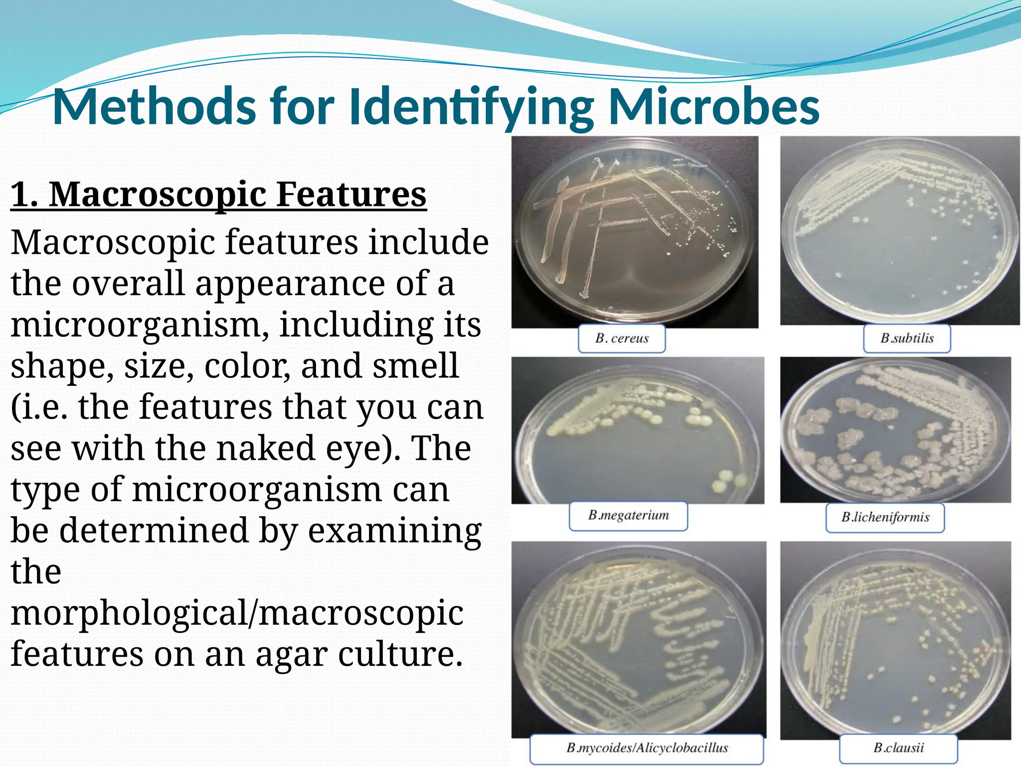
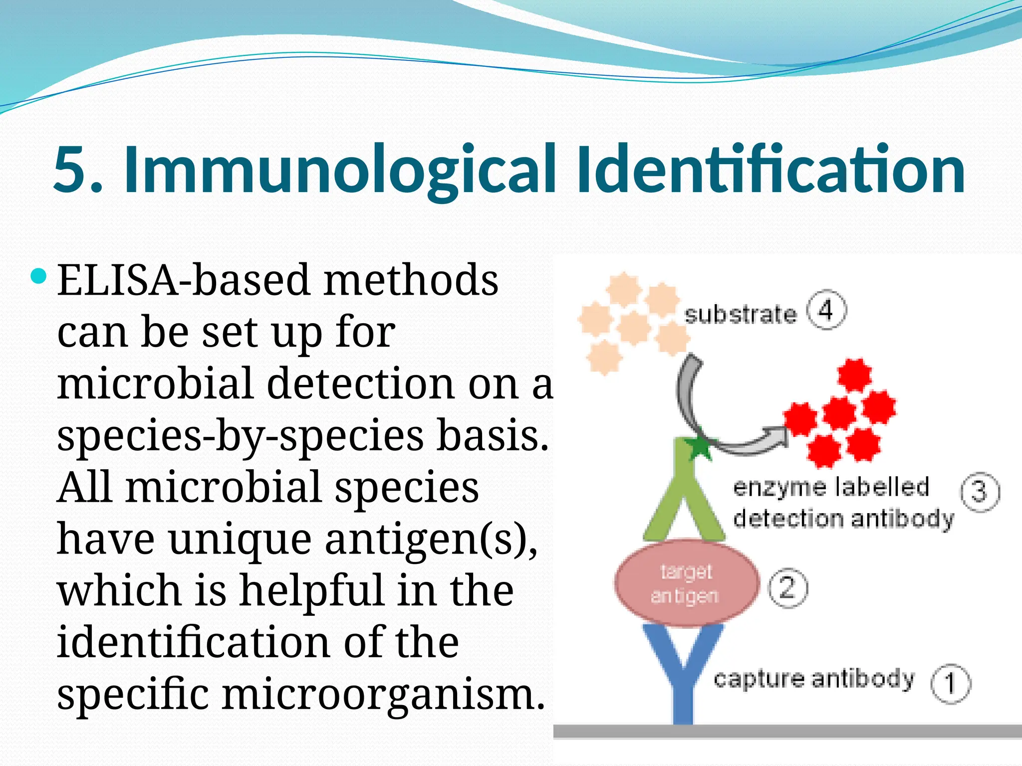

IDENTIFICATION OF MICROORGANISMS **Methods for identifying microbes 1. Macroscopic Features 2. Staining and Microscopy (Gram staining, Endospore staining, Ziehl-Neelsen Staining, Stains for Fungi and Yeast) 3. Simple Biochemical Tests (Catalase Testing, Oxidase Testing) 4. Identifying Microbes Using PCR 5. Immunological Identification