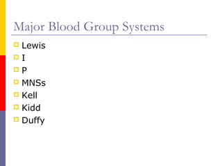
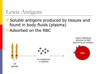
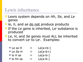
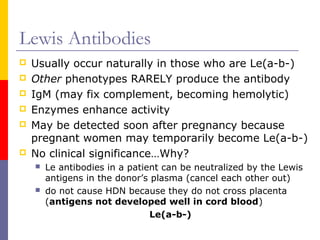
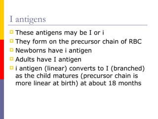
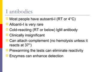
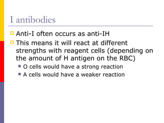
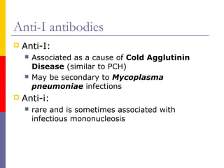
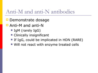
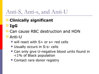
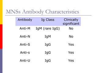
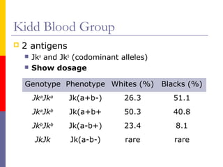
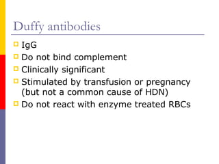
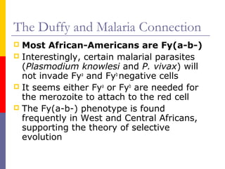
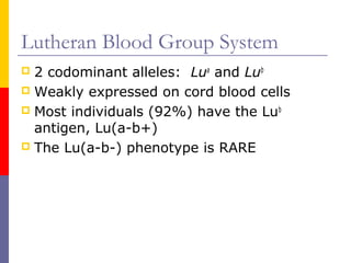
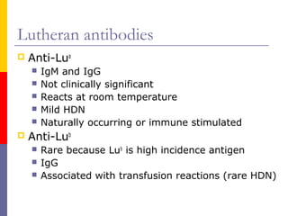
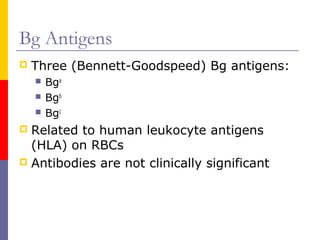
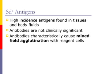
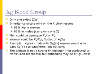
Other Blood Group
     Systems


       BUDANG
Facts
 Over 200 blood antigens exist!
 Unfortunately, we only get to review the
  most relevant antigens
 We will discuss each of these major
  antigens, their antibodies, and the clinical
  significance of each
Major Blood Group Systems
 Lewis
I
P
 MNSs
 Kell
 Kidd
 Duffy
Basic terms to remember
   Clinical significance: antibodies that are
    associated with decreased RBC survival
       Transfusion reactions
       HDN
   Not clinically significant: antibodies that do
    not cause red cell destruction
   Cold reacting antibodies: agglutination best
    observed at or below room temp.
   Warm reacting antibodies: agglutination best
    observed at 37°C
Systems that Produce
Cold-Reacting Antibodies
Lewis Antigens
 Soluble antigens produced by tissues and
  found in body fluids (plasma)
 Adsorbed on the RBC

                                   Lewis substance
                                   adheres to RBC
                                 becoming an antigen


                                       RBC

               Le substance
                in plasma
     Le
    genes
Lewis inheritance
 Lewis system depends on Hh, Se, and Le
  genes
 le, h, and se do not produce products
 If the Le gene is inherited, Lea substance is
  produced
 Le, H, and Se genes must ALL be inherited
  to convert Lea to Leb. Examples:

       Le se H       Le(a+b-)
       Le Se H       Le(a-b+)
       le H se       Le(a-b-)
       le hh se      Le(a-b-)
Lewis Antibodies
   Usually occur naturally in those who are Le(a-b-)
   Other phenotypes RARELY produce the antibody
   IgM (may fix complement, becoming hemolytic)
   Enzymes enhance activity
   May be detected soon after pregnancy because
    pregnant women may temporarily become Le(a-b-)
   No clinical significance…Why?
       Le antibodies in a patient can be neutralized by the Lewis
        antigens in the donor’s plasma (cancel each other out)
       do not cause HDN because they do not cross placenta
        (antigens not developed well in cord blood)
                                Le(a-b-)
I antigens
 These antigens may be I or i
 They form on the precursor chain of RBC
 Newborns have i antigen
 Adults have I antigen
 i antigen (linear) converts to I (branched)
  as the child matures (precursor chain is
  more linear at birth) at about 18 months
I antibodies
 Most people have autoanti-I (RT or 4°C)
 Alloanti-I is very rare
 Cold-reacting (RT or below) IgM antibody
 Clinically insignificant
 Can attach complement (no hemolysis unless it
  reacts at 37°)
 Prewarming the tests can eliminate reactivity
 Enzymes can enhance detection
I antibodies
 Anti-I often occurs as anti-IH
 This means it will react at different
  strengths with reagent cells (depending on
  the amount of H antigen on the RBC)
       O cells would have a strong reaction
       A cells would have a weaker reaction
Anti-I antibodies
   Anti-I:
       Associated as a cause of Cold Agglutinin
        Disease (similar to PCH)
       May be secondary to Mycoplasma
        pneumoniae infections
   Anti-i:
       rare and is sometimes associated with
        infectious mononucleosis
P Antigen
 Similar to the ABO system
 The most common phenotypes are P1 and
  P2
       P1 – consists of P1 and P antigens
       P2 – consists of only P antigens
   Like the A2 subgroup, P2 groups can
    produce anti-P1
   75% of adults have P1
P1 Antigen
 Strength of the antigen decreases upon
  storage
 Found in secretions like plasma and
  hydatid cyst fluid
       Cyst of a dog tapeworm
P antibodies
   Anti-P1
       Naturally occurring IgM
       Not clinically significant
       Can be neutralized by hydatid cyst fluid to reveal more
        clinically significant antibodies
   Anti-P
       Produced in individuals with paroxysmal cold
        hemoglobinuria (PCH)
       PCH – IgG auto-anti-P attaches complement when cold
        (fingers, toes). As the red cells circulate, they begin to
        lyse (releasing Hgb)
       This PCH antibody is also called the Donath-
        Landsteiner antibody
MNSs Blood System
   4 important antigens (more exist):
       M
       N
       S
       s
       U (ALWAYS present when S & s are inherited)
 M & N located on Glycophorin A
 S & s and U located on Glycophorin B
 Remember: Glycophorin is a protein that
  carries many RBC antigens
MNSs Antigens

                                                   M & N only differ in
                               M                    their amino acid
  Glycophorin A                    N              sequence at positions
                                                         1 and 5


     RBC


                      S                            S & s only differ in
                  U
  Glycophorin B           s                         their amino acid
                                                  sequence at position
                                                           29


 COOH end …..         ….5, 4, 3, 2, 1 (NH2 end)
MNSs antigens
 all show dosage
 M & N give a stronger reaction when
  homozygous, (M+N-) or (M-N+)
 Weaker reactions occur when in the
  heterozygous state (M+N+)
 Antigens are destroyed by enzymes (i.e.
  ficin, papain)
U (Su) antigen
 The U antigen is ALWAYS present when S
  & s are inherited
 About 85% of S-s- individuals are U-
  negative (RARE)
 U-negative cells are only found in the
  Black population
Frequency of MNSs antigens
   Phenotypes     Blacks            Whites
                   (%)               (%)
      M+            74                78

      N+             75               72

      S+            30.5              55

      s+             94               89

      U+             99              99.9

           High-incidence antigen
Thought…..
   Can a person have NO MNSs antigens?
       Yes, the Mk allele produces no M, N, S, or s
        antigens
       Frequency of 0.00064 or .064%
Anti-M and anti-N antibodies
 Demonstrate dosage
 Anti-M and anti-N
       IgM (rarely IgG)
       Clinically insignificant
       If IgG, could be implicated in HDN (RARE)
       Will not react with enzyme treated cells
Anti-S, Anti-s, and Anti-U
 Clinically significant
 IgG
 Can cause RBC destruction and HDN
 Anti-U
       will react with S+ or s+ red cells
       Usually occurs in S-s- cells
       Can only give U-negative blood units found in
        <1% of Black population
       Contact rare donor registry
MNSs Antibody Characteristics
    Antibody      Ig Class     Clinically
                              significant
     Anti-M    IgM (rare IgG)     No

     Anti-N         IgM           No

     Anti-S         IgG           Yes

     Anti-s         IgG           Yes

     Anti-U         IgG           Yes
Systems that Produce Warm-
    Reacting Antibodies
Kell System
 Similar to the Rh system
 2 major antigens (over 20 exist)
       K (Kell), <9% of population
       k (cellano), >90% of population
 The K and k genes are codominant alleles
  on chromosome 7 that code for the
  antigens
 Well developed at birth
 The K antigen is very immunogenic (2nd to
  the D antigen) in stimulating antibody
  production
Other Kell antigens
 Other sets of alleles also exist in the Kell
  system:
 Analogous to the Rh system: C/c and E/e
 Kp antigens
       Kpa is a low frequency antigen (only 2%)
       Kpb is a high frequency antigen (99.9%)
   Js antigens
       Jsa (20% in Blacks, 0.1% in Whites)
       Jsb is high frequency (80-100%)
Kell antigens
 Kell antigens have disulfide-bonded
  regions on the glycoproteins
 This makes them sensitive to sulfhydryl
  reagents:
       2-mercaptoethanol (2-ME)
       Dithiothreitol (DTT)
       2-aminoethylisothiouronium bromide (AET)
Kellnull or K0
 No expression of Kell antigens except a
  related antigen called Kx
 As a result of transfusion, K0 individuals
  can develop anti-Ku (Ku is on RBCs that
  have Kell antigens)
 Rare Kell negative units should be given
Kell antibodies
   IgG (react well at AHG)
   Produced as a result of immune stimulation
    (transfusion, pregnancy)
   Clinically significant
   Anti-K is most common because the K antigen
    is extremely immunogenic
   k, Kpb, and Jsb antibodies are rare (many
    individuals have these antigens and won’t
    develop an antibody)
   The other antibodies are also rare since few
    donors have the antigen
Kx antigen
   Not a part of the Kell system, but is
    related
       Kx antigens are present in small amounts in
        individuals with normal Kell antigens
       Kx antigens are increased in those who are K0
McLeod Syndrome
   The XK1 gene (on the X chromosome) codes for
    the Kx antigen
   When the gene is not inherited, Kx is absent
    (almost exclusive in White males)
   Causes abnormal red cell morphologies and
    decreased red cell survival:
       Acanthocytes – spur cells (defected cell membrane)
       Reticulocytes – immature red cells
   Associated with chronic granulomatous
    disease
       WBCs engulf microorganisms, but cannot kill (normal
        flora)
Kidd Blood Group
   2 antigens
       Jka and Jkb (codominant alleles)
       Show dosage

    Genotype Phenotype Whites (%) Blacks (%)
        JkaJka    Jk(a+b-)       26.3      51.1
        JkaJkb    Jk(a+b+        50.3      40.8
        JkbJkb    Jk(a-b+)       23.4      8.1
        JkJk      Jk(a-b-)       rare      rare
Kidd Antigens
 Well developed at birth
 Enhanced by enzymes
 Not very acessible on the RBC membrane
Kidd antibodies
   Anti-Jka and Anti-Jkb
       IgG
       Clinically significant
       Implicated in HTR and HDN
       Common cause of delayed HTR
       Usually appears with other antibodies when
        detected
Kidd antibodies
   Anti-Jk3
       Found in some individuals who are Jk(a-b-)
       Far East and Pacific Islanders (RARE)
Duffy Blood Group
   Predominant genes (codominant alleles):
       Fya and Fyb code for antigens that are well
        developed at birth
       Antigens are destroyed by enzymes
       Show dosage

         Phenotypes        Blacks         Whites
           Fy(a+b-)           9              17
           Fy(a+b+)           1              49
           Fy(a-b+)           22             34
           Fy(a-b-)           68           RARE
Duffy antibodies
 IgG
 Do not bind complement
 Clinically significant
 Stimulated by transfusion or pregnancy
  (but not a common cause of HDN)
 Do not react with enzyme treated RBCs
The Duffy and Malaria Connection
 Most African-Americans are Fy(a-b-)
 Interestingly, certain malarial parasites
  (Plasmodium knowlesi and P. vivax) will
  not invade Fya and Fyb negative cells
 It seems either Fya or Fyb are needed for
  the merozoite to attach to the red cell
 The Fy(a-b-) phenotype is found
  frequently in West and Central Africans,
  supporting the theory of selective
  evolution
Other Blood Group
   Antigens…
Lutheran Blood Group System
 2 codominant alleles: Lua and Lub
 Weakly expressed on cord blood cells
 Most individuals (92%) have the Lub
  antigen, Lu(a-b+)
 The Lu(a-b-) phenotype is RARE
Lutheran antibodies
   Anti-Lua
       IgM and IgG
       Not clinically significant
       Reacts at room temperature
       Mild HDN
       Naturally occurring or immune stimulated
   Anti-Lub
       Rare because Lub is high incidence antigen
       IgG
       Associated with transfusion reactions (rare HDN)
Bg Antigens
   Three (Bennett-Goodspeed) Bg antigens:
       Bga
       Bgb
       Bgc
 Related to human leukocyte antigens
  (HLA) on RBCs
 Antibodies are not clinically significant
Sda Antigens
 High incidence antigens found in tissues
  and body fluids
 Antibodies are not clinically significant
 Antibodies characteristically cause mixed
  field agglutination with reagent cells
Xg Blood Group
   Only one exists (Xga)
   Inheritance occurs only on the X chromosome
      89% Xga in women
      66% in males (carry only one X)
   Men could be genotype Xga or Xg
   Women could be XgaXga, XgaXg, or XgXg
   Example: Xg(a+) male with Xg(a-) woman would only
    pass Xg(a+) to daughters, but not sons
   The antigen is not a strong immunogen (not attributed to
    transfusion reactions); but antibodies may be of IgG class
HTLA Antigens
 High Titer Low Avidity (HTLA)
 Occur with high frequency
 Antibodies are VERY weak and are not
  clinically significant
 Do not cause HDN or HTR
Review
Cold Antibodies (IgM)
   Anti-Lea
   Anti-Leb
   Anti-I
   Anti-P1
   Anti-M
   Anti-A, -B, -H
   Anti-N


     LIiPMABHN
       Naturally Occurring
Warm antibodies (IgG)

     Rh antibodies
     Kell
     Duffy
     Kidd
     S,s
Remember enzyme activity:

  Papain, bromelin,    Enhanced by    Destroyed
  ficin, and trypsin     enzymes     by enzymes

                          Kidd       Fya and Fyb
                           Rh            M, N
                          Lewis          S, s
                            I
                            P
Remembering Dosage:
   Kidds and Duffy the Monkey (Rh) eat
    lots of M&Ns


                                      M&Ns
                                      M&Ns


    Jka, Jkb,   Fya, Fyb,   C, c, E, e (no D),   M, N, S, s
                                                      MNSs
      Kidd        Duffy          Rh

OTHER BLOOD GROUP SYSTEMS

  • 1.
    Other Blood Group Systems BUDANG
  • 2.
    Facts  Over 200blood antigens exist!  Unfortunately, we only get to review the most relevant antigens  We will discuss each of these major antigens, their antibodies, and the clinical significance of each
  • 3.
    Major Blood GroupSystems  Lewis I P  MNSs  Kell  Kidd  Duffy
  • 4.
    Basic terms toremember  Clinical significance: antibodies that are associated with decreased RBC survival  Transfusion reactions  HDN  Not clinically significant: antibodies that do not cause red cell destruction  Cold reacting antibodies: agglutination best observed at or below room temp.  Warm reacting antibodies: agglutination best observed at 37°C
  • 5.
  • 6.
    Lewis Antigens  Solubleantigens produced by tissues and found in body fluids (plasma)  Adsorbed on the RBC Lewis substance adheres to RBC becoming an antigen RBC Le substance in plasma Le genes
  • 7.
    Lewis inheritance  Lewissystem depends on Hh, Se, and Le genes  le, h, and se do not produce products  If the Le gene is inherited, Lea substance is produced  Le, H, and Se genes must ALL be inherited to convert Lea to Leb. Examples:  Le se H  Le(a+b-)  Le Se H  Le(a-b+)  le H se  Le(a-b-)  le hh se  Le(a-b-)
  • 8.
    Lewis Antibodies  Usually occur naturally in those who are Le(a-b-)  Other phenotypes RARELY produce the antibody  IgM (may fix complement, becoming hemolytic)  Enzymes enhance activity  May be detected soon after pregnancy because pregnant women may temporarily become Le(a-b-)  No clinical significance…Why?  Le antibodies in a patient can be neutralized by the Lewis antigens in the donor’s plasma (cancel each other out)  do not cause HDN because they do not cross placenta (antigens not developed well in cord blood) Le(a-b-)
  • 9.
    I antigens  Theseantigens may be I or i  They form on the precursor chain of RBC  Newborns have i antigen  Adults have I antigen  i antigen (linear) converts to I (branched) as the child matures (precursor chain is more linear at birth) at about 18 months
  • 10.
    I antibodies  Mostpeople have autoanti-I (RT or 4°C)  Alloanti-I is very rare  Cold-reacting (RT or below) IgM antibody  Clinically insignificant  Can attach complement (no hemolysis unless it reacts at 37°)  Prewarming the tests can eliminate reactivity  Enzymes can enhance detection
  • 11.
    I antibodies  Anti-Ioften occurs as anti-IH  This means it will react at different strengths with reagent cells (depending on the amount of H antigen on the RBC)  O cells would have a strong reaction  A cells would have a weaker reaction
  • 12.
    Anti-I antibodies  Anti-I:  Associated as a cause of Cold Agglutinin Disease (similar to PCH)  May be secondary to Mycoplasma pneumoniae infections  Anti-i:  rare and is sometimes associated with infectious mononucleosis
  • 13.
    P Antigen  Similarto the ABO system  The most common phenotypes are P1 and P2  P1 – consists of P1 and P antigens  P2 – consists of only P antigens  Like the A2 subgroup, P2 groups can produce anti-P1  75% of adults have P1
  • 14.
    P1 Antigen  Strengthof the antigen decreases upon storage  Found in secretions like plasma and hydatid cyst fluid  Cyst of a dog tapeworm
  • 15.
    P antibodies  Anti-P1  Naturally occurring IgM  Not clinically significant  Can be neutralized by hydatid cyst fluid to reveal more clinically significant antibodies  Anti-P  Produced in individuals with paroxysmal cold hemoglobinuria (PCH)  PCH – IgG auto-anti-P attaches complement when cold (fingers, toes). As the red cells circulate, they begin to lyse (releasing Hgb)  This PCH antibody is also called the Donath- Landsteiner antibody
  • 16.
    MNSs Blood System  4 important antigens (more exist):  M  N  S  s  U (ALWAYS present when S & s are inherited)  M & N located on Glycophorin A  S & s and U located on Glycophorin B  Remember: Glycophorin is a protein that carries many RBC antigens
  • 17.
    MNSs Antigens M & N only differ in M their amino acid Glycophorin A N sequence at positions 1 and 5 RBC S S & s only differ in U Glycophorin B s their amino acid sequence at position 29 COOH end ….. ….5, 4, 3, 2, 1 (NH2 end)
  • 18.
    MNSs antigens  allshow dosage  M & N give a stronger reaction when homozygous, (M+N-) or (M-N+)  Weaker reactions occur when in the heterozygous state (M+N+)  Antigens are destroyed by enzymes (i.e. ficin, papain)
  • 19.
    U (Su) antigen The U antigen is ALWAYS present when S & s are inherited  About 85% of S-s- individuals are U- negative (RARE)  U-negative cells are only found in the Black population
  • 20.
    Frequency of MNSsantigens Phenotypes Blacks Whites (%) (%) M+ 74 78 N+ 75 72 S+ 30.5 55 s+ 94 89 U+ 99 99.9 High-incidence antigen
  • 21.
    Thought…..  Can a person have NO MNSs antigens?  Yes, the Mk allele produces no M, N, S, or s antigens  Frequency of 0.00064 or .064%
  • 22.
    Anti-M and anti-Nantibodies  Demonstrate dosage  Anti-M and anti-N  IgM (rarely IgG)  Clinically insignificant  If IgG, could be implicated in HDN (RARE)  Will not react with enzyme treated cells
  • 23.
    Anti-S, Anti-s, andAnti-U  Clinically significant  IgG  Can cause RBC destruction and HDN  Anti-U  will react with S+ or s+ red cells  Usually occurs in S-s- cells  Can only give U-negative blood units found in <1% of Black population  Contact rare donor registry
  • 24.
    MNSs Antibody Characteristics Antibody Ig Class Clinically significant Anti-M IgM (rare IgG) No Anti-N IgM No Anti-S IgG Yes Anti-s IgG Yes Anti-U IgG Yes
  • 25.
    Systems that ProduceWarm- Reacting Antibodies
  • 26.
    Kell System  Similarto the Rh system  2 major antigens (over 20 exist)  K (Kell), <9% of population  k (cellano), >90% of population  The K and k genes are codominant alleles on chromosome 7 that code for the antigens  Well developed at birth  The K antigen is very immunogenic (2nd to the D antigen) in stimulating antibody production
  • 27.
    Other Kell antigens Other sets of alleles also exist in the Kell system:  Analogous to the Rh system: C/c and E/e  Kp antigens  Kpa is a low frequency antigen (only 2%)  Kpb is a high frequency antigen (99.9%)  Js antigens  Jsa (20% in Blacks, 0.1% in Whites)  Jsb is high frequency (80-100%)
  • 28.
    Kell antigens  Kellantigens have disulfide-bonded regions on the glycoproteins  This makes them sensitive to sulfhydryl reagents:  2-mercaptoethanol (2-ME)  Dithiothreitol (DTT)  2-aminoethylisothiouronium bromide (AET)
  • 29.
    Kellnull or K0 No expression of Kell antigens except a related antigen called Kx  As a result of transfusion, K0 individuals can develop anti-Ku (Ku is on RBCs that have Kell antigens)  Rare Kell negative units should be given
  • 30.
    Kell antibodies  IgG (react well at AHG)  Produced as a result of immune stimulation (transfusion, pregnancy)  Clinically significant  Anti-K is most common because the K antigen is extremely immunogenic  k, Kpb, and Jsb antibodies are rare (many individuals have these antigens and won’t develop an antibody)  The other antibodies are also rare since few donors have the antigen
  • 31.
    Kx antigen  Not a part of the Kell system, but is related  Kx antigens are present in small amounts in individuals with normal Kell antigens  Kx antigens are increased in those who are K0
  • 32.
    McLeod Syndrome  The XK1 gene (on the X chromosome) codes for the Kx antigen  When the gene is not inherited, Kx is absent (almost exclusive in White males)  Causes abnormal red cell morphologies and decreased red cell survival:  Acanthocytes – spur cells (defected cell membrane)  Reticulocytes – immature red cells  Associated with chronic granulomatous disease  WBCs engulf microorganisms, but cannot kill (normal flora)
  • 33.
    Kidd Blood Group  2 antigens  Jka and Jkb (codominant alleles)  Show dosage Genotype Phenotype Whites (%) Blacks (%) JkaJka Jk(a+b-) 26.3 51.1 JkaJkb Jk(a+b+ 50.3 40.8 JkbJkb Jk(a-b+) 23.4 8.1 JkJk Jk(a-b-) rare rare
  • 34.
    Kidd Antigens  Welldeveloped at birth  Enhanced by enzymes  Not very acessible on the RBC membrane
  • 35.
    Kidd antibodies  Anti-Jka and Anti-Jkb  IgG  Clinically significant  Implicated in HTR and HDN  Common cause of delayed HTR  Usually appears with other antibodies when detected
  • 36.
    Kidd antibodies  Anti-Jk3  Found in some individuals who are Jk(a-b-)  Far East and Pacific Islanders (RARE)
  • 37.
    Duffy Blood Group  Predominant genes (codominant alleles):  Fya and Fyb code for antigens that are well developed at birth  Antigens are destroyed by enzymes  Show dosage Phenotypes Blacks Whites Fy(a+b-) 9 17 Fy(a+b+) 1 49 Fy(a-b+) 22 34 Fy(a-b-) 68 RARE
  • 38.
    Duffy antibodies  IgG Do not bind complement  Clinically significant  Stimulated by transfusion or pregnancy (but not a common cause of HDN)  Do not react with enzyme treated RBCs
  • 39.
    The Duffy andMalaria Connection  Most African-Americans are Fy(a-b-)  Interestingly, certain malarial parasites (Plasmodium knowlesi and P. vivax) will not invade Fya and Fyb negative cells  It seems either Fya or Fyb are needed for the merozoite to attach to the red cell  The Fy(a-b-) phenotype is found frequently in West and Central Africans, supporting the theory of selective evolution
  • 40.
    Other Blood Group Antigens…
  • 41.
    Lutheran Blood GroupSystem  2 codominant alleles: Lua and Lub  Weakly expressed on cord blood cells  Most individuals (92%) have the Lub antigen, Lu(a-b+)  The Lu(a-b-) phenotype is RARE
  • 42.
    Lutheran antibodies  Anti-Lua  IgM and IgG  Not clinically significant  Reacts at room temperature  Mild HDN  Naturally occurring or immune stimulated  Anti-Lub  Rare because Lub is high incidence antigen  IgG  Associated with transfusion reactions (rare HDN)
  • 43.
    Bg Antigens  Three (Bennett-Goodspeed) Bg antigens:  Bga  Bgb  Bgc  Related to human leukocyte antigens (HLA) on RBCs  Antibodies are not clinically significant
  • 44.
    Sda Antigens  Highincidence antigens found in tissues and body fluids  Antibodies are not clinically significant  Antibodies characteristically cause mixed field agglutination with reagent cells
  • 45.
    Xg Blood Group  Only one exists (Xga)  Inheritance occurs only on the X chromosome  89% Xga in women  66% in males (carry only one X)  Men could be genotype Xga or Xg  Women could be XgaXga, XgaXg, or XgXg  Example: Xg(a+) male with Xg(a-) woman would only pass Xg(a+) to daughters, but not sons  The antigen is not a strong immunogen (not attributed to transfusion reactions); but antibodies may be of IgG class
  • 46.
    HTLA Antigens  HighTiter Low Avidity (HTLA)  Occur with high frequency  Antibodies are VERY weak and are not clinically significant  Do not cause HDN or HTR
  • 47.
  • 48.
    Cold Antibodies (IgM)  Anti-Lea  Anti-Leb  Anti-I  Anti-P1  Anti-M  Anti-A, -B, -H  Anti-N LIiPMABHN Naturally Occurring
  • 49.
    Warm antibodies (IgG)  Rh antibodies  Kell  Duffy  Kidd  S,s
  • 50.
    Remember enzyme activity: Papain, bromelin, Enhanced by Destroyed ficin, and trypsin enzymes by enzymes Kidd Fya and Fyb Rh M, N Lewis S, s I P
  • 51.
    Remembering Dosage:  Kidds and Duffy the Monkey (Rh) eat lots of M&Ns M&Ns M&Ns Jka, Jkb, Fya, Fyb, C, c, E, e (no D), M, N, S, s MNSs Kidd Duffy Rh

Editor's Notes

  • #9 May become hemolytic in vitro (in the tube) If pregnant women have Le antibodies detected, they need to first be neutralized so that HDN antibodies may be detected
  • #13 PCH- paroxysmal cold hemoglobinuria
  • #28 Kpa = Penney Kpb = Rautenberg Jsa = Sutter Jsb = Matthews