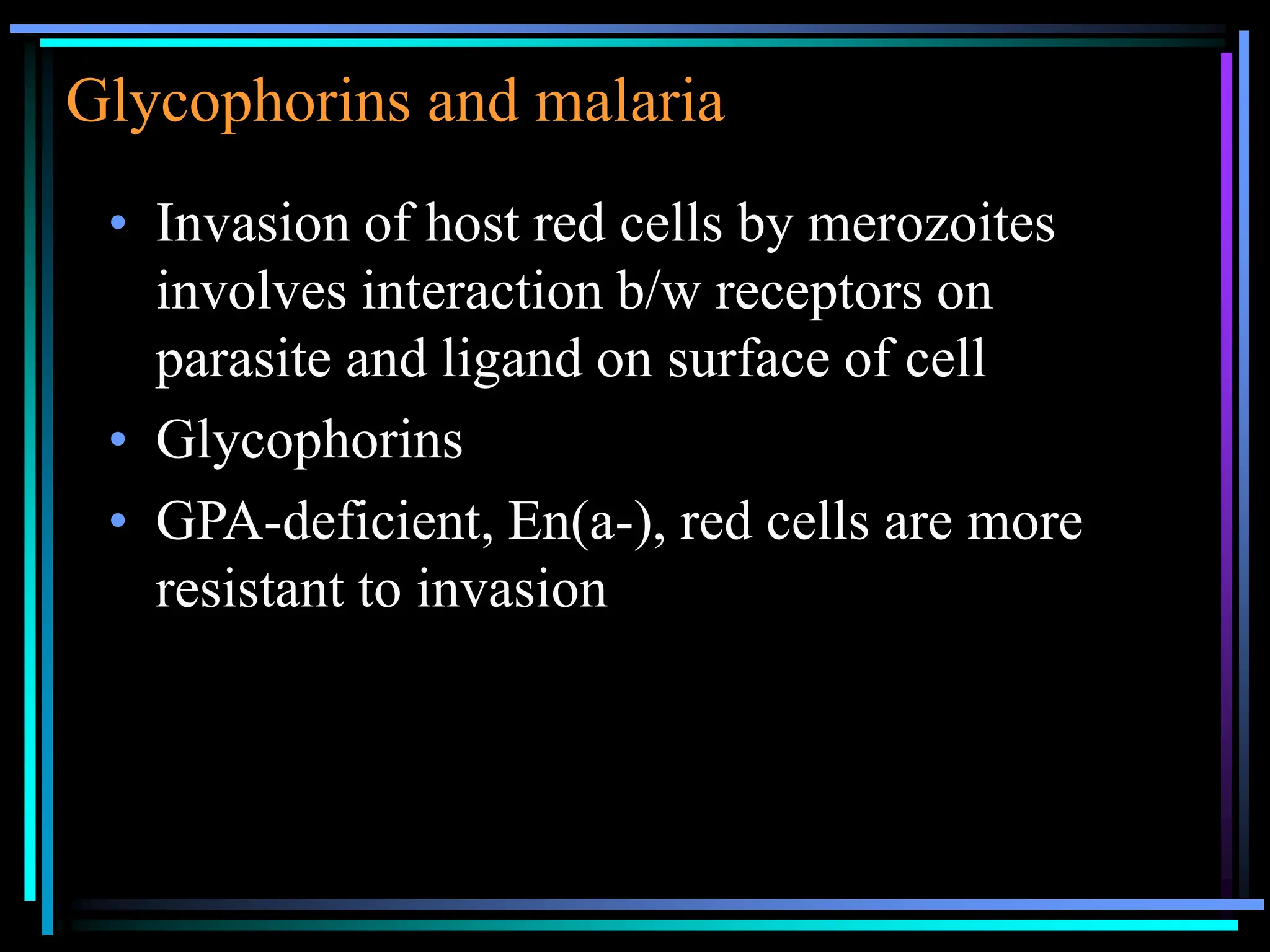
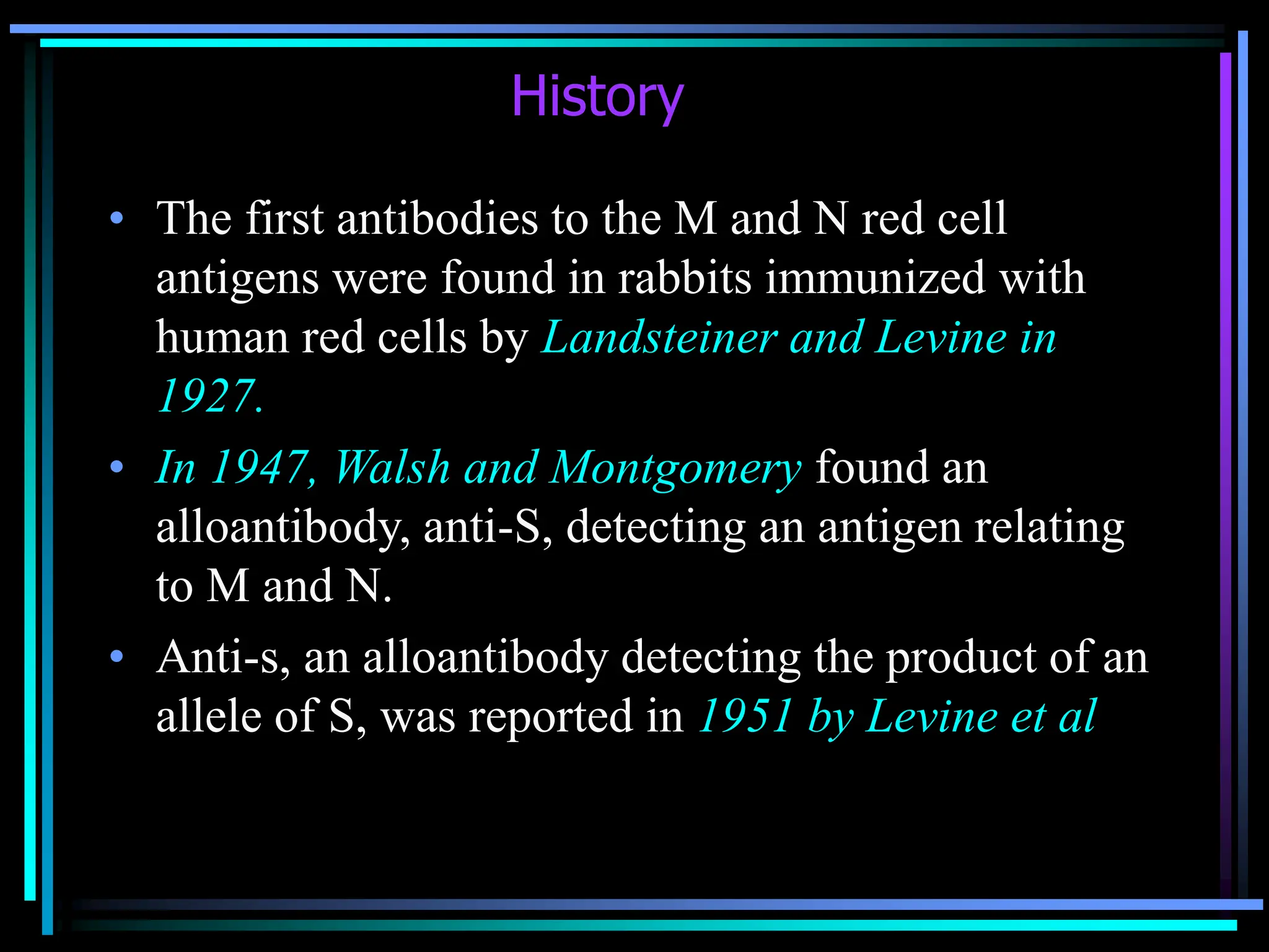
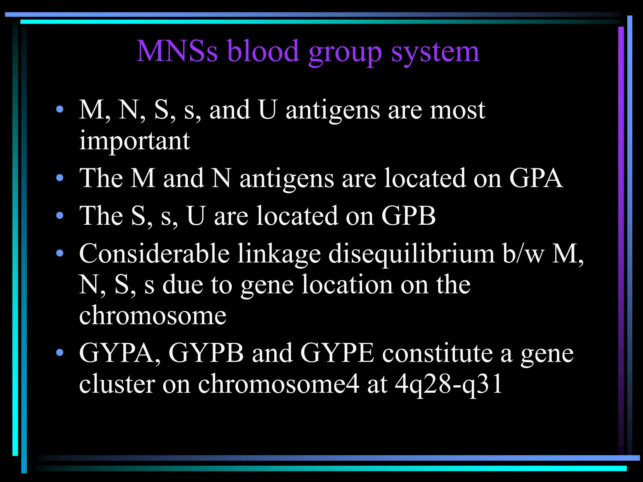
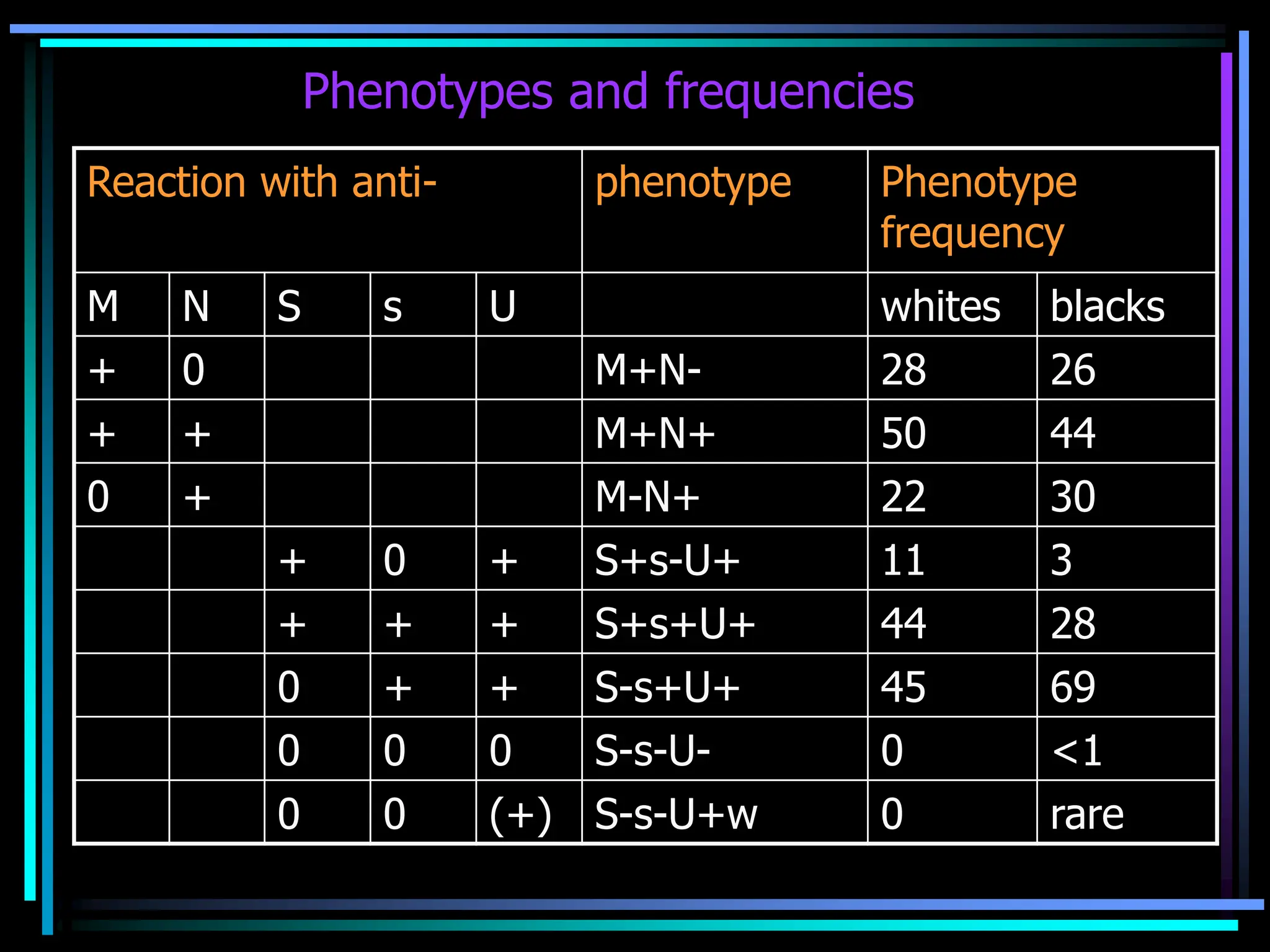
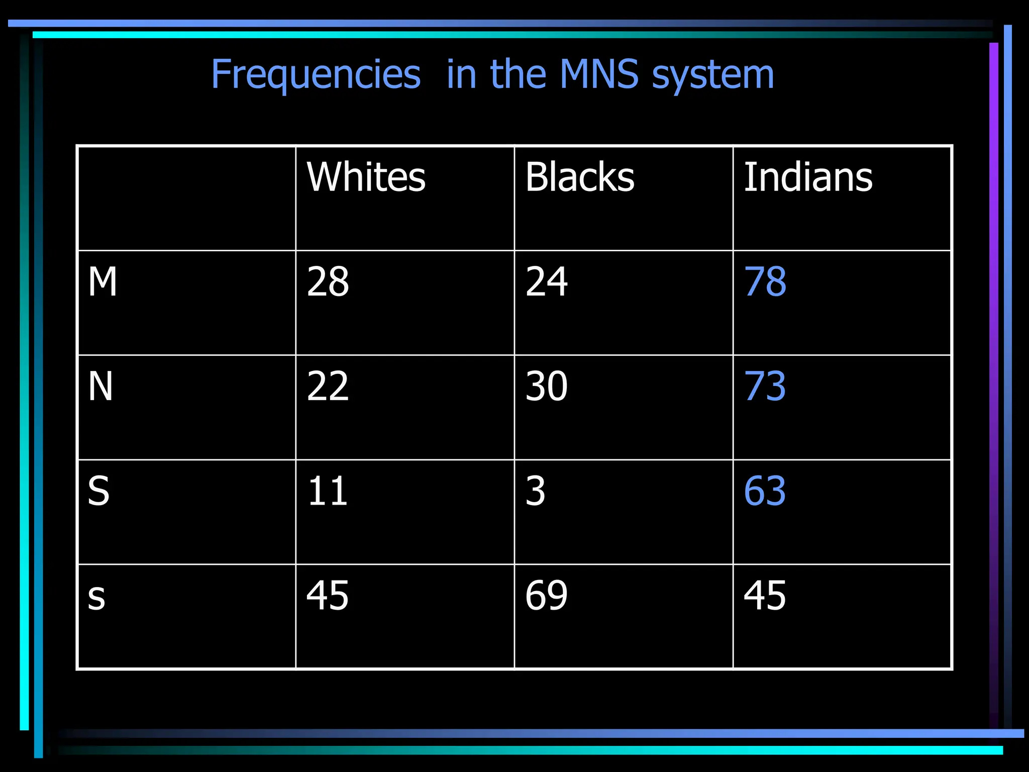
Introduction:
• MNS, the second blood group system
discovered
• Second only to Rh in its complexity
• 43 antigens are known
• the first blood group polymorphisms to be
defined at the level of protein sequence
History
• The first antibodies to the M and N red cell
antigens were found in rabbits immunized with
human red cells by Landsteiner and Levine in
1927.
• In 1947, Walsh and Montgomery found an
alloantibody, anti-S, detecting an antigen relating
to M and N.
• Anti-s, an alloantibody detecting the product of an
allele of S, was reported in 1951 by Levine et al
MNSs blood group system
• M, N, S, s, and U antigens are most
important
• The M and N antigens are located on GPA
• The S, s, U are located on GPB
• Considerable linkage disequilibrium b/w M,
N, S, s due to gene location on the
chromosome
• GYPA, GYPB and GYPE constitute a gene
cluster on chromosome4 at 4q28-q31
Development and distribution of
MNS antigens
• Well developed at birth
• GPA restricted to blood cells of erythroid
lineage and is often used as an erythroid
marker
• NOT present on lymphocytes, granulocytes,
megakaryocytes or platelets
• GPA –has been detected on renal
endothelium (incompletely sialylated)
MNSs antigens
Common
antigens
encoded by
MNS loci
M
N
S
s
Low frquency
antigens
<1%
He
Mia
Mc
Vw
Mur
Mg
Vr
Me
Mta
Sta
Ria
Cla
Nya
Hut
Hil
Mv
Far
sD
Mit
Dantu
Hop
Nob
EnaKT
Or
DANE
TSEN
MINY
High
frequency
antigens
>99%
U
Ena
‘N’
Glycophorin
• Heavily glycosylated
glycoproteins rich in sialic
acid (N- acetyl neuraminic
acid)
• C-terminus & N-terminus
• N-glycans & O- glycans
• GPB is not N-glycosylated
• O-glycans contain most of
the sialic acid
GPA & GPB
MN antigens
• The M and N antigens are found on glycophorin-
A(CD235A) , the major red cell sialic acid-rich
glycoprotein ( α-sialoglycoprotein 36 kD,131 AA)
• Defined by the 1st and 5th AA on this structure
• About 106 copies of MN-SGP/ red cell.
• The antigens exhibit dosage
• The antigens can be detected as early as 9 weeks
GA.
• M differs from N in the amino acid composition of
the extracellular tip of GPA:
– M has serine at position 1 and glycine at
position 5;
– N has leucine at position 1 and glutamic acid at
position 5.
• Carbohydrate , sialic acid also plays a part in the
expression of M&N antigens.
• M and N antigens are primarily red cell antigens
• Not detected on lymphocytes, monocytes,
granulocytes, or platelets
• MN antigens have been detected on renal capillary
endothelium
• Because MN antigens are at the outer end of
the GPA, they are easily destroyed or
removed by the routine blood bank enzymes
ficin, papain, and bromelin, and by less
common enzymes trypsin and pronase
• The antigens are also destroyed by ZZAP, a
solution of dithiothreitol (DTT) and papain
Ss antigens
• Located on a glycoprotein- Ss-SGP, δ-SGP, or
glycophorin-B (CD235B)
• AA on position 29 on GPB is critical to antigen
expression-
• S has methionine, whereas s has threonine
• Ss-SGP complexes with Rh protein, which
helps Ss-SGP stabilize and incorporate into
the red cell membrane
• Rh null red cells have greatly reduced Ss
expression
• There are 2.5x10⁵ copies of Ss-SGP/red cell
• Ss antigens show dosage
• Well developed at birth and appear on red cells at
an early GA(12 wks)
• Less easily degraded by enzymes
• Like MN, Ss are considered red cell antigens
Phenotypes and frequencies
Reaction with anti- phenotype Phenotype
frequency
M N S s U whites blacks
+ 0 M+N- 28 26
+ + M+N+ 50 44
0 + M-N+ 22 30
+ 0 + S+s-U+ 11 3
+ + + S+s+U+ 44 28
0 + + S-s+U+ 45 69
0 0 0 S-s-U- 0 <1
0 0 (+) S-s-U+w 0 rare
Frequencies in the MNS system
Whites Blacks Indians
M 28 24 78
N 22 30 73
S 11 3 63
s 45 69 45
‘N’
• The first 26 aa residues from the extracellular
terminus of GPB are identical to those of N-active
GPA ( GPAN ).
• Consequently, GPB also demonstrates N activity
(‘N’), detected in homozygous M/M individuals
by some anti-N.
• Trypsin resistant N antigen
U (universal)
• Weiner et al. 1953 -A high frequency blood group ag.
• Located on Ss-SGP very close to the red cell membrane
• Found in all individuals except about 1% of american
blacks, who lack Ss-SGP.
• U- cells are almost always S-s-, but S-s- cells are often U+
• Resistant to denaturation by sialidase, trypsin,
chymotrypsin, papain, and ficin.
Anti-U –
• A non-complement binding IgG antibodies (IgG1)
• Greater activity at temp <22° C than at body temperature
En(a-)
• Lack GPA and, consequently, MN antigen
expression (apart from the ‘N’ antigen carried on
GPB)
• En(a-) cells express normal Ss antigens but lack a
variety of GPA-borne high frequency antigens
collectively named Ena.
• En(a-) cells also lack Wrb, expression of which
results from an interaction between GPA and red
cell glycoprotein band 3
Mk
• A new allele of M and N that lack all MNS antigens
• Have no GPA or GPB
• Red cells are M-N-S-s-U-En(a)Wr(a-b-)
• Showed all reactions characteristic of reduced sialic acid
• The Miltenberger subsystem-
A group of very low frequency antigens in the MNS
group, related to each other through the overlapping
specificity of a no. of low frequency alloantigens
Hybrid glycophorins- LFAs
• Lepore type-
Loss of GYPA and GYPB and the
formation of a novel gene that produces a
GP (A-B) hybrid. N terminal of GPA and C-
terminal of GPB
• Anti-lepore type-
GP(B-A) hybrid
Anti-M
• Naturally occuring, cold reactive saline
agglutinins
• Mostly IgM, 50 to 80 % are IgG or have an
IgG component
• Usually do not bind complement, regardless
of their of their Ig class
• Do not react with enzyme treated red cells
• The frequency of finding saline reactive
anti-M in routine blood donors- 1 in 2500 to
5000
• More common in infants than adults
• Because of antigen dosage, M antibodies
react better with M+N- red cells (MM
genotype) than with M+N+ red cells
• Glucose dependent
• Some examples of anti-M are pH
dependent, reacting best at pH 6.5
• As long as anti-M does not react at 37 C, it
is not clinically significant in transfusion
• Anti-M rarely causes –hemolytic
transfusion reactions, decreased cell
survival, or hemolytic disease of newborn
Anti-N
• Cold reactive IgM or IgG saline agglutinin
that does not bind complement or react with
enzyme-treated red cells
• Demonstrates dosage
• Not clinically significant unless reacts at 37
C
• More rare than anti-M
Anti-Nf
• Anti-N also seen in kidney transplant
patients maintained on chronic
hemodialysis , regardless of their MN type,
who are dialized on equipment sterilized
with formaldehyde
• Ab disappeared after transplantation
• 21 to 27 % of dialysis patients
[Am J Nephrol 1995]
Anti-S and Anti-s
• Mostly IgG, reactive at 37 C in the
antiglobulin phase
• Although seen less often than anti-M, more
likely to be clinically significant
• May bind complement
• Have been implicated with severe HTR
with hbnuria
• Have also caused HDN
Blood groups and their specific lectin.
Lectins with anti-N activity
• Anti-N lectins are common and highly specific,
while anti-M is rare
• Anti-N lectin B. purpurea has been found to be a
good typing reagent .
• Anti-N lectin from Vicia graminea, is easily
available & shows excellent specificity and
avidity.
• Saliva extracts of Vicia unijuga agglutinate both M
& N cells, but adsorption with pooled human M
cells gives a potent anti-N lectin
• A+ N reactivity - Molucella laevis
Bandeiraea simpicifolia
Lectins with anti-M activity
Iberis amara, I. umbellata,I. semperivens
• The best lectins are isolated from Iberis
amara.
• Other lectins with anti-M activity include
Japanese radishes and Escherichia coli .
• anti-M lectin from non-fimbrial E. coli is
the most specific
Glycophorins and malaria
• Invasion of host red cells by merozoites
involves interaction b/w receptors on
parasite and ligand on surface of cell
• Glycophorins
• GPA-deficient, En(a-), red cells are more
resistant to invasion
Phenotype Deficient
structure
% invasion
Normal 100
En(a-)GW GPA 8
En(a-)RL GPA 14
S-s-U- GPB 72
Ge:-2,-3,-4 leach GPC 57
Tn Gal+ sialic acid 8
Trypsin treated
normal
GPA-T1,GPCT1 38
Trypsin treated S-
s-U-
GPA-T1, GPCT1,
GPB
5
Other pathogens:
• GPAM –uropathogenic E.coli strain 1H11165
• GP acts as a receptors for the bac. toxins that lyse
red cells
• Coating of red cells with antibodies to GPA &
GPB protects from lysis by hemolysins from E.
coli and vibrio cholerae respectively
• GPA also acts as a receptor for influenza virus

MNS Blood group system powerpoint in.ppt

  • 2.
    Introduction: • MNS, thesecond blood group system discovered • Second only to Rh in its complexity • 43 antigens are known • the first blood group polymorphisms to be defined at the level of protein sequence
  • 3.
    History • The firstantibodies to the M and N red cell antigens were found in rabbits immunized with human red cells by Landsteiner and Levine in 1927. • In 1947, Walsh and Montgomery found an alloantibody, anti-S, detecting an antigen relating to M and N. • Anti-s, an alloantibody detecting the product of an allele of S, was reported in 1951 by Levine et al
  • 4.
    MNSs blood groupsystem • M, N, S, s, and U antigens are most important • The M and N antigens are located on GPA • The S, s, U are located on GPB • Considerable linkage disequilibrium b/w M, N, S, s due to gene location on the chromosome • GYPA, GYPB and GYPE constitute a gene cluster on chromosome4 at 4q28-q31
  • 5.
    Development and distributionof MNS antigens • Well developed at birth • GPA restricted to blood cells of erythroid lineage and is often used as an erythroid marker • NOT present on lymphocytes, granulocytes, megakaryocytes or platelets • GPA –has been detected on renal endothelium (incompletely sialylated)
  • 6.
    MNSs antigens Common antigens encoded by MNSloci M N S s Low frquency antigens <1% He Mia Mc Vw Mur Mg Vr Me Mta Sta Ria Cla Nya Hut Hil Mv Far sD Mit Dantu Hop Nob EnaKT Or DANE TSEN MINY High frequency antigens >99% U Ena ‘N’
  • 7.
    Glycophorin • Heavily glycosylated glycoproteinsrich in sialic acid (N- acetyl neuraminic acid) • C-terminus & N-terminus • N-glycans & O- glycans • GPB is not N-glycosylated • O-glycans contain most of the sialic acid
  • 8.
  • 9.
    MN antigens • TheM and N antigens are found on glycophorin- A(CD235A) , the major red cell sialic acid-rich glycoprotein ( α-sialoglycoprotein 36 kD,131 AA) • Defined by the 1st and 5th AA on this structure • About 106 copies of MN-SGP/ red cell. • The antigens exhibit dosage • The antigens can be detected as early as 9 weeks GA.
  • 10.
    • M differsfrom N in the amino acid composition of the extracellular tip of GPA: – M has serine at position 1 and glycine at position 5; – N has leucine at position 1 and glutamic acid at position 5. • Carbohydrate , sialic acid also plays a part in the expression of M&N antigens.
  • 11.
    • M andN antigens are primarily red cell antigens • Not detected on lymphocytes, monocytes, granulocytes, or platelets • MN antigens have been detected on renal capillary endothelium
  • 12.
    • Because MNantigens are at the outer end of the GPA, they are easily destroyed or removed by the routine blood bank enzymes ficin, papain, and bromelin, and by less common enzymes trypsin and pronase • The antigens are also destroyed by ZZAP, a solution of dithiothreitol (DTT) and papain
  • 13.
    Ss antigens • Locatedon a glycoprotein- Ss-SGP, δ-SGP, or glycophorin-B (CD235B) • AA on position 29 on GPB is critical to antigen expression- • S has methionine, whereas s has threonine • Ss-SGP complexes with Rh protein, which helps Ss-SGP stabilize and incorporate into the red cell membrane • Rh null red cells have greatly reduced Ss expression
  • 14.
    • There are2.5x105 copies of Ss-SGP/red cell • Ss antigens show dosage • Well developed at birth and appear on red cells at an early GA(12 wks) • Less easily degraded by enzymes • Like MN, Ss are considered red cell antigens
  • 15.
    Phenotypes and frequencies Reactionwith anti- phenotype Phenotype frequency M N S s U whites blacks + 0 M+N- 28 26 + + M+N+ 50 44 0 + M-N+ 22 30 + 0 + S+s-U+ 11 3 + + + S+s+U+ 44 28 0 + + S-s+U+ 45 69 0 0 0 S-s-U- 0 <1 0 0 (+) S-s-U+w 0 rare
  • 16.
    Frequencies in theMNS system Whites Blacks Indians M 28 24 78 N 22 30 73 S 11 3 63 s 45 69 45
  • 17.
    ‘N’ • The first26 aa residues from the extracellular terminus of GPB are identical to those of N-active GPA ( GPAN ). • Consequently, GPB also demonstrates N activity (‘N’), detected in homozygous M/M individuals by some anti-N. • Trypsin resistant N antigen
  • 18.
    U (universal) • Weineret al. 1953 -A high frequency blood group ag. • Located on Ss-SGP very close to the red cell membrane • Found in all individuals except about 1% of american blacks, who lack Ss-SGP. • U- cells are almost always S-s-, but S-s- cells are often U+ • Resistant to denaturation by sialidase, trypsin, chymotrypsin, papain, and ficin. Anti-U – • A non-complement binding IgG antibodies (IgG1) • Greater activity at temp <22° C than at body temperature
  • 19.
    En(a-) • Lack GPAand, consequently, MN antigen expression (apart from the ‘N’ antigen carried on GPB) • En(a-) cells express normal Ss antigens but lack a variety of GPA-borne high frequency antigens collectively named Ena. • En(a-) cells also lack Wrb, expression of which results from an interaction between GPA and red cell glycoprotein band 3
  • 20.
    Mk • A newallele of M and N that lack all MNS antigens • Have no GPA or GPB • Red cells are M-N-S-s-U-En(a)Wr(a-b-) • Showed all reactions characteristic of reduced sialic acid • The Miltenberger subsystem- A group of very low frequency antigens in the MNS group, related to each other through the overlapping specificity of a no. of low frequency alloantigens
  • 21.
    Hybrid glycophorins- LFAs •Lepore type- Loss of GYPA and GYPB and the formation of a novel gene that produces a GP (A-B) hybrid. N terminal of GPA and C- terminal of GPB • Anti-lepore type- GP(B-A) hybrid
  • 22.
    Anti-M • Naturally occuring,cold reactive saline agglutinins • Mostly IgM, 50 to 80 % are IgG or have an IgG component • Usually do not bind complement, regardless of their of their Ig class • Do not react with enzyme treated red cells
  • 23.
    • The frequencyof finding saline reactive anti-M in routine blood donors- 1 in 2500 to 5000 • More common in infants than adults • Because of antigen dosage, M antibodies react better with M+N- red cells (MM genotype) than with M+N+ red cells • Glucose dependent
  • 24.
    • Some examplesof anti-M are pH dependent, reacting best at pH 6.5 • As long as anti-M does not react at 37 C, it is not clinically significant in transfusion • Anti-M rarely causes –hemolytic transfusion reactions, decreased cell survival, or hemolytic disease of newborn
  • 25.
    Anti-N • Cold reactiveIgM or IgG saline agglutinin that does not bind complement or react with enzyme-treated red cells • Demonstrates dosage • Not clinically significant unless reacts at 37 C • More rare than anti-M
  • 26.
    Anti-Nf • Anti-N alsoseen in kidney transplant patients maintained on chronic hemodialysis , regardless of their MN type, who are dialized on equipment sterilized with formaldehyde • Ab disappeared after transplantation • 21 to 27 % of dialysis patients [Am J Nephrol 1995]
  • 27.
    Anti-S and Anti-s •Mostly IgG, reactive at 37 C in the antiglobulin phase • Although seen less often than anti-M, more likely to be clinically significant • May bind complement • Have been implicated with severe HTR with hbnuria • Have also caused HDN
  • 28.
    Blood groups andtheir specific lectin.
  • 29.
    Lectins with anti-Nactivity • Anti-N lectins are common and highly specific, while anti-M is rare • Anti-N lectin B. purpurea has been found to be a good typing reagent . • Anti-N lectin from Vicia graminea, is easily available & shows excellent specificity and avidity. • Saliva extracts of Vicia unijuga agglutinate both M & N cells, but adsorption with pooled human M cells gives a potent anti-N lectin • A+ N reactivity - Molucella laevis Bandeiraea simpicifolia
  • 30.
    Lectins with anti-Mactivity Iberis amara, I. umbellata,I. semperivens • The best lectins are isolated from Iberis amara. • Other lectins with anti-M activity include Japanese radishes and Escherichia coli . • anti-M lectin from non-fimbrial E. coli is the most specific
  • 31.
    Glycophorins and malaria •Invasion of host red cells by merozoites involves interaction b/w receptors on parasite and ligand on surface of cell • Glycophorins • GPA-deficient, En(a-), red cells are more resistant to invasion
  • 32.
    Phenotype Deficient structure % invasion Normal100 En(a-)GW GPA 8 En(a-)RL GPA 14 S-s-U- GPB 72 Ge:-2,-3,-4 leach GPC 57 Tn Gal+ sialic acid 8 Trypsin treated normal GPA-T1,GPCT1 38 Trypsin treated S- s-U- GPA-T1, GPCT1, GPB 5
  • 33.
    Other pathogens: • GPAM–uropathogenic E.coli strain 1H11165 • GP acts as a receptors for the bac. toxins that lyse red cells • Coating of red cells with antibodies to GPA & GPB protects from lysis by hemolysins from E. coli and vibrio cholerae respectively • GPA also acts as a receptor for influenza virus