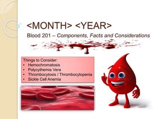
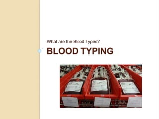
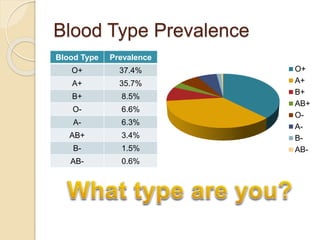
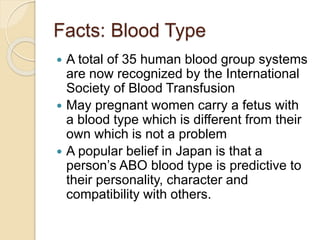
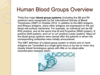
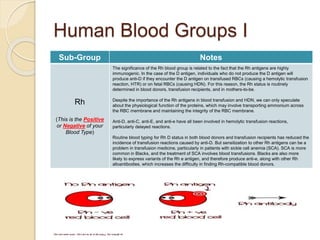
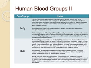
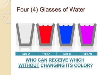
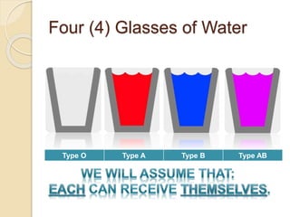
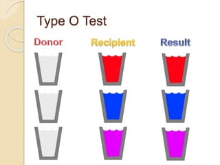
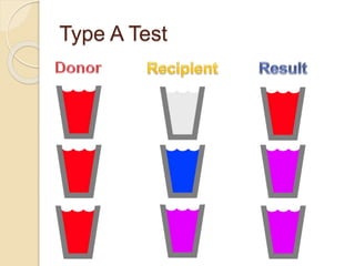
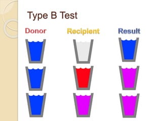
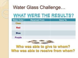
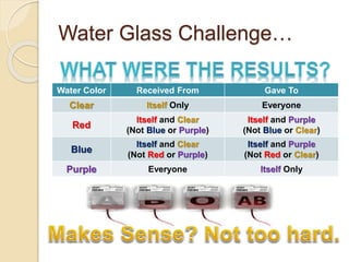
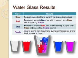
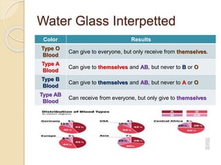
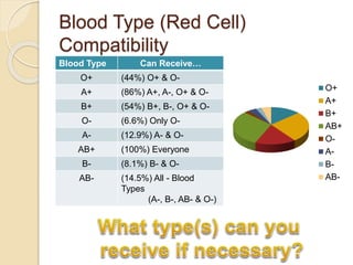
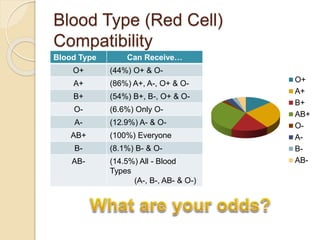
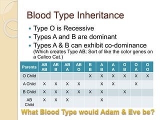
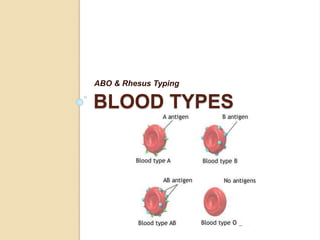
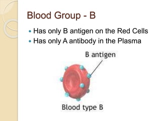
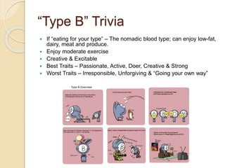
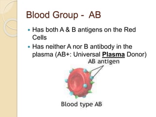
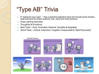
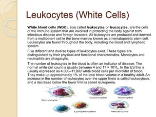
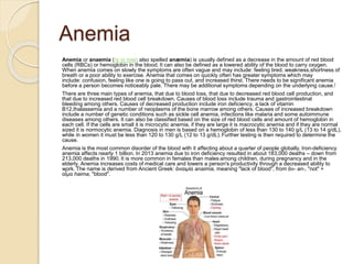
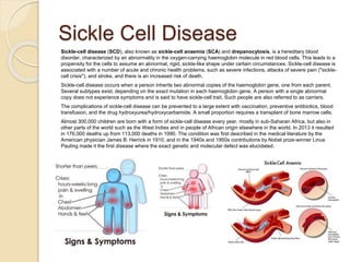
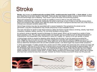
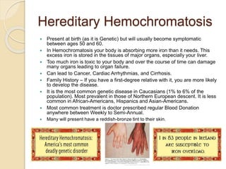
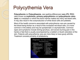
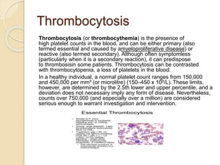
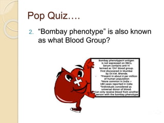
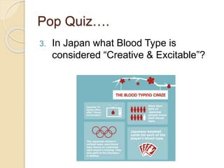
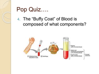
SlideShare a Scribd company logo
WFAC Drill Series
Waynesboro First Aid Crew
Prepared by:
Christopher Bayonet, EMT-B
Emergency Medical Technician, Waynesboro First Aid Crew
Blood Collections Specialist II / Patient Services, Virginia Blood Services
<MONTH> <YEAR>
Blood 201 – Components, Facts and Considerations
Things to Consider:
• Hemochromatosis
• Polycythemia Vera
• Thrombocytosis / Thrombocytopenia
• Sickle Cell Anemia
BLOOD TYPING
What are the Blood Types?
Blood Type Prevalence
O+ 37.4%
A+ 35.7%
B+ 8.5%
O- 6.6%
A- 6.3%
AB+ 3.4%
B- 1.5%
AB- 0.6%
O+
A+
B+
AB+
O-
A-
B-
AB-
Facts: Blood Type
 A total of 35 human blood group systems
are now recognized by the International
Society of Blood Transfusion
 May pregnant women carry a fetus with
a blood type which is different from their
own which is not a problem
 A popular belief in Japan is that a
person’s ABO blood type is predictive to
their personality, character and
compatibility with others.
Human Blood Groups Overview
Thirty-five major blood group systems (including the AB and Rh
systems) were recognized by the International Society of Blood
Transfusion (ISBT) in October 2012. In addition to the ABO antigens
and Rhesus antigens, many other antigens are expressed on the red
blood cell surface membrane. For example, an individual can be AB
RhD positive, and at the same time M and N positive (MNS system), K
positive (Kell system), and Lea or Leb positive (Lewis system). Many of
the blood group systems were named after the patients in whom the
corresponding antibodies were initially encountered.
The ISBT definition of a blood group system is where one or more
antigens are "controlled at a single gene locus or by two or more very
closely linked homologous genes with little or no observable
recombination between them".
Human Blood Groups I
Sub-Group Notes
Rh
(This is the Positive
or Negative of your
Blood Type)
The significance of the Rh blood group is related to the fact that the Rh antigens are highly
immunogenic. In the case of the D antigen, individuals who do not produce the D antigen will
produce anti-D if they encounter the D antigen on transfused RBCs (causing a hemolytic transfusion
reaction, HTR) or on fetal RBCs (causing HDN). For this reason, the Rh status is routinely
determined in blood donors, transfusion recipients, and in mothers-to-be.
Despite the importance of the Rh antigens in blood transfusion and HDN, we can only speculate
about the physiological function of the proteins, which may involve transporting ammonium across
the RBC membrane and maintaining the integrity of the RBC membrane.
Anti-D, anti-C, anti-E, and anti-e have all been involved in hemolytic transfusion reactions,
particularly delayed reactions.
Routine blood typing for Rh D status in both blood donors and transfusion recipients has reduced the
incidence of transfusion reactions caused by anti-D. But sensitization to other Rh antigens can be a
problem in transfusion medicine, particularly in patients with sickle cell anemia (SCA). SCA is more
common in Blacks, and the treatment of SCA involves blood transfusions. Blacks are also more
likely to express variants of the Rh e antigen, and therefore produce anti-e, along with other Rh
alloantibodies, which increases the difficulty in finding Rh-compatible blood donors.
Human Blood Groups II
Sub-Group Notes
Duffy
The Duffy glycoprotein is a receptor for chemicals that are secreted by blood cells during
inflammation. It also happens to be a receptor for Plasmodium vivax, a parasite that invades red
blood cells (RBCs) and causes malaria. RBCs that lack the Duffy antigens are relatively resistant to
invasion by P. vivax. This has influenced the variation in Duffy blood types seen in populations
where malaria is common.
Antibodies formed against the Duffy antigens are a cause of both transfusion reactions and
hemolytic disease of the newborn.
Antibodies against the Duffy antigens Fya, Fyb, Fy3, and Fy5 have all been implicated as the cause
of a transfusion reaction. Anti-Fya is more commonly found in patients who are of African descent (in
whom the Duffy null phenotype is more common) and have sickle cell anemia (and therefore may
require multiple blood transfusions).
Kidd
The Kidd (JK) glycoprotein is the red blood cell (RBC) urea transporter. Situated in the membrane it
rapidly transports urea into and out of RBCs, maintaining the osmotic stability and shape of the RBC
in the process. The Kidd glycoprotein is also expressed in the kidney, where it enables the kidney to
build up a high concentration of urea which is needed for the kidney to produce concentrated urine.
People who do not produce the Kidd glycoprotein tend not to be able to maximally concentrate urine,
but despite this, they are healthy and their RBCs have a normal shape and lifespan.
Antibodies that target Kidd antigens are a significant cause of delayed hemolytic transfusion
reactions. Anti-Kidd antibodies are also a cause of hemolytic disease of the newborn (HDN), the
severity of the disease varies but tends to be mild in nature.
Anti-Jka can cause severe and fatal hemolytic transfusion reactions but is more commonly
associated with less severe DHTRs. It has been estimated that over one-third of DHTRs are caused
by anti-Jka. Case studies have also pointed to anti-Jkb as being responsible for severe DHTR. Anti-
Jk3 has also been responsible for causing severe hemolytic transfusion reactions, both immediate
and delayed
Human Blood Groups III
Sub-Group Notes
Diego
The antigens of the Diego blood group are carried on an important protein, called the band 3 protein,
which lies in the red blood cell (RBC) membrane. This protein is a chloride/bicarbonate exchanger
involved in carbon dioxide transport from tissues to lungs. It also is found in the kidney, where it is
involved in acid secretion.
Many mutations in the gene that encodes the Diego antigens, SLC4A1, are known. These mutations
can result in RBCs with an abnormal membrane (hereditary ovalocytosis and spherocytosis) and
kidneys that are defective in secreting acid (renal tubule acidosis). Other SLC4A1 mutations that do
not give rise to disease may result in new blood group antigens that belong to the Diego blood group
system
Anti-Dia and anti-Dib are more commonly associated with HDN than transfusion reactions. However,
these antibodies are capable of causing immediate and delayed hemolytic transfusion reactions .
MNS
The antigens of the MNS blood group are carried on sugar-bearing proteins called glycophorins.
These lie in the red blood cell (RBC) membrane. One end of a glycophorin is attached to the
underlying cell, and the other end bears the sugars and determines a person's MNS blood type.
Anti-M and anti-N are not considered to be a cause of transfusion reactions, although rare cases of
delayed transfusion reactions have occurred as a result of anti-M. Anti-M is fairly common and is
thought to mostly be naturally occurring because it is frequently found in children who have never
received a blood transfusion.
Mild to moderate transfusion reactions can be caused by the presence of anti-S and anti-s in the
patient's serum.
Severe transfusion reactions have been attributed to anti-U, anti-Vw, anti-Mur, and anti-En
Human Blood Groups IV
Sub-Group Notes
Hh
H antigen deficiency is known as the "Bombay phenotype" (h/h, also known as Oh) and is found in 1
of 10,000 individuals in India and 1 in a million people in Europe. There is no ill effect with being H
deficient, but if a blood transfusion is ever needed, people with this blood type can receive blood only
from other donors who are also H deficient. (A transfusion of "normal" group O blood can trigger a
severe transfusion reaction.)
If patients with anti-H in their circulation receive transfusions of blood that contains the H antigen
(e.g., blood group O), they are at risk of suffering an acute hemolytic transfusion reaction.
Kell
The Kell blood group system is complex and contains many antigens that are highly immunogenic.
These antigens are the third most potent, after those of the ABO and Rh blood groups, at triggering
an immune reaction.
Antibodies that target Kell antigens can cause transfusion reactions and hemolytic disease of the
newborn (HDN). In the case of HDN, ABO and Rh incompatibility are more common causes.
However, disease caused by maternal anti-ABO tends to be mild, and disease caused by maternal
anti-Rh can largely be prevented. The infrequent cases of HDN caused by Kell immunization tend to
result in severe fetal anemia because maternal anti-Kell target fetal red blood cell (RBC) precursors,
suppressing the fetal production of RBCs.
COMPATIBILITY
Who can receive what type of blood?
Blood Transfusion History
Picture if you will…
 Four Glasses of Water
 One of which is dyed Blue for Type B
 One of which is dyed Red for Type A
 One of which is dyed Purple for Type AB
 One of which is kept clear for Type O
Four (4) Glasses of Water
Type O Type A Type B Type AB
Four (4) Glasses of Water
Type O Type A Type B Type AB
Type O Test
Type A Test
Type B Test
Type AB Test
Water Glass Challenge…
Water Color Received From Gave To
Clear
Red
Blue
Purple
Water Glass Challenge…
Water Color Received From Gave To
Clear Itself Only Everyone
Red
Itself and Clear
(Not Blue or Purple)
Itself and Purple
(Not Blue or Clear)
Blue
Itself and Clear
(Not Red or Purple)
Itself and Purple
(Not Red or Clear)
Purple Everyone Itself Only
Water Glass Results
Color Results
Clear Forever giving to others, but only relying on themselves
Red
Forever at war with Blue, but taking support from Clear
and supporting Purple.
Blue
Forever at war with Red, and likewise taking support from
Clear and supporting Purple as well.
Purple
Always taking from the others, but never themselves giving
back to them in return.
Water Glass Interpetted
Color Results
Type O
Blood
Can give to everyone, but only receive from themselves.
Type A
Blood
Can give to themselves and AB, but never to B or O
Type B
Blood
Can give to themselves and AB, but never to A or O
Type AB
Blood
Can receive from everyone, but only give to themselves
Blood Type (Red Cell)
Compatibility
Blood Type Can Receive…
O+ (44%) O+ & O-
A+ (86%) A+, A-, O+ & O-
B+ (54%) B+, B-, O+ & O-
O- (6.6%) Only O-
A- (12.9%) A- & O-
AB+ (100%) Everyone
B- (8.1%) B- & O-
AB- (14.5%) All - Blood
Types
(A-, B-, AB- & O-)
O+
A+
B+
AB+
O-
A-
B-
AB-
Blood Type (Red Cell)
Compatibility
Blood Type Can Receive…
O+ (44%) O+ & O-
A+ (86%) A+, A-, O+ & O-
B+ (54%) B+, B-, O+ & O-
O- (6.6%) Only O-
A- (12.9%) A- & O-
AB+ (100%) Everyone
B- (8.1%) B- & O-
AB- (14.5%) All - Blood
Types
(A-, B-, AB- & O-)
O+
A+
B+
AB+
O-
A-
B-
AB-
Blood Type Inheritance
 Type O is Recessive
 Types A and B are dominant
 Types A & B can exhibit co-dominance
(Which creates Type AB; Sort of like the color genes on
a Calico Cat.)
Parents
AB
AB
AB
B
AB
A
AB
O
B
B
A
B
A
A
O
B
O
A
O
O
O Child X X X X X X
A Child X X X X X X X
B Child X X X X X X X
AB
Child
X X X X
BLOOD TYPES
ABO & Rhesus Typing
Blood Type Overview
Antigens
Antigens: Any substance that causes your immune
system to produce antibodies against it. An antigen
may be a foreign substance from the environment,
such as chemicals, bacteria, viruses, or pollen. An
antigen may also be formed inside the body, as
with bacterial toxins or tissue cells.
Antibodies
Antibody: An immunoglobulin, a specialized immune protein, produced because of
the introduction of an antigen into the body, and which possesses the remarkable
ability to combine with the very antigen that triggered its production.
The production of antibodies is a major function of the immune system and is carried
out by a type of white blood cell called a B cell (B lymphocyte). Antibodies can be
triggered by and directed at foreign proteins, microorganisms, or toxins. Some
antibodies are autoantibodies and home in against our own tissues.
The term "antibody" dates to 1901. Prior to that time, an "antibody" referred to any of
a host of different substances that served as "bodies" (foot soldiers) in the fight
against infection and its ill effects.
Blood Group - O
 Has neither A nor B antigens on the Red
Cells (O-: Universal Red Cell Donor)
 Has both A and B antibodies in the
plasma
“Type O” Trivia
 If “eating for your type” – The digestive tract retains the memory of ancient times.
Enjoy lean meats, poultry and fish; restrict grains, breads and legumes
 The “Oldest” Blood Type – Recessive yet is the most prevalent Blood Type
 Enjoy vigorous exercise
 Confident Leader
 Best Traits – Confident, Self-Determined, Optimistic, Strong-Willed & Intuitive
 Worst Traits – Self-Centered, Cold, Doubtful, Unpredictable & “Workaholic”
Blood Group - A
 Has only A antigen on Red Cells
 Has only B antibody in the Plasma
“Type A” Trivia
 If “eating for your type” – Flourishes on vegetarian diets. The
inheritance of their more settled and less warlike farmer ancestors
 Encourage gentle exercise
 Calm & Trustworthy
 Best Traits – Earnest, Sensible, Reserved, Patient & Responsible
 Worst Traits – Fastidious, Overearnest, Stubborn & Tense
Blood Group - B
 Has only B antigen on the Red Cells
 Has only A antibody in the Plasma
“Type B” Trivia
 If “eating for your type” – The nomadic blood type; can enjoy low-fat,
dairy, meat and produce.
 Enjoy moderate exercise
 Creative & Excitable
 Best Traits – Passionate, Active, Doer, Creative & Strong
 Worst Traits – Irresponsible, Unforgiving & “Going your own way”
Blood Group - AB
 Has both A & B antigens on the Red
Cells
 Has neither A nor B antibody in the
plasma (AB+: Universal Plasma Donor)
“Type AB” Trivia
 If “eating for your type” – Has a sensitive digestive tract and should avoid chicken,
beef and pork but enjoy seafood, tofu, dairy and most produce.
 Enjoy calming exercises
 Thoughtful & Emotional
 Best Traits – Cool, Controlled, Rational, Sociable & Adaptable
 Worst Traits – Critical, Indecisive, Forgetful, Irresponsible & “Split Personality”
Someone told me I have:
“Baby Blood,” what is that?
 Generally “Baby Blood” are types A+,
A- and O- that is also CMV Negative
and Sickle Cell Trait Negative.
Cytomeglovirus
 What is CMV?
CMV (Cytomegalovirus) CMV is a complex flu-like virus that most adults are exposed to at
sometime in their lives. It is a double stranded DNA virus belonging to the herpes virus family.
The virus is harmless to adults.
 Who gets CMV?
Almost everyone is susceptible to the virus, although males ages 18-26 seem to be the least
likely to get it. As with other viruses, once you've had them, your body retains the antibodies.
Many adults are exposed to CMV and don’t know it.
 Why is CMV Negative Blood Preferred for Pediatric Transfusions?
CMV can persist in infected donor white cells and is often transmitted by a blood transfusion,
but rarely causes disease. However, in the case of low birth weight infants the consequences
of such infection may be severe or even fatal. Because the immune systems in these infants
are not fully developed, every precaution must be taken to avoid infection. Scientific studies
have shown blood lacking this virus (CMV negative blood) is safer for pediatric patients.
Sickle Cell Trait
Sickle cell trait (or sicklemia) describes a condition in which a person has one abnormal allele of
the hemoglobin beta gene (is heterozygous), but does not display the severe symptoms of sickle
cell disease that occur in a person who has two copies of that allele (is homozygous). Those who
are heterozygous for the sickle cell allele produce both normal and abnormal hemoglobin (the two
alleles are codominant with respect to the actual concentration of hemoglobin in the circulating
cells).
Sickle cell trait prevalence is highest in West Africa (25% of the population). It also has a high
prevalence in South and Central Americans, especially those in Panama. However, it also very
infrequently appears in Mediterranean countries such as Italy, Greece, and Spain, where it most
likely expanded via the selective pressure of malaria, a disease that was endemic to the region. It
has been described in Indians, Middle Easterners (such as Arabs and Iranians), Native American
peoples, North Africans, and Turks.
Sickle cell trait is a hemoglobin genotype AS and is generally regarded as a benign condition.
However, individuals with sickle cell trait may have rare complications. For example, in November
2010, Dr. Jeffery K. Taubenberger of the National Institutes of Health discovered the earliest proof
of Sickle-cell disease while looking for the virus of the 1918 flu during the autopsy of an African-
American soldier. Taubenberger autopsy results show that the soldier suffered a sickle-cell crisis
that contributed to his death even though he had only one copy of the gene. There have been calls
to reclassify sickle cell trait as a disease state, based on its malignant clinical presentations.
Significance may be greater during exercise.
Someone told me I have:
“Rare Blood,” what is that?
A blood type is classified as rare when more than 200 donors have to
be screened to find one compatible donor with blood of that type. In the
"ABO" system, all blood belongs to one of four major group: A, B, AB,
or O. But there are more than two hundred minor blood groups that can
complicate blood transfusions.
These are known as rare blood types.
About one person in 1,000 will inherit a rare blood type. Whereas
common blood types are expressed in a letter or two, with maybe a
plus or a minus, a fewer number of people express their blood type in
an extensive series of letters in addition to their 'ABO' type designation.
For example, AB +ve, O-ve, and A1 -ve are rare types
BLOOD COMPONENTS
Red Cells, Leukocytes, Plasma and Platelets? What’s
the deal?
Blood Component Review
Red Cells
Red blood cells (RBCs), also called erythrocytes, are the most common type of blood cell and
the vertebrate organism's principal means of delivering oxygen (O2) to the body tissues—via blood
flow through the circulatory system. RBCs take up oxygen in the lungs or gills and release it into
tissues while squeezing through the body's capillaries.
The cytoplasm of erythrocytes is rich in hemoglobin, an iron-containing biomolecule that can bind
oxygen and is responsible for the red color of the cells. The cell membrane is composed of
proteins and lipids, and this structure provides properties essential for physiological cell function
such as deformability and stability while traversing the circulatory system and specifically the
capillary network.
In humans, mature red blood cells are flexible and oval biconcave disks. They lack a cell nucleus
and most organelles, in order to accommodate maximum space for hemoglobin. Approximately 2.4
million new erythrocytes are produced per second in human adults. The cells develop in the bone
marrow and circulate for about 100–120 days in the body before their components are recycled by
macrophages. Each circulation takes about 20 seconds. Approximately a quarter of the cells in the
human body are red blood cells.
Red blood cells are also known as RBCs, red cells, red blood corpuscles (an archaic term),
haematids, erythroid cells or erythrocytes (from Greek erythros for "red" and kytos for "hollow
vessel", with -cyte translated as "cell" in modern usage). Packed red blood cells (pRBC) are red
blood cells that have been donated, processed, and stored in a blood bank for blood transfusion.
Plasma
Blood plasma is the pale yellow liquid component of blood that normally holds the
blood cells in whole blood in suspension; this makes plasma the extracellular matrix
of blood cells. It makes up about 55% of the body's total blood volume. It is the
intravascular fluid part of extracellular fluid (all body fluid outside of cells). It is mostly
water (up to 95% by volume), and contains dissolved proteins (6–8%) (i.e.—serum
albumins, globulins, and fibrinogen), glucose, clotting factors, electrolytes (Na+, Ca2+,
Mg2+, HCO3
−, Cl−, etc.), hormones, and carbon dioxide (plasma being the main
medium for excretory product transportation). Plasma also serves as the protein
reserve of the human body. It plays a vital role in an intravascular osmotic effect that
keeps electrolytes in balanced form and protects the body from infection and other
blood disorders.
Blood plasma is prepared by spinning a tube of fresh blood containing an
anticoagulant in a centrifuge until the blood cells fall to the bottom of the tube. The
blood plasma is then poured or drawn off. Blood plasma has a density of
approximately 1025 kg/m3, or 1.025 g/ml.
Blood serum is blood plasma without clotting factors; in other words, "pure" blood.
Plasmapheresis is a medical therapy that involves blood plasma extraction,
treatment, and reintegration.
Platelets
Platelets, also called "thrombocytes", are a component of blood whose function (along with the coagulation
factors) is to stop bleeding by clumping and clogging blood vessel injuries. Platelets have no cell nucleus:
they are fragments of cytoplasm which are derived from the megakaryocytes of the bone marrow, and then
enter the circulation. These unactivated platelets are biconvex discoid (lens-shaped) structures, 2–3 µm in
greatest diameter. Platelets are found only in mammals, whereas in other animals (e.g. birds, amphibians)
thrombocytes circulate as intact mononuclear cells.
The main function of platelets is to contribute to hemostasis: the process of stopping bleeding at the site of
interrupted endothelium. They gather at the site and unless the interruption is physically too large, they plug
the hole. First, platelets attach to substances outside the interrupted endothelium: adhesion. Second, they
change shape, turn on receptors and secrete chemical messengers: activation. Third, they connect to each
other through receptor bridges: aggregation. Formation of this platelet plug (primary hemostasis) is
associated with activation of the coagulation cascade with resultant fibrin deposition and linking (secondary
hemostasis). These processes may overlap: the spectrum is from a predominantly platelet plug, or "white
clot" to a predominantly fibrin clot, or "red clot" or the more typical mixture. The final result is the clot. Some
would add the subsequent clot retraction and platelet inhibition as fourth and fifth steps to the completion of
the process and still others a sixth step wound repair.
Normal platelets can respond to an abnormality on the vessel wall rather than to hemorrhage, resulting in
inappropriate platelet adhesion/activation and thrombosis: the formation of a clot within an intact vessel.
These arise by different mechanisms than a normal clot. Examples are: extending the fibrin clot of venous
thrombosis; extending an unstable or ruptured arterial plaque, causing arterial thrombosis; and
microcirculatory thrombosis. An arterial thrombus may partially obstruct blood flow, causing downstream
ischemia; or completely obstruct it, causing downstream tissue death.
Leukocytes (White Cells)
White blood cells (WBC), also called leukocytes or leucocytes, are the cells
of the immune system that are involved in protecting the body against both
infectious disease and foreign invaders. All leukocytes are produced and derived
from a multipotent cell in the bone marrow known as a hematopoietic stem cell.
Leukocytes are found throughout the body, including the blood and lymphatic
system.
Five different and diverse types of leukocytes exist. These types are
distinguished by their physical and functional characteristics. Monocytes and
neutrophils are phagocytic.
The number of leukocytes in the blood is often an indicator of disease. The
normal white cell count is usually between 4 and 11 × 109/L. In the US this is
usually expressed as 4,000–11,000 white blood cells per microliter of blood.
They make up approximately 1% of the total blood volume in a healthy adult. An
increase in the number of leukocytes over the upper limits is called leukocytosis,
and a decrease below the lower limit is called leukopenia.
BLOOD AILMENTS
Not all that ails can be tested with what we have on hand.
You have a patient….
 History (Hx) of:
◦ Diabetes
◦ Heart Failure
You have a patient….
 History (Hx) of:
◦ Diabetes
◦ Heart Failure
You have a patient….
 History (Hx) of:
◦ Diabetes
◦ Heart Failure
You have a patient….
 History (Hx) of:
◦ Diabetes
◦ Heart Failure
For the record:
Waynesboro First Aid Crew saw
<Insert Number> cases with these signs/symptoms
between 2014 and 2015.
Anemia
Anemia or anaemia (/əˈniːmiə/; also spelled anæmia) is usually defined as a decrease in the amount of red blood
cells (RBCs) or hemoglobin in the blood. It can also be defined as a lowered ability of the blood to carry oxygen.
When anemia comes on slowly the symptoms are often vague and may include: feeling tired, weakness,shortness of
breath or a poor ability to exercise. Anemia that comes on quickly often has greater symptoms which may
include: confusion, feeling like one is going to pass out, and increased thirst. There needs to be significant anemia
before a person becomes noticeably pale. There may be additional symptoms depending on the underlying cause.[
There are three main types of anemia, that due to blood loss, that due to decreased red blood cell production, and
that due to increased red blood cell breakdown. Causes of blood loss include trauma and gastrointestinal
bleeding among others. Causes of decreased production include iron deficiency, a lack of vitamin
B12,thalassemia and a number of neoplasms of the bone marrow among others. Causes of increased breakdown
include a number of genetic conditions such as sickle cell anemia, infections like malaria and some autoimmune
diseases among others. It can also be classified based on the size of red blood cells and amount of hemoglobin in
each cell. If the cells are small it is microcytic anemia, if they are large it is macrocytic anemia and if they are normal
sized it is normocytic anemia. Diagnosis in men is based on a hemoglobin of less than 130 to 140 g/L (13 to 14 g/dL),
while in women it must be less than 120 to 130 g/L (12 to 13 g/dL).Further testing is then required to determine the
cause.
Anemia is the most common disorder of the blood with it affecting about a quarter of people globally. Iron-deficiency
anemia affects nearly 1 billion. In 2013 anemia due to iron deficiency resulted in about 183,000 deaths – down from
213,000 deaths in 1990. It is more common in females than males among children, during pregnancy and in the
elderly. Anemia increases costs of medical care and lowers a person's productivity through a decreased ability to
work. The name is derived from Ancient Greek: ἀναιμία anaimia, meaning "lack of blood", from ἀν- an-, "not" +
αἷμα haima, "blood".
Sickle Cell Disease
Sickle-cell disease (SCD), also known as sickle-cell anaemia (SCA) and drepanocytosis, is a hereditary blood
disorder, characterized by an abnormality in the oxygen-carrying haemoglobin molecule in red blood cells. This leads to a
propensity for the cells to assume an abnormal, rigid, sickle-like shape under certain circumstances. Sickle-cell disease is
associated with a number of acute and chronic health problems, such as severe infections, attacks of severe pain ("sickle-
cell crisis"), and stroke, and there is an increased risk of death.
Sickle-cell disease occurs when a person inherits two abnormal copies of the haemoglobin gene, one from each parent.
Several subtypes exist, depending on the exact mutation in each haemoglobin gene. A person with a single abnormal
copy does not experience symptoms and is said to have sickle-cell trait. Such people are also referred to as carriers.
The complications of sickle-cell disease can be prevented to a large extent with vaccination, preventive antibiotics, blood
transfusion, and the drug hydroxyurea/hydroxycarbamide. A small proportion requires a transplant of bone marrow cells.
Almost 300,000 children are born with a form of sickle-cell disease every year, mostly in sub-Saharan Africa, but also in
other parts of the world such as the West Indies and in people of African origin elsewhere in the world. In 2013 it resulted
in 176,000 deaths up from 113,000 deaths in 1990. The condition was first described in the medical literature by the
American physician James B. Herrick in 1910, and in the 1940s and 1950s contributions by Nobel prize-winner Linus
Pauling made it the first disease where the exact genetic and molecular defect was elucidated.
Stroke
Stroke, also known as cerebrovascular accident (CVA), cerebrovascular insult (CVI), or brain attack, is when
poor blood flow to the brain results in cell death. There are two main types of stroke: ischemic due to lack of blood
flow and hemorrhagic due to bleeding. They result in part of the brain not functioning properly.
Signs and symptoms of a stroke may include an inability to move or feel on one side of the body,
problems understanding or speaking, feeling like the world is spinning, or loss of vision to one side among others.
Signs and symptoms often appear soon after the stroke has occurred. If symptoms last less than one or two hours it
is known as a transient ischemic attack (TIA).
Hemorrhagic strokes may also be associated with a severe headache. The symptoms of a stroke can be
permanent. Long term complications may include pneumonia or loss of bladder control.
The main risk factor for stroke is high blood pressure. Other risk factors include tobacco smoking, obesity, high blood
cholesterol, diabetes mellitus, previous TIA, and atrial fibrillation among others.
An ischemic stroke is typically caused by blockage of a blood vessel. Which can be caused by a variety of other
factors besides a clot. This can also include excess iron brought upon by conditions such as Hemochromatosis or
Polycythemia Vera (especially if their Hematocrit is about 50, roughly 16.7 grams of iron per deciliter of blood)
A hemorrhagic stroke is caused by bleeding either directly into the brain or into the space surrounding the
brain. Bleeding may occur due to a brain aneurysm. Diagnosis is typically with medical imaging such as a CT
scan or MRI scan along with a physical exam. Other tests such as an electrocardiogram (ECG) and blood tests are
done to determine risk factors and rule out other possible causes. Low blood sugar may cause similar symptoms.
In 2010 approximately 17 million people had a stroke and 33 million people had previously had a stroke and were still
alive. Between 1990 and 2010 the number of strokes which occurred each year decreased by approximately 10% in
the developed world and increased by 10% in the developing world. In 2013, stroke was the second most frequent
cause of death after coronary artery disease, accounting for 6.4 million deaths (12% of the total). About 3.3 million
deaths resulted from ischemic stroke while 3.2 million deaths resulted from hemorrhagic stroke.About half of people
who have had a stroke live less than one year. Overall, two thirds of strokes occurred in those over 65 years old.
Hereditary Hemochromatosis
 Present at birth (as it is Genetic) but will usually become symptomatic
between ages 50 and 60.
 In Hemochromatosis your body is absorbing more iron than it needs. This
excess iron is stored in the tissues of major organs, especially your liver.
 Too much iron is toxic to your body and over the course of time can damage
many organs leading to organ failure.
 Can lead to Cancer, Cardiac Arrhythmias, and Cirrhosis.
 Family History – If you have a first-degree relative with it, you are more likely
to develop the disease.
 It is the most common genetic disease in Caucasians (1% to 6% of the
population). Most prevalent in those of Northern European descent. It is less
common in African-Americans, Hispanics and Asian-Americans.
 Most common treatment is doctor prescribed regular Blood Donation
anywhere between Weekly to Semi-Annual.
 Many will present have a reddish-bronze tint to their skin.
Polycythemia Vera
Polycythemia (or Polycythaemia, see spelling differences) vera (PV, PCV)
(also known as erythremia, primary polycythemia and polycythemia rubra
vera) is a neoplasm in which the bone marrow makes too many red blood cells.
It may also result in the overproduction of white blood cells and platelets.
Most of the health concerns associated with polycythemia vera are caused by
the blood being thicker as a result of the increased red blood cells. It is more
common in the elderly and may be symptomatic or asymptomatic. Common
signs and symptoms include itching (pruritus), and severe burning pain in the
hands or feet that is usually accompanied by a reddish or bluish coloration of the
skin. Patients with polycythemia vera are more likely to have gouty arthritis.
Treatment consists primarily of phlebotomy.
Thrombocytosis
Thrombocytosis (or thrombocythemia) is the presence of
high platelet counts in the blood, and can be either primary (also
termed essential and caused by amyeloproliferative disease) or
reactive (also termed secondary). Although often symptomless
(particularly when it is a secondary reaction), it can predispose
to thrombosisin some patients. Thrombocytosis can be contrasted
with thrombocytopenia, a loss of platelets in the blood.
In a healthy individual, a normal platelet count ranges from 150,000
and 450,000 per mm³ (or microlitre) (150–450 x 109/L). These limits,
however, are determined by the 2.5th lower and upper percentile, and a
deviation does not necessary imply any form of disease. Nevertheless,
counts over 750,000 (and especially over a million) are considered
serious enough to warrant investigation and intervention.
Idiopathic Thrombocytopenia Purpura
Many cases of ITP can be left untreated, and spontaneous remission
(especially in children) is not uncommon. However, counts of under
50,000 are usually monitored with regular blood tests, and those with
counts of under 10,000 are usually treated, as the risk of serious
spontaneous bleeding is high with a platelet count this low. Any patient
experiencing severe bleeding symptoms is also usually treated. The
threshold for treating ITP has decreased since the 1990s,
and hematologists recognize that patients rarely spontaneously bleed
with platelet counts greater than 10,000—though there are documented
exceptions to this observation.
Pop Quiz!
Pop Quiz!
Pop Quiz….
1. What are the odds of people in
Ireland that have Hemochromatosis?
Pop Quiz….
1. What are the odds of people in
Ireland that have Hemochromatosis?
1 in 83 People
Pop Quiz….
2. “Bombay phenotype” is also known
as what Blood Group?
Pop Quiz….
2. “Bombay phenotype” is also known
as what Blood Group?
Hh
Pop Quiz….
3. In Japan what Blood Type is
considered “Creative & Excitable”?
Pop Quiz….
3. In Japan what Blood Type is
considered “Creative & Excitable”?
Type B
Pop Quiz….
4. The “Buffy Coat” of Blood is
composed of what components?
Pop Quiz….
4. The “Buffy Coat” of Blood is
composed of what components?
Leukocytes (White Cells) and Platelets
Pop Quiz Answers
5. When does the term “Antibody” date
back to?
Pop Quiz Answers
5. When does the term “Antibody” date
back to?
1901
Where you paying attention?
Hidden throughout this presentation
where various “Easter Eggs”
referencing both Movies and Television.
Where you paying attention?
Hidden throughout this presentation
where various “Easter Eggs”
referencing both Movies and Television.
Did you find them all?
Where you paying attention?
Hidden throughout this presentation where
various “Easter Eggs” referencing both
Movies and Television.
Did you find them all?
What were they??
Where you paying attention?
Hidden throughout this presentation where
various “Easter Eggs” referencing both
Movies and Television.
Did you find them all?
What were they??
How many were there???
How many references did you catch?
 Movies:
 Television:
How many references did you catch?
 Movies:
◦
◦
 Television:
◦
How many references did you catch?
 Movies:
◦ Osmosis Jones
◦
 Television:
◦
How many references did you catch?
 Movies:
◦ Osmosis Jones
◦ Brahms Stoker's Dracula
 Television:
◦
How many references did you catch?
 Movies:
◦ Osmosis Jones
◦ Brahms Stoker's Dracula
 Television:
◦ The Magic School Bus
How many references did you catch?
 Movies:
◦ Osmosis Jones (2)
◦ Brahms Stoker's Dracula (1)
 Television:
◦ The Magic School Bus (1)
How many references did you catch?
 Movies:
◦ Osmosis Jones (2)
◦ Brahms Stoker's Dracula (1)
 Television:
◦ The Magic School Bus (1)
How many references did you catch?
Christopher Bayonet – bayonetc@gmail.com

More Related Content

What's hot

Hemophilia presentation
Hemophilia presentationHemophilia presentation
Hemophilia presentation
Muhammad Nabeel
 
Clotting Disorders
Clotting DisordersClotting Disorders
Clotting Disorderscphcosu
 
Genetics of hemophilia A
Genetics of hemophilia AGenetics of hemophilia A
Genetics of hemophilia A
Ahmad Qudah
 
Management of haemophilia
Management of haemophiliaManagement of haemophilia
Management of haemophilia
Dr Pooja Hurkat
 
Hemophilia talk
Hemophilia talkHemophilia talk
Hemophilia talk
Muhammad Aizat Sofian
 
Hemophilia
HemophiliaHemophilia
7 haemostasis
7 haemostasis7 haemostasis
7 haemostasis
Lama K Banna
 
Anticoagulants
AnticoagulantsAnticoagulants
Anticoagulants
Sowmya Srinivas
 
Haemophilia
HaemophiliaHaemophilia
Haemophilia
Helao Silas
 
Hemophilia ppt
Hemophilia pptHemophilia ppt
Hemophilia ppt
zuhatariq1
 
Cardiac biomarkers and lab services
Cardiac biomarkers and lab servicesCardiac biomarkers and lab services
Cardiac biomarkers and lab servicesWinton Gibbons
 
Blood safety
Blood safetyBlood safety
Blood safety
piyush9220
 
Hemophilia,Clinical Presentation, Types,molecular Basis And Inheritance,overv...
Hemophilia,Clinical Presentation, Types,molecular Basis And Inheritance,overv...Hemophilia,Clinical Presentation, Types,molecular Basis And Inheritance,overv...
Hemophilia,Clinical Presentation, Types,molecular Basis And Inheritance,overv...
imam univarsity , college of medicine .
 
Hemophilia
HemophiliaHemophilia
Hemophilia
Lydia Qiao
 
Hemophilia.ppt
Hemophilia.pptHemophilia.ppt
Hemophilia.ppt
AbdulKaderSouid
 
Sinan abdulhammed hemophilia
Sinan abdulhammed hemophiliaSinan abdulhammed hemophilia
Sinan abdulhammed hemophilia
Sinan Abdulhammed
 

What's hot (20)

Hemophilia presentation
Hemophilia presentationHemophilia presentation
Hemophilia presentation
 
Clotting Disorders
Clotting DisordersClotting Disorders
Clotting Disorders
 
Genetics of hemophilia A
Genetics of hemophilia AGenetics of hemophilia A
Genetics of hemophilia A
 
Dyserythropoietic anaemia
Dyserythropoietic anaemiaDyserythropoietic anaemia
Dyserythropoietic anaemia
 
Management of haemophilia
Management of haemophiliaManagement of haemophilia
Management of haemophilia
 
Hemophilia talk
Hemophilia talkHemophilia talk
Hemophilia talk
 
Hemophilia
HemophiliaHemophilia
Hemophilia
 
7 haemostasis
7 haemostasis7 haemostasis
7 haemostasis
 
Anticoagulants
AnticoagulantsAnticoagulants
Anticoagulants
 
Haemophilia
HaemophiliaHaemophilia
Haemophilia
 
Hemophilia ppt
Hemophilia pptHemophilia ppt
Hemophilia ppt
 
Hemophilia
HemophiliaHemophilia
Hemophilia
 
Hemophilia
HemophiliaHemophilia
Hemophilia
 
Cardiac biomarkers and lab services
Cardiac biomarkers and lab servicesCardiac biomarkers and lab services
Cardiac biomarkers and lab services
 
Blood safety
Blood safetyBlood safety
Blood safety
 
Hemophilia,Clinical Presentation, Types,molecular Basis And Inheritance,overv...
Hemophilia,Clinical Presentation, Types,molecular Basis And Inheritance,overv...Hemophilia,Clinical Presentation, Types,molecular Basis And Inheritance,overv...
Hemophilia,Clinical Presentation, Types,molecular Basis And Inheritance,overv...
 
Hemophilia
HemophiliaHemophilia
Hemophilia
 
Hemophilia.ppt
Hemophilia.pptHemophilia.ppt
Hemophilia.ppt
 
Hemophilia
HemophiliaHemophilia
Hemophilia
 
Sinan abdulhammed hemophilia
Sinan abdulhammed hemophiliaSinan abdulhammed hemophilia
Sinan abdulhammed hemophilia
 

Similar to Blood Types & Ailments

Blood transfusion
Blood transfusionBlood transfusion
Blood transfusion
Amith W A
 
Blood transfusion in OMFS
Blood transfusion in OMFSBlood transfusion in OMFS
Blood transfusion in OMFS
Dr Rayan Malick
 
Blood group.pptx
Blood group.pptxBlood group.pptx
Blood group.pptx
Arfi12
 
Blood grouping
Blood grouping Blood grouping
Blood grouping
MynameisHabib1
 
Immune hematology(abo)
Immune hematology(abo)Immune hematology(abo)
Immune hematology(abo)eman youssif
 
Immunohematology
ImmunohematologyImmunohematology
Immunohematology
MANISH TIWARI
 
Blood transfusion 2.pdf
Blood transfusion 2.pdfBlood transfusion 2.pdf
Blood transfusion 2.pdf
MohammadBakari
 
Tranfusion therapy 2012
Tranfusion therapy 2012Tranfusion therapy 2012
Tranfusion therapy 2012
mostafa hegazy
 
1569612387419 physiology[1]
1569612387419 physiology[1]1569612387419 physiology[1]
1569612387419 physiology[1]
SanaYaseen8
 
BLOOD GROUPS
BLOOD GROUPSBLOOD GROUPS
Blood component therapy
Blood component therapyBlood component therapy
Blood component therapyStevenP302
 
讲课2 Blood Component
讲课2  Blood Component讲课2  Blood Component
讲课2 Blood Component
Deep Deep
 
blood component therapy
blood component therapyblood component therapy
blood component therapyDeep Deep
 
ISO IMMUNE DISEASE.pptx
ISO IMMUNE DISEASE.pptxISO IMMUNE DISEASE.pptx
ISO IMMUNE DISEASE.pptx
SANCHAYEETA2
 
Blood group and transfusion
Blood group and transfusionBlood group and transfusion
Blood group and transfusion
abhiraj83
 
Iso immune disease
Iso immune diseaseIso immune disease
Iso immune disease
Santosh Kumari
 

Similar to Blood Types & Ailments (20)

Blood transfusion
Blood transfusionBlood transfusion
Blood transfusion
 
Blood transfusion in OMFS
Blood transfusion in OMFSBlood transfusion in OMFS
Blood transfusion in OMFS
 
Blood group
Blood groupBlood group
Blood group
 
Blood group
Blood groupBlood group
Blood group
 
Blood group.pptx
Blood group.pptxBlood group.pptx
Blood group.pptx
 
Blood grouping
Blood grouping Blood grouping
Blood grouping
 
Immune hematology(abo)
Immune hematology(abo)Immune hematology(abo)
Immune hematology(abo)
 
Blood group
Blood groupBlood group
Blood group
 
Immunohematology
ImmunohematologyImmunohematology
Immunohematology
 
Blood transfusion 2.pdf
Blood transfusion 2.pdfBlood transfusion 2.pdf
Blood transfusion 2.pdf
 
Blood 2
Blood 2Blood 2
Blood 2
 
Tranfusion therapy 2012
Tranfusion therapy 2012Tranfusion therapy 2012
Tranfusion therapy 2012
 
1569612387419 physiology[1]
1569612387419 physiology[1]1569612387419 physiology[1]
1569612387419 physiology[1]
 
BLOOD GROUPS
BLOOD GROUPSBLOOD GROUPS
BLOOD GROUPS
 
Blood component therapy
Blood component therapyBlood component therapy
Blood component therapy
 
讲课2 Blood Component
讲课2  Blood Component讲课2  Blood Component
讲课2 Blood Component
 
blood component therapy
blood component therapyblood component therapy
blood component therapy
 
ISO IMMUNE DISEASE.pptx
ISO IMMUNE DISEASE.pptxISO IMMUNE DISEASE.pptx
ISO IMMUNE DISEASE.pptx
 
Blood group and transfusion
Blood group and transfusionBlood group and transfusion
Blood group and transfusion
 
Iso immune disease
Iso immune diseaseIso immune disease
Iso immune disease
 

Recently uploaded

ARTIFICIAL INTELLIGENCE IN HEALTHCARE.pdf
ARTIFICIAL INTELLIGENCE IN  HEALTHCARE.pdfARTIFICIAL INTELLIGENCE IN  HEALTHCARE.pdf
ARTIFICIAL INTELLIGENCE IN HEALTHCARE.pdf
Anujkumaranit
 
Superficial & Deep Fascia of the NECK.pptx
Superficial & Deep Fascia of the NECK.pptxSuperficial & Deep Fascia of the NECK.pptx
Superficial & Deep Fascia of the NECK.pptx
Dr. Rabia Inam Gandapore
 
Surat @ℂall @Girls ꧁❤8527049040❤꧂@ℂall @Girls Service Vip Top Model Safe
Surat @ℂall @Girls ꧁❤8527049040❤꧂@ℂall @Girls Service Vip Top Model SafeSurat @ℂall @Girls ꧁❤8527049040❤꧂@ℂall @Girls Service Vip Top Model Safe
Surat @ℂall @Girls ꧁❤8527049040❤꧂@ℂall @Girls Service Vip Top Model Safe
Savita Shen $i11
 
How STIs Influence the Development of Pelvic Inflammatory Disease.pptx
How STIs Influence the Development of Pelvic Inflammatory Disease.pptxHow STIs Influence the Development of Pelvic Inflammatory Disease.pptx
How STIs Influence the Development of Pelvic Inflammatory Disease.pptx
FFragrant
 
Lung Cancer: Artificial Intelligence, Synergetics, Complex System Analysis, S...
Lung Cancer: Artificial Intelligence, Synergetics, Complex System Analysis, S...Lung Cancer: Artificial Intelligence, Synergetics, Complex System Analysis, S...
Lung Cancer: Artificial Intelligence, Synergetics, Complex System Analysis, S...
Oleg Kshivets
 
The Normal Electrocardiogram - Part I of II
The Normal Electrocardiogram - Part I of IIThe Normal Electrocardiogram - Part I of II
The Normal Electrocardiogram - Part I of II
MedicoseAcademics
 
Charaka Samhita Sutra sthana Chapter 15 Upakalpaniyaadhyaya
Charaka Samhita Sutra sthana Chapter 15 UpakalpaniyaadhyayaCharaka Samhita Sutra sthana Chapter 15 Upakalpaniyaadhyaya
Charaka Samhita Sutra sthana Chapter 15 Upakalpaniyaadhyaya
Dr KHALID B.M
 
Pharynx and Clinical Correlations BY Dr.Rabia Inam Gandapore.pptx
Pharynx and Clinical Correlations BY Dr.Rabia Inam Gandapore.pptxPharynx and Clinical Correlations BY Dr.Rabia Inam Gandapore.pptx
Pharynx and Clinical Correlations BY Dr.Rabia Inam Gandapore.pptx
Dr. Rabia Inam Gandapore
 
HOT NEW PRODUCT! BIG SALES FAST SHIPPING NOW FROM CHINA!! EU KU DB BK substit...
HOT NEW PRODUCT! BIG SALES FAST SHIPPING NOW FROM CHINA!! EU KU DB BK substit...HOT NEW PRODUCT! BIG SALES FAST SHIPPING NOW FROM CHINA!! EU KU DB BK substit...
HOT NEW PRODUCT! BIG SALES FAST SHIPPING NOW FROM CHINA!! EU KU DB BK substit...
GL Anaacs
 
Physiology of Chemical Sensation of smell.pdf
Physiology of Chemical Sensation of smell.pdfPhysiology of Chemical Sensation of smell.pdf
Physiology of Chemical Sensation of smell.pdf
MedicoseAcademics
 
Cervical & Brachial Plexus By Dr. RIG.pptx
Cervical & Brachial Plexus By Dr. RIG.pptxCervical & Brachial Plexus By Dr. RIG.pptx
Cervical & Brachial Plexus By Dr. RIG.pptx
Dr. Rabia Inam Gandapore
 
For Better Surat #ℂall #Girl Service ❤85270-49040❤ Surat #ℂall #Girls
For Better Surat #ℂall #Girl Service ❤85270-49040❤ Surat #ℂall #GirlsFor Better Surat #ℂall #Girl Service ❤85270-49040❤ Surat #ℂall #Girls
For Better Surat #ℂall #Girl Service ❤85270-49040❤ Surat #ℂall #Girls
Savita Shen $i11
 
ACUTE SCROTUM.....pdf. ACUTE SCROTAL CONDITIOND
ACUTE SCROTUM.....pdf. ACUTE SCROTAL CONDITIONDACUTE SCROTUM.....pdf. ACUTE SCROTAL CONDITIOND
ACUTE SCROTUM.....pdf. ACUTE SCROTAL CONDITIOND
DR SETH JOTHAM
 
Prix Galien International 2024 Forum Program
Prix Galien International 2024 Forum ProgramPrix Galien International 2024 Forum Program
Prix Galien International 2024 Forum Program
Levi Shapiro
 
NVBDCP.pptx Nation vector borne disease control program
NVBDCP.pptx Nation vector borne disease control programNVBDCP.pptx Nation vector borne disease control program
NVBDCP.pptx Nation vector borne disease control program
Sapna Thakur
 
Couples presenting to the infertility clinic- Do they really have infertility...
Couples presenting to the infertility clinic- Do they really have infertility...Couples presenting to the infertility clinic- Do they really have infertility...
Couples presenting to the infertility clinic- Do they really have infertility...
Sujoy Dasgupta
 
Ocular injury ppt Upendra pal optometrist upums saifai etawah
Ocular injury  ppt  Upendra pal  optometrist upums saifai etawahOcular injury  ppt  Upendra pal  optometrist upums saifai etawah
Ocular injury ppt Upendra pal optometrist upums saifai etawah
pal078100
 
Phone Us ❤85270-49040❤ #ℂall #gIRLS In Surat By Surat @ℂall @Girls Hotel With...
Phone Us ❤85270-49040❤ #ℂall #gIRLS In Surat By Surat @ℂall @Girls Hotel With...Phone Us ❤85270-49040❤ #ℂall #gIRLS In Surat By Surat @ℂall @Girls Hotel With...
Phone Us ❤85270-49040❤ #ℂall #gIRLS In Surat By Surat @ℂall @Girls Hotel With...
Savita Shen $i11
 
Flu Vaccine Alert in Bangalore Karnataka
Flu Vaccine Alert in Bangalore KarnatakaFlu Vaccine Alert in Bangalore Karnataka
Flu Vaccine Alert in Bangalore Karnataka
addon Scans
 
New Directions in Targeted Therapeutic Approaches for Older Adults With Mantl...
New Directions in Targeted Therapeutic Approaches for Older Adults With Mantl...New Directions in Targeted Therapeutic Approaches for Older Adults With Mantl...
New Directions in Targeted Therapeutic Approaches for Older Adults With Mantl...
i3 Health
 

Recently uploaded (20)

ARTIFICIAL INTELLIGENCE IN HEALTHCARE.pdf
ARTIFICIAL INTELLIGENCE IN  HEALTHCARE.pdfARTIFICIAL INTELLIGENCE IN  HEALTHCARE.pdf
ARTIFICIAL INTELLIGENCE IN HEALTHCARE.pdf
 
Superficial & Deep Fascia of the NECK.pptx
Superficial & Deep Fascia of the NECK.pptxSuperficial & Deep Fascia of the NECK.pptx
Superficial & Deep Fascia of the NECK.pptx
 
Surat @ℂall @Girls ꧁❤8527049040❤꧂@ℂall @Girls Service Vip Top Model Safe
Surat @ℂall @Girls ꧁❤8527049040❤꧂@ℂall @Girls Service Vip Top Model SafeSurat @ℂall @Girls ꧁❤8527049040❤꧂@ℂall @Girls Service Vip Top Model Safe
Surat @ℂall @Girls ꧁❤8527049040❤꧂@ℂall @Girls Service Vip Top Model Safe
 
How STIs Influence the Development of Pelvic Inflammatory Disease.pptx
How STIs Influence the Development of Pelvic Inflammatory Disease.pptxHow STIs Influence the Development of Pelvic Inflammatory Disease.pptx
How STIs Influence the Development of Pelvic Inflammatory Disease.pptx
 
Lung Cancer: Artificial Intelligence, Synergetics, Complex System Analysis, S...
Lung Cancer: Artificial Intelligence, Synergetics, Complex System Analysis, S...Lung Cancer: Artificial Intelligence, Synergetics, Complex System Analysis, S...
Lung Cancer: Artificial Intelligence, Synergetics, Complex System Analysis, S...
 
The Normal Electrocardiogram - Part I of II
The Normal Electrocardiogram - Part I of IIThe Normal Electrocardiogram - Part I of II
The Normal Electrocardiogram - Part I of II
 
Charaka Samhita Sutra sthana Chapter 15 Upakalpaniyaadhyaya
Charaka Samhita Sutra sthana Chapter 15 UpakalpaniyaadhyayaCharaka Samhita Sutra sthana Chapter 15 Upakalpaniyaadhyaya
Charaka Samhita Sutra sthana Chapter 15 Upakalpaniyaadhyaya
 
Pharynx and Clinical Correlations BY Dr.Rabia Inam Gandapore.pptx
Pharynx and Clinical Correlations BY Dr.Rabia Inam Gandapore.pptxPharynx and Clinical Correlations BY Dr.Rabia Inam Gandapore.pptx
Pharynx and Clinical Correlations BY Dr.Rabia Inam Gandapore.pptx
 
HOT NEW PRODUCT! BIG SALES FAST SHIPPING NOW FROM CHINA!! EU KU DB BK substit...
HOT NEW PRODUCT! BIG SALES FAST SHIPPING NOW FROM CHINA!! EU KU DB BK substit...HOT NEW PRODUCT! BIG SALES FAST SHIPPING NOW FROM CHINA!! EU KU DB BK substit...
HOT NEW PRODUCT! BIG SALES FAST SHIPPING NOW FROM CHINA!! EU KU DB BK substit...
 
Physiology of Chemical Sensation of smell.pdf
Physiology of Chemical Sensation of smell.pdfPhysiology of Chemical Sensation of smell.pdf
Physiology of Chemical Sensation of smell.pdf
 
Cervical & Brachial Plexus By Dr. RIG.pptx
Cervical & Brachial Plexus By Dr. RIG.pptxCervical & Brachial Plexus By Dr. RIG.pptx
Cervical & Brachial Plexus By Dr. RIG.pptx
 
For Better Surat #ℂall #Girl Service ❤85270-49040❤ Surat #ℂall #Girls
For Better Surat #ℂall #Girl Service ❤85270-49040❤ Surat #ℂall #GirlsFor Better Surat #ℂall #Girl Service ❤85270-49040❤ Surat #ℂall #Girls
For Better Surat #ℂall #Girl Service ❤85270-49040❤ Surat #ℂall #Girls
 
ACUTE SCROTUM.....pdf. ACUTE SCROTAL CONDITIOND
ACUTE SCROTUM.....pdf. ACUTE SCROTAL CONDITIONDACUTE SCROTUM.....pdf. ACUTE SCROTAL CONDITIOND
ACUTE SCROTUM.....pdf. ACUTE SCROTAL CONDITIOND
 
Prix Galien International 2024 Forum Program
Prix Galien International 2024 Forum ProgramPrix Galien International 2024 Forum Program
Prix Galien International 2024 Forum Program
 
NVBDCP.pptx Nation vector borne disease control program
NVBDCP.pptx Nation vector borne disease control programNVBDCP.pptx Nation vector borne disease control program
NVBDCP.pptx Nation vector borne disease control program
 
Couples presenting to the infertility clinic- Do they really have infertility...
Couples presenting to the infertility clinic- Do they really have infertility...Couples presenting to the infertility clinic- Do they really have infertility...
Couples presenting to the infertility clinic- Do they really have infertility...
 
Ocular injury ppt Upendra pal optometrist upums saifai etawah
Ocular injury  ppt  Upendra pal  optometrist upums saifai etawahOcular injury  ppt  Upendra pal  optometrist upums saifai etawah
Ocular injury ppt Upendra pal optometrist upums saifai etawah
 
Phone Us ❤85270-49040❤ #ℂall #gIRLS In Surat By Surat @ℂall @Girls Hotel With...
Phone Us ❤85270-49040❤ #ℂall #gIRLS In Surat By Surat @ℂall @Girls Hotel With...Phone Us ❤85270-49040❤ #ℂall #gIRLS In Surat By Surat @ℂall @Girls Hotel With...
Phone Us ❤85270-49040❤ #ℂall #gIRLS In Surat By Surat @ℂall @Girls Hotel With...
 
Flu Vaccine Alert in Bangalore Karnataka
Flu Vaccine Alert in Bangalore KarnatakaFlu Vaccine Alert in Bangalore Karnataka
Flu Vaccine Alert in Bangalore Karnataka
 
New Directions in Targeted Therapeutic Approaches for Older Adults With Mantl...
New Directions in Targeted Therapeutic Approaches for Older Adults With Mantl...New Directions in Targeted Therapeutic Approaches for Older Adults With Mantl...
New Directions in Targeted Therapeutic Approaches for Older Adults With Mantl...
 

Blood Types & Ailments

  • 1. WFAC Drill Series Waynesboro First Aid Crew Prepared by: Christopher Bayonet, EMT-B Emergency Medical Technician, Waynesboro First Aid Crew Blood Collections Specialist II / Patient Services, Virginia Blood Services
  • 2. <MONTH> <YEAR> Blood 201 – Components, Facts and Considerations Things to Consider: • Hemochromatosis • Polycythemia Vera • Thrombocytosis / Thrombocytopenia • Sickle Cell Anemia
  • 3. BLOOD TYPING What are the Blood Types?
  • 4. Blood Type Prevalence Blood Type Prevalence O+ 37.4% A+ 35.7% B+ 8.5% O- 6.6% A- 6.3% AB+ 3.4% B- 1.5% AB- 0.6% O+ A+ B+ AB+ O- A- B- AB-
  • 5. Facts: Blood Type  A total of 35 human blood group systems are now recognized by the International Society of Blood Transfusion  May pregnant women carry a fetus with a blood type which is different from their own which is not a problem  A popular belief in Japan is that a person’s ABO blood type is predictive to their personality, character and compatibility with others.
  • 6. Human Blood Groups Overview Thirty-five major blood group systems (including the AB and Rh systems) were recognized by the International Society of Blood Transfusion (ISBT) in October 2012. In addition to the ABO antigens and Rhesus antigens, many other antigens are expressed on the red blood cell surface membrane. For example, an individual can be AB RhD positive, and at the same time M and N positive (MNS system), K positive (Kell system), and Lea or Leb positive (Lewis system). Many of the blood group systems were named after the patients in whom the corresponding antibodies were initially encountered. The ISBT definition of a blood group system is where one or more antigens are "controlled at a single gene locus or by two or more very closely linked homologous genes with little or no observable recombination between them".
  • 7. Human Blood Groups I Sub-Group Notes Rh (This is the Positive or Negative of your Blood Type) The significance of the Rh blood group is related to the fact that the Rh antigens are highly immunogenic. In the case of the D antigen, individuals who do not produce the D antigen will produce anti-D if they encounter the D antigen on transfused RBCs (causing a hemolytic transfusion reaction, HTR) or on fetal RBCs (causing HDN). For this reason, the Rh status is routinely determined in blood donors, transfusion recipients, and in mothers-to-be. Despite the importance of the Rh antigens in blood transfusion and HDN, we can only speculate about the physiological function of the proteins, which may involve transporting ammonium across the RBC membrane and maintaining the integrity of the RBC membrane. Anti-D, anti-C, anti-E, and anti-e have all been involved in hemolytic transfusion reactions, particularly delayed reactions. Routine blood typing for Rh D status in both blood donors and transfusion recipients has reduced the incidence of transfusion reactions caused by anti-D. But sensitization to other Rh antigens can be a problem in transfusion medicine, particularly in patients with sickle cell anemia (SCA). SCA is more common in Blacks, and the treatment of SCA involves blood transfusions. Blacks are also more likely to express variants of the Rh e antigen, and therefore produce anti-e, along with other Rh alloantibodies, which increases the difficulty in finding Rh-compatible blood donors.
  • 8. Human Blood Groups II Sub-Group Notes Duffy The Duffy glycoprotein is a receptor for chemicals that are secreted by blood cells during inflammation. It also happens to be a receptor for Plasmodium vivax, a parasite that invades red blood cells (RBCs) and causes malaria. RBCs that lack the Duffy antigens are relatively resistant to invasion by P. vivax. This has influenced the variation in Duffy blood types seen in populations where malaria is common. Antibodies formed against the Duffy antigens are a cause of both transfusion reactions and hemolytic disease of the newborn. Antibodies against the Duffy antigens Fya, Fyb, Fy3, and Fy5 have all been implicated as the cause of a transfusion reaction. Anti-Fya is more commonly found in patients who are of African descent (in whom the Duffy null phenotype is more common) and have sickle cell anemia (and therefore may require multiple blood transfusions). Kidd The Kidd (JK) glycoprotein is the red blood cell (RBC) urea transporter. Situated in the membrane it rapidly transports urea into and out of RBCs, maintaining the osmotic stability and shape of the RBC in the process. The Kidd glycoprotein is also expressed in the kidney, where it enables the kidney to build up a high concentration of urea which is needed for the kidney to produce concentrated urine. People who do not produce the Kidd glycoprotein tend not to be able to maximally concentrate urine, but despite this, they are healthy and their RBCs have a normal shape and lifespan. Antibodies that target Kidd antigens are a significant cause of delayed hemolytic transfusion reactions. Anti-Kidd antibodies are also a cause of hemolytic disease of the newborn (HDN), the severity of the disease varies but tends to be mild in nature. Anti-Jka can cause severe and fatal hemolytic transfusion reactions but is more commonly associated with less severe DHTRs. It has been estimated that over one-third of DHTRs are caused by anti-Jka. Case studies have also pointed to anti-Jkb as being responsible for severe DHTR. Anti- Jk3 has also been responsible for causing severe hemolytic transfusion reactions, both immediate and delayed
  • 9. Human Blood Groups III Sub-Group Notes Diego The antigens of the Diego blood group are carried on an important protein, called the band 3 protein, which lies in the red blood cell (RBC) membrane. This protein is a chloride/bicarbonate exchanger involved in carbon dioxide transport from tissues to lungs. It also is found in the kidney, where it is involved in acid secretion. Many mutations in the gene that encodes the Diego antigens, SLC4A1, are known. These mutations can result in RBCs with an abnormal membrane (hereditary ovalocytosis and spherocytosis) and kidneys that are defective in secreting acid (renal tubule acidosis). Other SLC4A1 mutations that do not give rise to disease may result in new blood group antigens that belong to the Diego blood group system Anti-Dia and anti-Dib are more commonly associated with HDN than transfusion reactions. However, these antibodies are capable of causing immediate and delayed hemolytic transfusion reactions . MNS The antigens of the MNS blood group are carried on sugar-bearing proteins called glycophorins. These lie in the red blood cell (RBC) membrane. One end of a glycophorin is attached to the underlying cell, and the other end bears the sugars and determines a person's MNS blood type. Anti-M and anti-N are not considered to be a cause of transfusion reactions, although rare cases of delayed transfusion reactions have occurred as a result of anti-M. Anti-M is fairly common and is thought to mostly be naturally occurring because it is frequently found in children who have never received a blood transfusion. Mild to moderate transfusion reactions can be caused by the presence of anti-S and anti-s in the patient's serum. Severe transfusion reactions have been attributed to anti-U, anti-Vw, anti-Mur, and anti-En
  • 10. Human Blood Groups IV Sub-Group Notes Hh H antigen deficiency is known as the "Bombay phenotype" (h/h, also known as Oh) and is found in 1 of 10,000 individuals in India and 1 in a million people in Europe. There is no ill effect with being H deficient, but if a blood transfusion is ever needed, people with this blood type can receive blood only from other donors who are also H deficient. (A transfusion of "normal" group O blood can trigger a severe transfusion reaction.) If patients with anti-H in their circulation receive transfusions of blood that contains the H antigen (e.g., blood group O), they are at risk of suffering an acute hemolytic transfusion reaction. Kell The Kell blood group system is complex and contains many antigens that are highly immunogenic. These antigens are the third most potent, after those of the ABO and Rh blood groups, at triggering an immune reaction. Antibodies that target Kell antigens can cause transfusion reactions and hemolytic disease of the newborn (HDN). In the case of HDN, ABO and Rh incompatibility are more common causes. However, disease caused by maternal anti-ABO tends to be mild, and disease caused by maternal anti-Rh can largely be prevented. The infrequent cases of HDN caused by Kell immunization tend to result in severe fetal anemia because maternal anti-Kell target fetal red blood cell (RBC) precursors, suppressing the fetal production of RBCs.
  • 11. COMPATIBILITY Who can receive what type of blood?
  • 13. Picture if you will…  Four Glasses of Water  One of which is dyed Blue for Type B  One of which is dyed Red for Type A  One of which is dyed Purple for Type AB  One of which is kept clear for Type O
  • 14. Four (4) Glasses of Water Type O Type A Type B Type AB
  • 15. Four (4) Glasses of Water Type O Type A Type B Type AB
  • 20. Water Glass Challenge… Water Color Received From Gave To Clear Red Blue Purple
  • 21. Water Glass Challenge… Water Color Received From Gave To Clear Itself Only Everyone Red Itself and Clear (Not Blue or Purple) Itself and Purple (Not Blue or Clear) Blue Itself and Clear (Not Red or Purple) Itself and Purple (Not Red or Clear) Purple Everyone Itself Only
  • 22. Water Glass Results Color Results Clear Forever giving to others, but only relying on themselves Red Forever at war with Blue, but taking support from Clear and supporting Purple. Blue Forever at war with Red, and likewise taking support from Clear and supporting Purple as well. Purple Always taking from the others, but never themselves giving back to them in return.
  • 23. Water Glass Interpetted Color Results Type O Blood Can give to everyone, but only receive from themselves. Type A Blood Can give to themselves and AB, but never to B or O Type B Blood Can give to themselves and AB, but never to A or O Type AB Blood Can receive from everyone, but only give to themselves
  • 24. Blood Type (Red Cell) Compatibility Blood Type Can Receive… O+ (44%) O+ & O- A+ (86%) A+, A-, O+ & O- B+ (54%) B+, B-, O+ & O- O- (6.6%) Only O- A- (12.9%) A- & O- AB+ (100%) Everyone B- (8.1%) B- & O- AB- (14.5%) All - Blood Types (A-, B-, AB- & O-) O+ A+ B+ AB+ O- A- B- AB-
  • 25. Blood Type (Red Cell) Compatibility Blood Type Can Receive… O+ (44%) O+ & O- A+ (86%) A+, A-, O+ & O- B+ (54%) B+, B-, O+ & O- O- (6.6%) Only O- A- (12.9%) A- & O- AB+ (100%) Everyone B- (8.1%) B- & O- AB- (14.5%) All - Blood Types (A-, B-, AB- & O-) O+ A+ B+ AB+ O- A- B- AB-
  • 26. Blood Type Inheritance  Type O is Recessive  Types A and B are dominant  Types A & B can exhibit co-dominance (Which creates Type AB; Sort of like the color genes on a Calico Cat.) Parents AB AB AB B AB A AB O B B A B A A O B O A O O O Child X X X X X X A Child X X X X X X X B Child X X X X X X X AB Child X X X X
  • 27. BLOOD TYPES ABO & Rhesus Typing
  • 29. Antigens Antigens: Any substance that causes your immune system to produce antibodies against it. An antigen may be a foreign substance from the environment, such as chemicals, bacteria, viruses, or pollen. An antigen may also be formed inside the body, as with bacterial toxins or tissue cells.
  • 30. Antibodies Antibody: An immunoglobulin, a specialized immune protein, produced because of the introduction of an antigen into the body, and which possesses the remarkable ability to combine with the very antigen that triggered its production. The production of antibodies is a major function of the immune system and is carried out by a type of white blood cell called a B cell (B lymphocyte). Antibodies can be triggered by and directed at foreign proteins, microorganisms, or toxins. Some antibodies are autoantibodies and home in against our own tissues. The term "antibody" dates to 1901. Prior to that time, an "antibody" referred to any of a host of different substances that served as "bodies" (foot soldiers) in the fight against infection and its ill effects.
  • 31. Blood Group - O  Has neither A nor B antigens on the Red Cells (O-: Universal Red Cell Donor)  Has both A and B antibodies in the plasma
  • 32. “Type O” Trivia  If “eating for your type” – The digestive tract retains the memory of ancient times. Enjoy lean meats, poultry and fish; restrict grains, breads and legumes  The “Oldest” Blood Type – Recessive yet is the most prevalent Blood Type  Enjoy vigorous exercise  Confident Leader  Best Traits – Confident, Self-Determined, Optimistic, Strong-Willed & Intuitive  Worst Traits – Self-Centered, Cold, Doubtful, Unpredictable & “Workaholic”
  • 33. Blood Group - A  Has only A antigen on Red Cells  Has only B antibody in the Plasma
  • 34. “Type A” Trivia  If “eating for your type” – Flourishes on vegetarian diets. The inheritance of their more settled and less warlike farmer ancestors  Encourage gentle exercise  Calm & Trustworthy  Best Traits – Earnest, Sensible, Reserved, Patient & Responsible  Worst Traits – Fastidious, Overearnest, Stubborn & Tense
  • 35. Blood Group - B  Has only B antigen on the Red Cells  Has only A antibody in the Plasma
  • 36. “Type B” Trivia  If “eating for your type” – The nomadic blood type; can enjoy low-fat, dairy, meat and produce.  Enjoy moderate exercise  Creative & Excitable  Best Traits – Passionate, Active, Doer, Creative & Strong  Worst Traits – Irresponsible, Unforgiving & “Going your own way”
  • 37. Blood Group - AB  Has both A & B antigens on the Red Cells  Has neither A nor B antibody in the plasma (AB+: Universal Plasma Donor)
  • 38. “Type AB” Trivia  If “eating for your type” – Has a sensitive digestive tract and should avoid chicken, beef and pork but enjoy seafood, tofu, dairy and most produce.  Enjoy calming exercises  Thoughtful & Emotional  Best Traits – Cool, Controlled, Rational, Sociable & Adaptable  Worst Traits – Critical, Indecisive, Forgetful, Irresponsible & “Split Personality”
  • 39. Someone told me I have: “Baby Blood,” what is that?  Generally “Baby Blood” are types A+, A- and O- that is also CMV Negative and Sickle Cell Trait Negative.
  • 40. Cytomeglovirus  What is CMV? CMV (Cytomegalovirus) CMV is a complex flu-like virus that most adults are exposed to at sometime in their lives. It is a double stranded DNA virus belonging to the herpes virus family. The virus is harmless to adults.  Who gets CMV? Almost everyone is susceptible to the virus, although males ages 18-26 seem to be the least likely to get it. As with other viruses, once you've had them, your body retains the antibodies. Many adults are exposed to CMV and don’t know it.  Why is CMV Negative Blood Preferred for Pediatric Transfusions? CMV can persist in infected donor white cells and is often transmitted by a blood transfusion, but rarely causes disease. However, in the case of low birth weight infants the consequences of such infection may be severe or even fatal. Because the immune systems in these infants are not fully developed, every precaution must be taken to avoid infection. Scientific studies have shown blood lacking this virus (CMV negative blood) is safer for pediatric patients.
  • 41. Sickle Cell Trait Sickle cell trait (or sicklemia) describes a condition in which a person has one abnormal allele of the hemoglobin beta gene (is heterozygous), but does not display the severe symptoms of sickle cell disease that occur in a person who has two copies of that allele (is homozygous). Those who are heterozygous for the sickle cell allele produce both normal and abnormal hemoglobin (the two alleles are codominant with respect to the actual concentration of hemoglobin in the circulating cells). Sickle cell trait prevalence is highest in West Africa (25% of the population). It also has a high prevalence in South and Central Americans, especially those in Panama. However, it also very infrequently appears in Mediterranean countries such as Italy, Greece, and Spain, where it most likely expanded via the selective pressure of malaria, a disease that was endemic to the region. It has been described in Indians, Middle Easterners (such as Arabs and Iranians), Native American peoples, North Africans, and Turks. Sickle cell trait is a hemoglobin genotype AS and is generally regarded as a benign condition. However, individuals with sickle cell trait may have rare complications. For example, in November 2010, Dr. Jeffery K. Taubenberger of the National Institutes of Health discovered the earliest proof of Sickle-cell disease while looking for the virus of the 1918 flu during the autopsy of an African- American soldier. Taubenberger autopsy results show that the soldier suffered a sickle-cell crisis that contributed to his death even though he had only one copy of the gene. There have been calls to reclassify sickle cell trait as a disease state, based on its malignant clinical presentations. Significance may be greater during exercise.
  • 42. Someone told me I have: “Rare Blood,” what is that? A blood type is classified as rare when more than 200 donors have to be screened to find one compatible donor with blood of that type. In the "ABO" system, all blood belongs to one of four major group: A, B, AB, or O. But there are more than two hundred minor blood groups that can complicate blood transfusions. These are known as rare blood types. About one person in 1,000 will inherit a rare blood type. Whereas common blood types are expressed in a letter or two, with maybe a plus or a minus, a fewer number of people express their blood type in an extensive series of letters in addition to their 'ABO' type designation. For example, AB +ve, O-ve, and A1 -ve are rare types
  • 43. BLOOD COMPONENTS Red Cells, Leukocytes, Plasma and Platelets? What’s the deal?
  • 45. Red Cells Red blood cells (RBCs), also called erythrocytes, are the most common type of blood cell and the vertebrate organism's principal means of delivering oxygen (O2) to the body tissues—via blood flow through the circulatory system. RBCs take up oxygen in the lungs or gills and release it into tissues while squeezing through the body's capillaries. The cytoplasm of erythrocytes is rich in hemoglobin, an iron-containing biomolecule that can bind oxygen and is responsible for the red color of the cells. The cell membrane is composed of proteins and lipids, and this structure provides properties essential for physiological cell function such as deformability and stability while traversing the circulatory system and specifically the capillary network. In humans, mature red blood cells are flexible and oval biconcave disks. They lack a cell nucleus and most organelles, in order to accommodate maximum space for hemoglobin. Approximately 2.4 million new erythrocytes are produced per second in human adults. The cells develop in the bone marrow and circulate for about 100–120 days in the body before their components are recycled by macrophages. Each circulation takes about 20 seconds. Approximately a quarter of the cells in the human body are red blood cells. Red blood cells are also known as RBCs, red cells, red blood corpuscles (an archaic term), haematids, erythroid cells or erythrocytes (from Greek erythros for "red" and kytos for "hollow vessel", with -cyte translated as "cell" in modern usage). Packed red blood cells (pRBC) are red blood cells that have been donated, processed, and stored in a blood bank for blood transfusion.
  • 46. Plasma Blood plasma is the pale yellow liquid component of blood that normally holds the blood cells in whole blood in suspension; this makes plasma the extracellular matrix of blood cells. It makes up about 55% of the body's total blood volume. It is the intravascular fluid part of extracellular fluid (all body fluid outside of cells). It is mostly water (up to 95% by volume), and contains dissolved proteins (6–8%) (i.e.—serum albumins, globulins, and fibrinogen), glucose, clotting factors, electrolytes (Na+, Ca2+, Mg2+, HCO3 −, Cl−, etc.), hormones, and carbon dioxide (plasma being the main medium for excretory product transportation). Plasma also serves as the protein reserve of the human body. It plays a vital role in an intravascular osmotic effect that keeps electrolytes in balanced form and protects the body from infection and other blood disorders. Blood plasma is prepared by spinning a tube of fresh blood containing an anticoagulant in a centrifuge until the blood cells fall to the bottom of the tube. The blood plasma is then poured or drawn off. Blood plasma has a density of approximately 1025 kg/m3, or 1.025 g/ml. Blood serum is blood plasma without clotting factors; in other words, "pure" blood. Plasmapheresis is a medical therapy that involves blood plasma extraction, treatment, and reintegration.
  • 47. Platelets Platelets, also called "thrombocytes", are a component of blood whose function (along with the coagulation factors) is to stop bleeding by clumping and clogging blood vessel injuries. Platelets have no cell nucleus: they are fragments of cytoplasm which are derived from the megakaryocytes of the bone marrow, and then enter the circulation. These unactivated platelets are biconvex discoid (lens-shaped) structures, 2–3 µm in greatest diameter. Platelets are found only in mammals, whereas in other animals (e.g. birds, amphibians) thrombocytes circulate as intact mononuclear cells. The main function of platelets is to contribute to hemostasis: the process of stopping bleeding at the site of interrupted endothelium. They gather at the site and unless the interruption is physically too large, they plug the hole. First, platelets attach to substances outside the interrupted endothelium: adhesion. Second, they change shape, turn on receptors and secrete chemical messengers: activation. Third, they connect to each other through receptor bridges: aggregation. Formation of this platelet plug (primary hemostasis) is associated with activation of the coagulation cascade with resultant fibrin deposition and linking (secondary hemostasis). These processes may overlap: the spectrum is from a predominantly platelet plug, or "white clot" to a predominantly fibrin clot, or "red clot" or the more typical mixture. The final result is the clot. Some would add the subsequent clot retraction and platelet inhibition as fourth and fifth steps to the completion of the process and still others a sixth step wound repair. Normal platelets can respond to an abnormality on the vessel wall rather than to hemorrhage, resulting in inappropriate platelet adhesion/activation and thrombosis: the formation of a clot within an intact vessel. These arise by different mechanisms than a normal clot. Examples are: extending the fibrin clot of venous thrombosis; extending an unstable or ruptured arterial plaque, causing arterial thrombosis; and microcirculatory thrombosis. An arterial thrombus may partially obstruct blood flow, causing downstream ischemia; or completely obstruct it, causing downstream tissue death.
  • 48. Leukocytes (White Cells) White blood cells (WBC), also called leukocytes or leucocytes, are the cells of the immune system that are involved in protecting the body against both infectious disease and foreign invaders. All leukocytes are produced and derived from a multipotent cell in the bone marrow known as a hematopoietic stem cell. Leukocytes are found throughout the body, including the blood and lymphatic system. Five different and diverse types of leukocytes exist. These types are distinguished by their physical and functional characteristics. Monocytes and neutrophils are phagocytic. The number of leukocytes in the blood is often an indicator of disease. The normal white cell count is usually between 4 and 11 × 109/L. In the US this is usually expressed as 4,000–11,000 white blood cells per microliter of blood. They make up approximately 1% of the total blood volume in a healthy adult. An increase in the number of leukocytes over the upper limits is called leukocytosis, and a decrease below the lower limit is called leukopenia.
  • 49. BLOOD AILMENTS Not all that ails can be tested with what we have on hand.
  • 50. You have a patient….
  • 51. You have a patient….  History (Hx) of: ◦ Diabetes ◦ Heart Failure
  • 52. You have a patient….  History (Hx) of: ◦ Diabetes ◦ Heart Failure
  • 53. You have a patient….  History (Hx) of: ◦ Diabetes ◦ Heart Failure
  • 54. You have a patient….  History (Hx) of: ◦ Diabetes ◦ Heart Failure For the record: Waynesboro First Aid Crew saw <Insert Number> cases with these signs/symptoms between 2014 and 2015.
  • 55. Anemia Anemia or anaemia (/əˈniːmiə/; also spelled anæmia) is usually defined as a decrease in the amount of red blood cells (RBCs) or hemoglobin in the blood. It can also be defined as a lowered ability of the blood to carry oxygen. When anemia comes on slowly the symptoms are often vague and may include: feeling tired, weakness,shortness of breath or a poor ability to exercise. Anemia that comes on quickly often has greater symptoms which may include: confusion, feeling like one is going to pass out, and increased thirst. There needs to be significant anemia before a person becomes noticeably pale. There may be additional symptoms depending on the underlying cause.[ There are three main types of anemia, that due to blood loss, that due to decreased red blood cell production, and that due to increased red blood cell breakdown. Causes of blood loss include trauma and gastrointestinal bleeding among others. Causes of decreased production include iron deficiency, a lack of vitamin B12,thalassemia and a number of neoplasms of the bone marrow among others. Causes of increased breakdown include a number of genetic conditions such as sickle cell anemia, infections like malaria and some autoimmune diseases among others. It can also be classified based on the size of red blood cells and amount of hemoglobin in each cell. If the cells are small it is microcytic anemia, if they are large it is macrocytic anemia and if they are normal sized it is normocytic anemia. Diagnosis in men is based on a hemoglobin of less than 130 to 140 g/L (13 to 14 g/dL), while in women it must be less than 120 to 130 g/L (12 to 13 g/dL).Further testing is then required to determine the cause. Anemia is the most common disorder of the blood with it affecting about a quarter of people globally. Iron-deficiency anemia affects nearly 1 billion. In 2013 anemia due to iron deficiency resulted in about 183,000 deaths – down from 213,000 deaths in 1990. It is more common in females than males among children, during pregnancy and in the elderly. Anemia increases costs of medical care and lowers a person's productivity through a decreased ability to work. The name is derived from Ancient Greek: ἀναιμία anaimia, meaning "lack of blood", from ἀν- an-, "not" + αἷμα haima, "blood".
  • 56. Sickle Cell Disease Sickle-cell disease (SCD), also known as sickle-cell anaemia (SCA) and drepanocytosis, is a hereditary blood disorder, characterized by an abnormality in the oxygen-carrying haemoglobin molecule in red blood cells. This leads to a propensity for the cells to assume an abnormal, rigid, sickle-like shape under certain circumstances. Sickle-cell disease is associated with a number of acute and chronic health problems, such as severe infections, attacks of severe pain ("sickle- cell crisis"), and stroke, and there is an increased risk of death. Sickle-cell disease occurs when a person inherits two abnormal copies of the haemoglobin gene, one from each parent. Several subtypes exist, depending on the exact mutation in each haemoglobin gene. A person with a single abnormal copy does not experience symptoms and is said to have sickle-cell trait. Such people are also referred to as carriers. The complications of sickle-cell disease can be prevented to a large extent with vaccination, preventive antibiotics, blood transfusion, and the drug hydroxyurea/hydroxycarbamide. A small proportion requires a transplant of bone marrow cells. Almost 300,000 children are born with a form of sickle-cell disease every year, mostly in sub-Saharan Africa, but also in other parts of the world such as the West Indies and in people of African origin elsewhere in the world. In 2013 it resulted in 176,000 deaths up from 113,000 deaths in 1990. The condition was first described in the medical literature by the American physician James B. Herrick in 1910, and in the 1940s and 1950s contributions by Nobel prize-winner Linus Pauling made it the first disease where the exact genetic and molecular defect was elucidated.
  • 57. Stroke Stroke, also known as cerebrovascular accident (CVA), cerebrovascular insult (CVI), or brain attack, is when poor blood flow to the brain results in cell death. There are two main types of stroke: ischemic due to lack of blood flow and hemorrhagic due to bleeding. They result in part of the brain not functioning properly. Signs and symptoms of a stroke may include an inability to move or feel on one side of the body, problems understanding or speaking, feeling like the world is spinning, or loss of vision to one side among others. Signs and symptoms often appear soon after the stroke has occurred. If symptoms last less than one or two hours it is known as a transient ischemic attack (TIA). Hemorrhagic strokes may also be associated with a severe headache. The symptoms of a stroke can be permanent. Long term complications may include pneumonia or loss of bladder control. The main risk factor for stroke is high blood pressure. Other risk factors include tobacco smoking, obesity, high blood cholesterol, diabetes mellitus, previous TIA, and atrial fibrillation among others. An ischemic stroke is typically caused by blockage of a blood vessel. Which can be caused by a variety of other factors besides a clot. This can also include excess iron brought upon by conditions such as Hemochromatosis or Polycythemia Vera (especially if their Hematocrit is about 50, roughly 16.7 grams of iron per deciliter of blood) A hemorrhagic stroke is caused by bleeding either directly into the brain or into the space surrounding the brain. Bleeding may occur due to a brain aneurysm. Diagnosis is typically with medical imaging such as a CT scan or MRI scan along with a physical exam. Other tests such as an electrocardiogram (ECG) and blood tests are done to determine risk factors and rule out other possible causes. Low blood sugar may cause similar symptoms. In 2010 approximately 17 million people had a stroke and 33 million people had previously had a stroke and were still alive. Between 1990 and 2010 the number of strokes which occurred each year decreased by approximately 10% in the developed world and increased by 10% in the developing world. In 2013, stroke was the second most frequent cause of death after coronary artery disease, accounting for 6.4 million deaths (12% of the total). About 3.3 million deaths resulted from ischemic stroke while 3.2 million deaths resulted from hemorrhagic stroke.About half of people who have had a stroke live less than one year. Overall, two thirds of strokes occurred in those over 65 years old.
  • 58. Hereditary Hemochromatosis  Present at birth (as it is Genetic) but will usually become symptomatic between ages 50 and 60.  In Hemochromatosis your body is absorbing more iron than it needs. This excess iron is stored in the tissues of major organs, especially your liver.  Too much iron is toxic to your body and over the course of time can damage many organs leading to organ failure.  Can lead to Cancer, Cardiac Arrhythmias, and Cirrhosis.  Family History – If you have a first-degree relative with it, you are more likely to develop the disease.  It is the most common genetic disease in Caucasians (1% to 6% of the population). Most prevalent in those of Northern European descent. It is less common in African-Americans, Hispanics and Asian-Americans.  Most common treatment is doctor prescribed regular Blood Donation anywhere between Weekly to Semi-Annual.  Many will present have a reddish-bronze tint to their skin.
  • 59. Polycythemia Vera Polycythemia (or Polycythaemia, see spelling differences) vera (PV, PCV) (also known as erythremia, primary polycythemia and polycythemia rubra vera) is a neoplasm in which the bone marrow makes too many red blood cells. It may also result in the overproduction of white blood cells and platelets. Most of the health concerns associated with polycythemia vera are caused by the blood being thicker as a result of the increased red blood cells. It is more common in the elderly and may be symptomatic or asymptomatic. Common signs and symptoms include itching (pruritus), and severe burning pain in the hands or feet that is usually accompanied by a reddish or bluish coloration of the skin. Patients with polycythemia vera are more likely to have gouty arthritis. Treatment consists primarily of phlebotomy.
  • 60. Thrombocytosis Thrombocytosis (or thrombocythemia) is the presence of high platelet counts in the blood, and can be either primary (also termed essential and caused by amyeloproliferative disease) or reactive (also termed secondary). Although often symptomless (particularly when it is a secondary reaction), it can predispose to thrombosisin some patients. Thrombocytosis can be contrasted with thrombocytopenia, a loss of platelets in the blood. In a healthy individual, a normal platelet count ranges from 150,000 and 450,000 per mm³ (or microlitre) (150–450 x 109/L). These limits, however, are determined by the 2.5th lower and upper percentile, and a deviation does not necessary imply any form of disease. Nevertheless, counts over 750,000 (and especially over a million) are considered serious enough to warrant investigation and intervention.
  • 61. Idiopathic Thrombocytopenia Purpura Many cases of ITP can be left untreated, and spontaneous remission (especially in children) is not uncommon. However, counts of under 50,000 are usually monitored with regular blood tests, and those with counts of under 10,000 are usually treated, as the risk of serious spontaneous bleeding is high with a platelet count this low. Any patient experiencing severe bleeding symptoms is also usually treated. The threshold for treating ITP has decreased since the 1990s, and hematologists recognize that patients rarely spontaneously bleed with platelet counts greater than 10,000—though there are documented exceptions to this observation.
  • 64. Pop Quiz…. 1. What are the odds of people in Ireland that have Hemochromatosis?
  • 65. Pop Quiz…. 1. What are the odds of people in Ireland that have Hemochromatosis? 1 in 83 People
  • 66. Pop Quiz…. 2. “Bombay phenotype” is also known as what Blood Group?
  • 67. Pop Quiz…. 2. “Bombay phenotype” is also known as what Blood Group? Hh
  • 68. Pop Quiz…. 3. In Japan what Blood Type is considered “Creative & Excitable”?
  • 69. Pop Quiz…. 3. In Japan what Blood Type is considered “Creative & Excitable”? Type B
  • 70. Pop Quiz…. 4. The “Buffy Coat” of Blood is composed of what components?
  • 71. Pop Quiz…. 4. The “Buffy Coat” of Blood is composed of what components? Leukocytes (White Cells) and Platelets
  • 72. Pop Quiz Answers 5. When does the term “Antibody” date back to?
  • 73. Pop Quiz Answers 5. When does the term “Antibody” date back to? 1901
  • 74. Where you paying attention? Hidden throughout this presentation where various “Easter Eggs” referencing both Movies and Television.
  • 75. Where you paying attention? Hidden throughout this presentation where various “Easter Eggs” referencing both Movies and Television. Did you find them all?
  • 76. Where you paying attention? Hidden throughout this presentation where various “Easter Eggs” referencing both Movies and Television. Did you find them all? What were they??
  • 77. Where you paying attention? Hidden throughout this presentation where various “Easter Eggs” referencing both Movies and Television. Did you find them all? What were they?? How many were there???
  • 78. How many references did you catch?
  • 79. How many references did you catch?  Movies:  Television:
  • 80. How many references did you catch?  Movies: ◦ ◦  Television: ◦
  • 81. How many references did you catch?  Movies: ◦ Osmosis Jones ◦  Television: ◦
  • 82. How many references did you catch?  Movies: ◦ Osmosis Jones ◦ Brahms Stoker's Dracula  Television: ◦
  • 83. How many references did you catch?  Movies: ◦ Osmosis Jones ◦ Brahms Stoker's Dracula  Television: ◦ The Magic School Bus
  • 84. How many references did you catch?  Movies: ◦ Osmosis Jones (2) ◦ Brahms Stoker's Dracula (1)  Television: ◦ The Magic School Bus (1)
  • 85. How many references did you catch?  Movies: ◦ Osmosis Jones (2) ◦ Brahms Stoker's Dracula (1)  Television: ◦ The Magic School Bus (1)
  • 86. How many references did you catch?
  • 87. Christopher Bayonet – bayonetc@gmail.com
  • 88. Christopher Bayonet – bayonetc@gmail.com