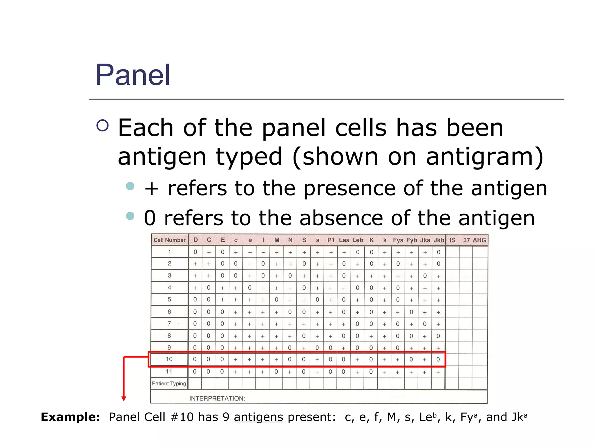
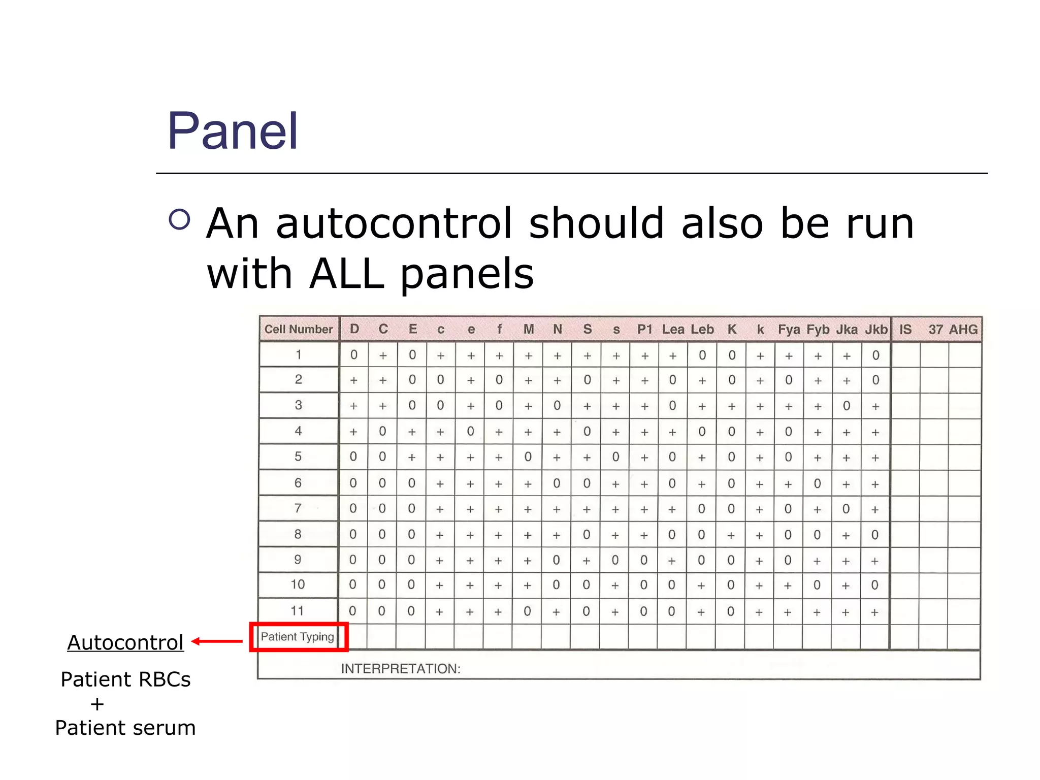
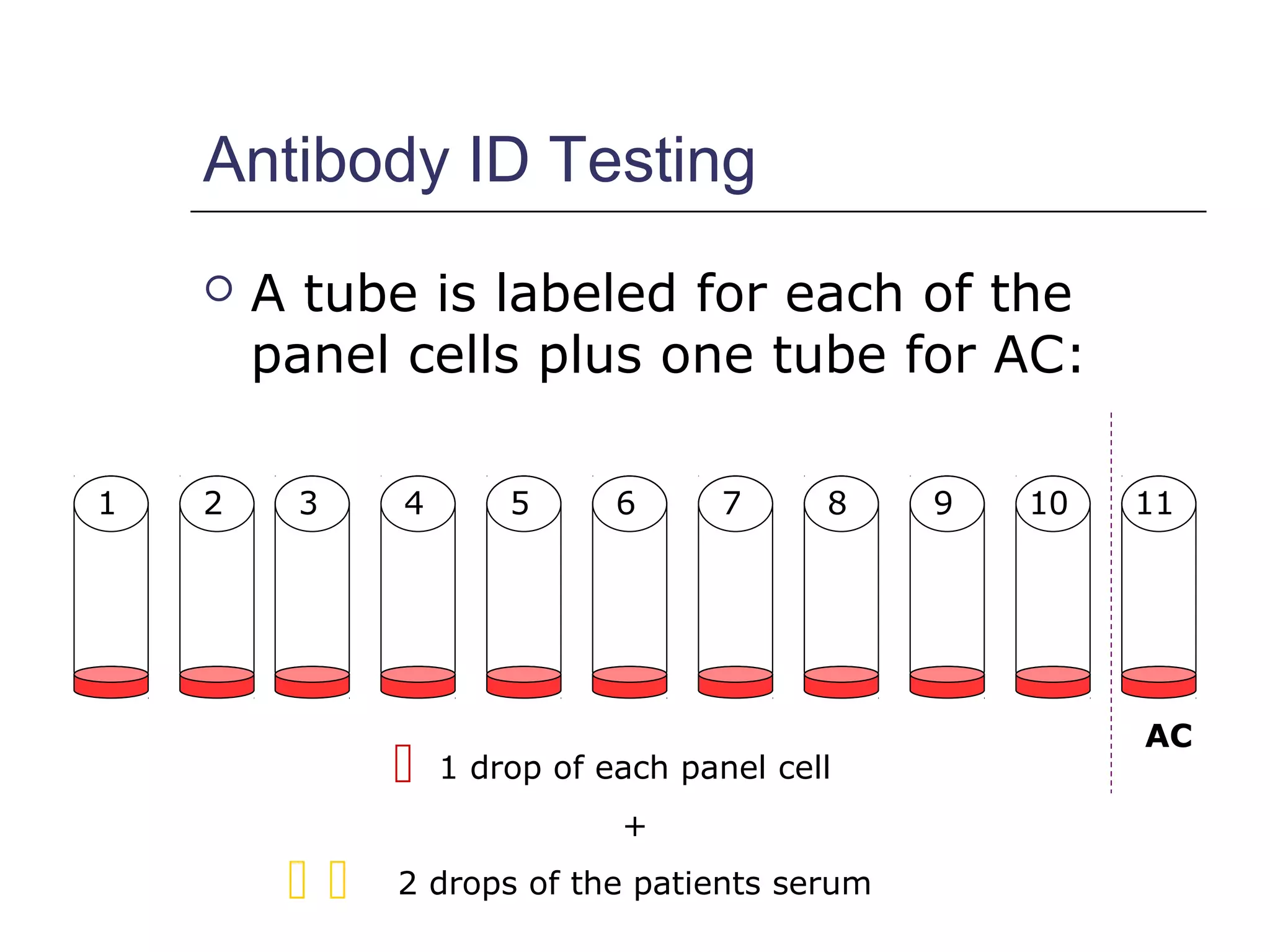
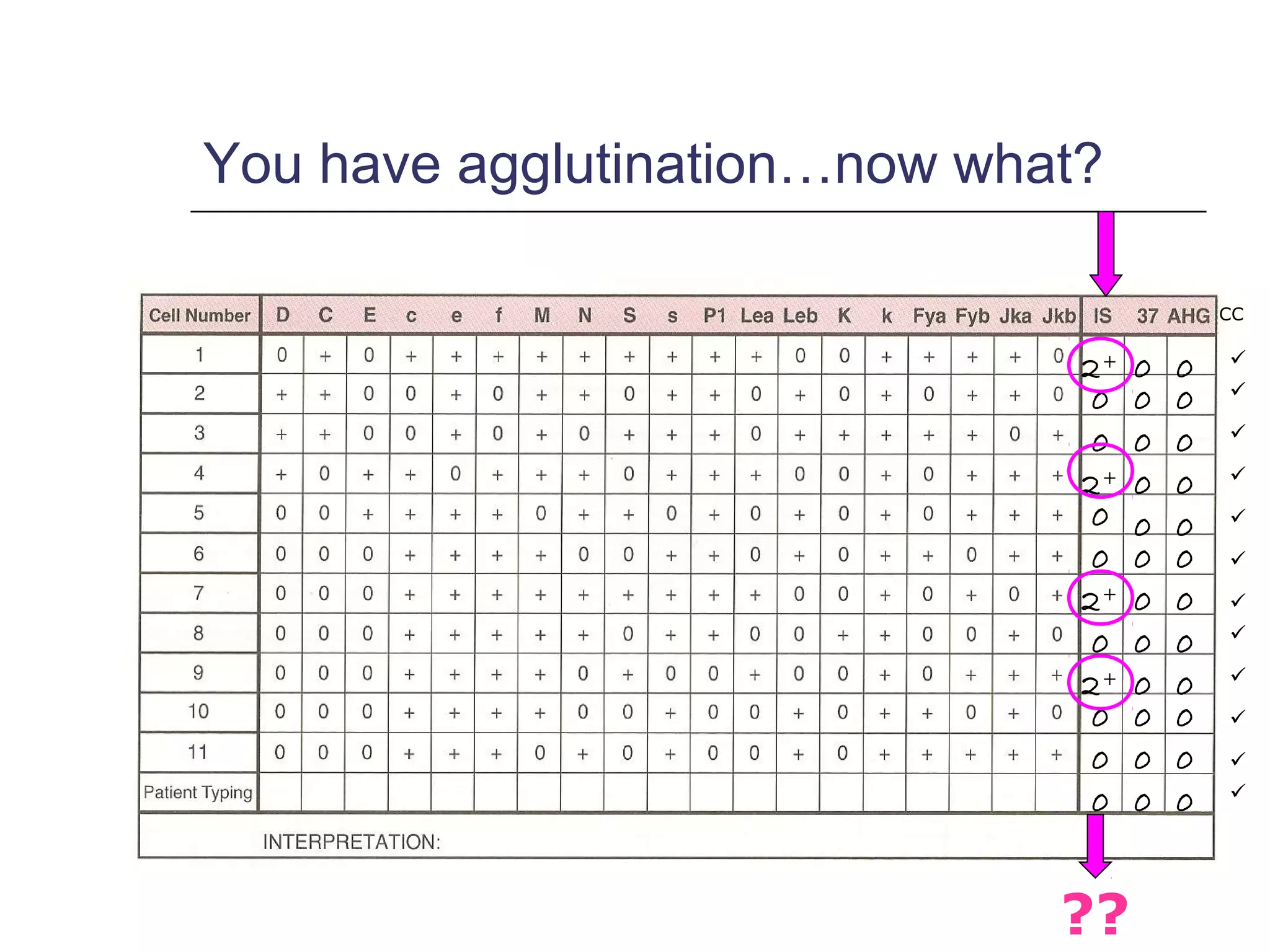
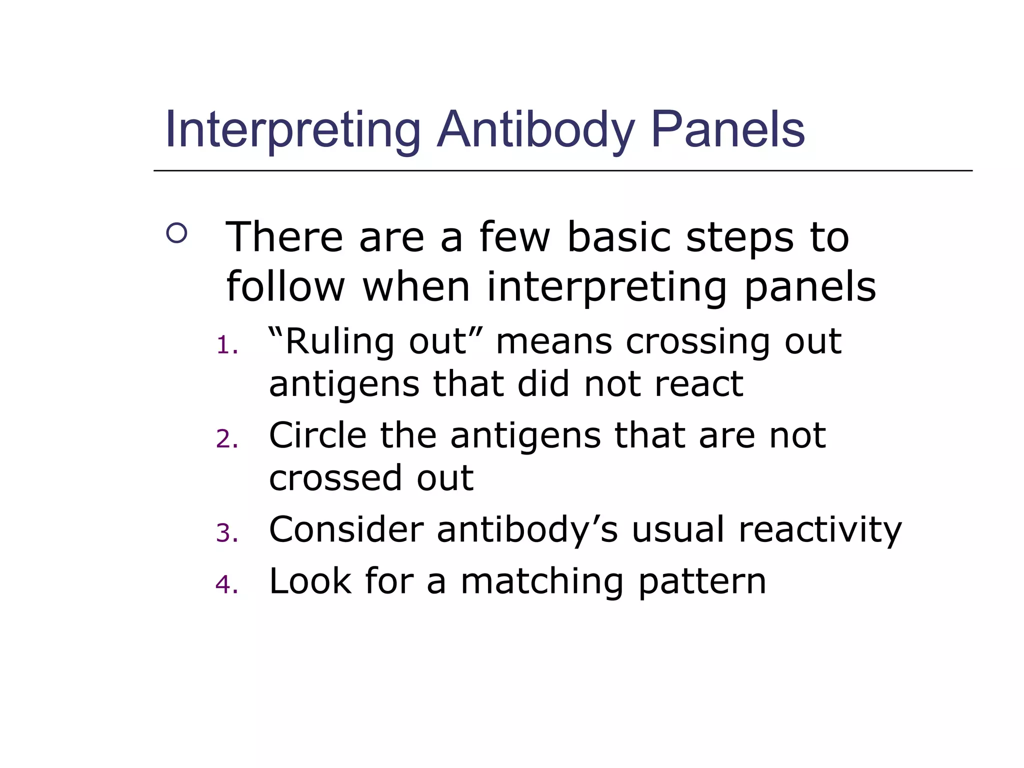
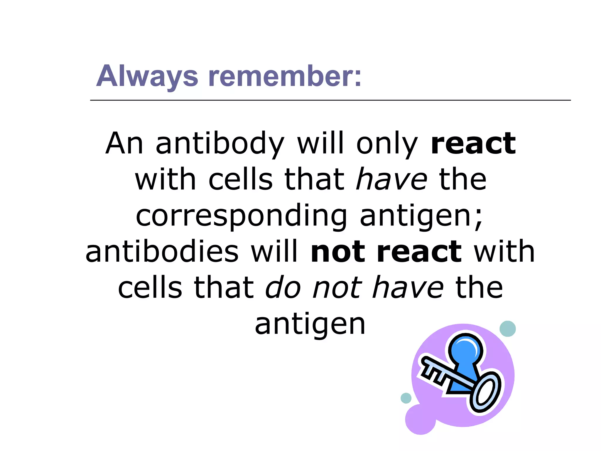
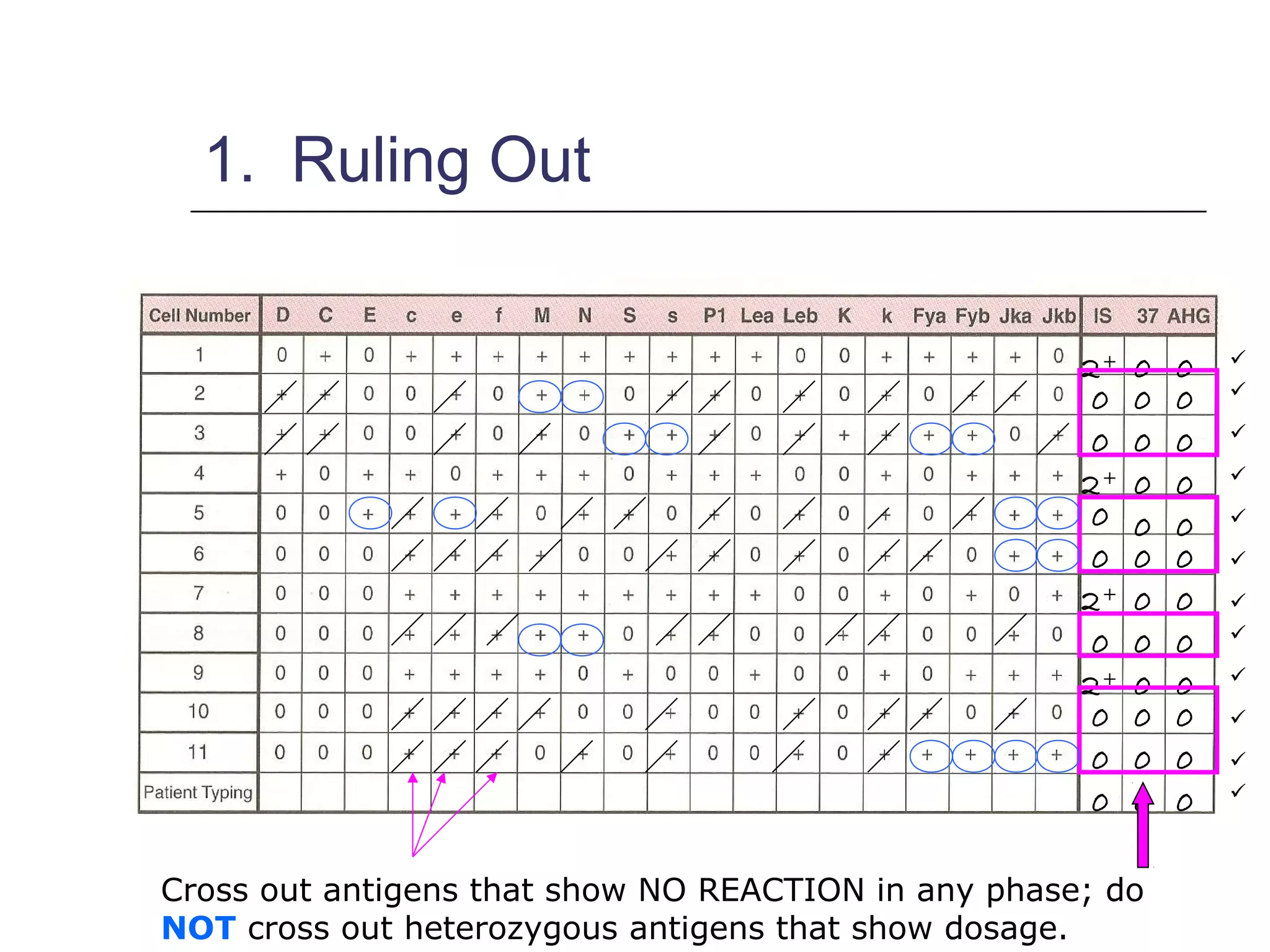
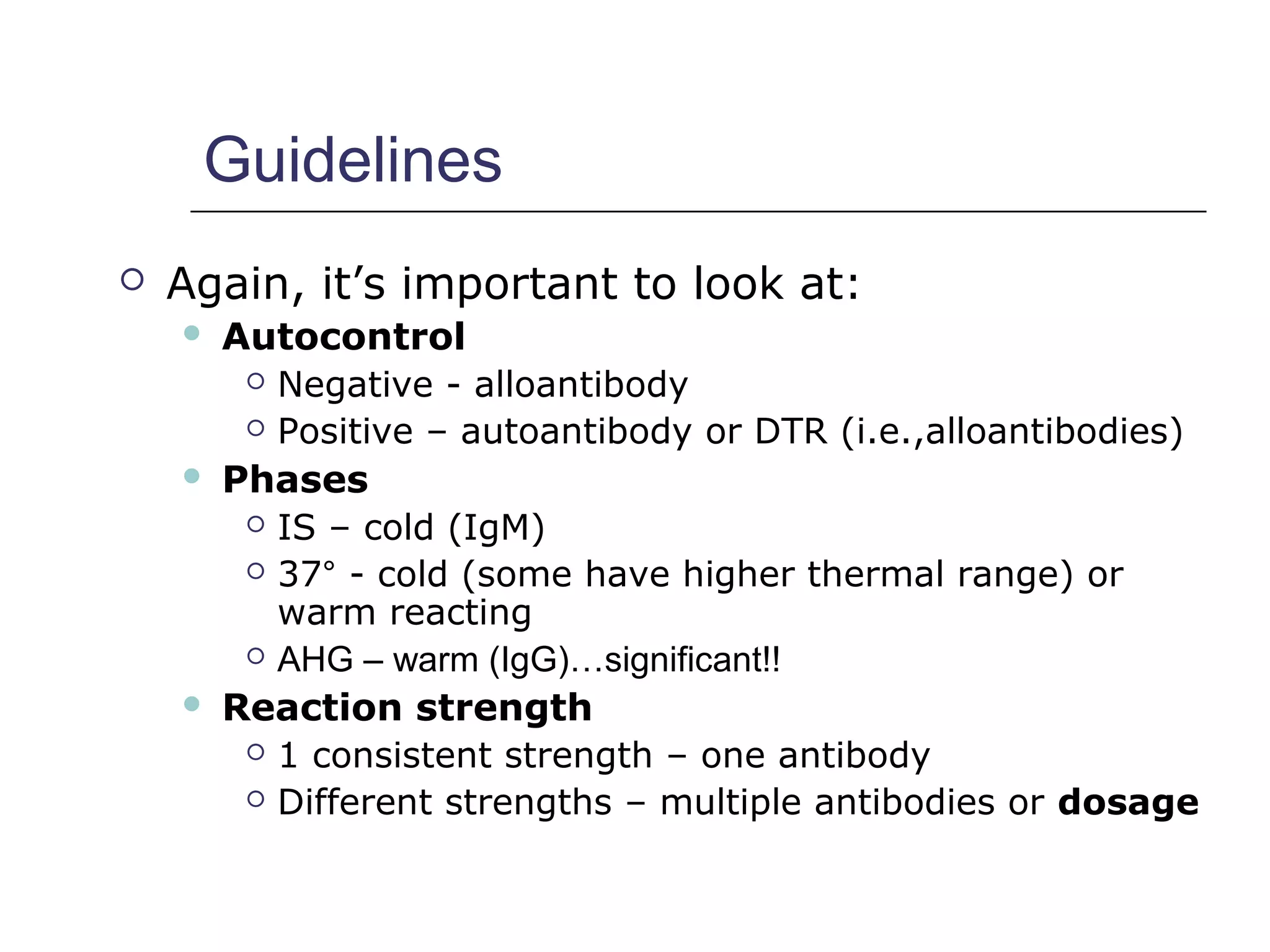
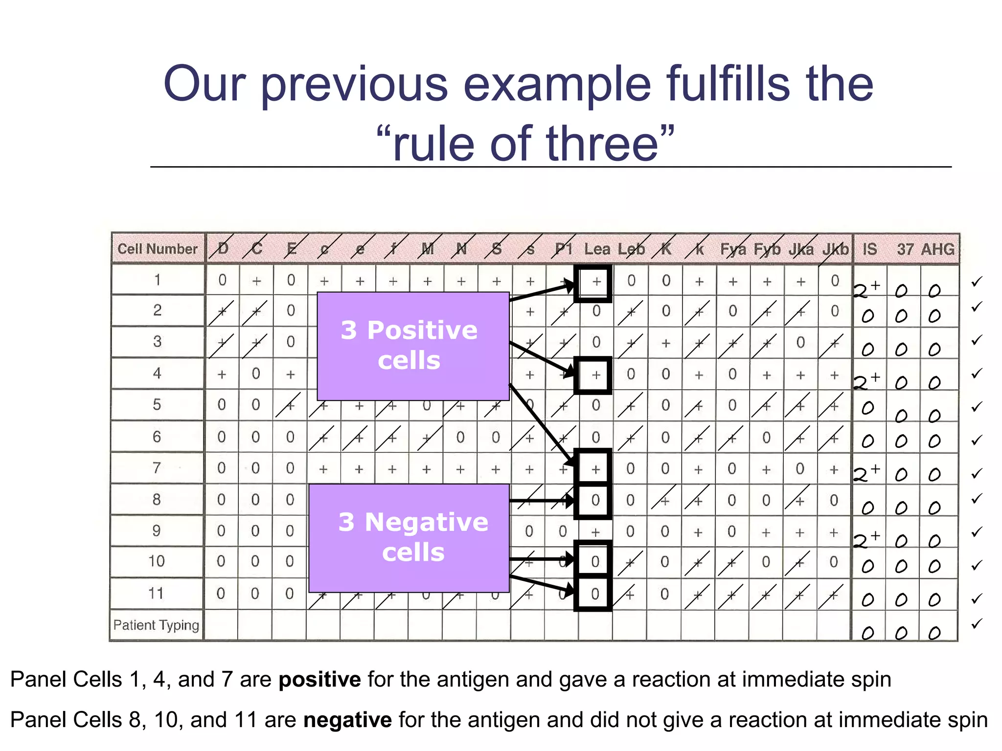
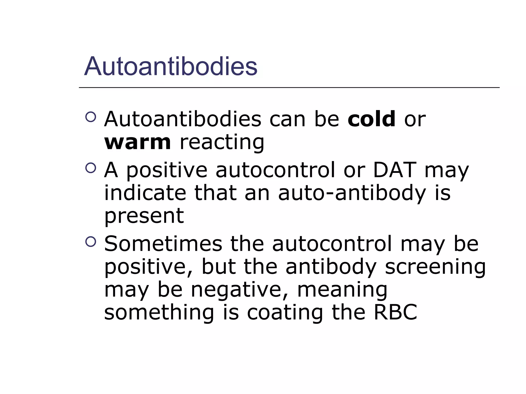
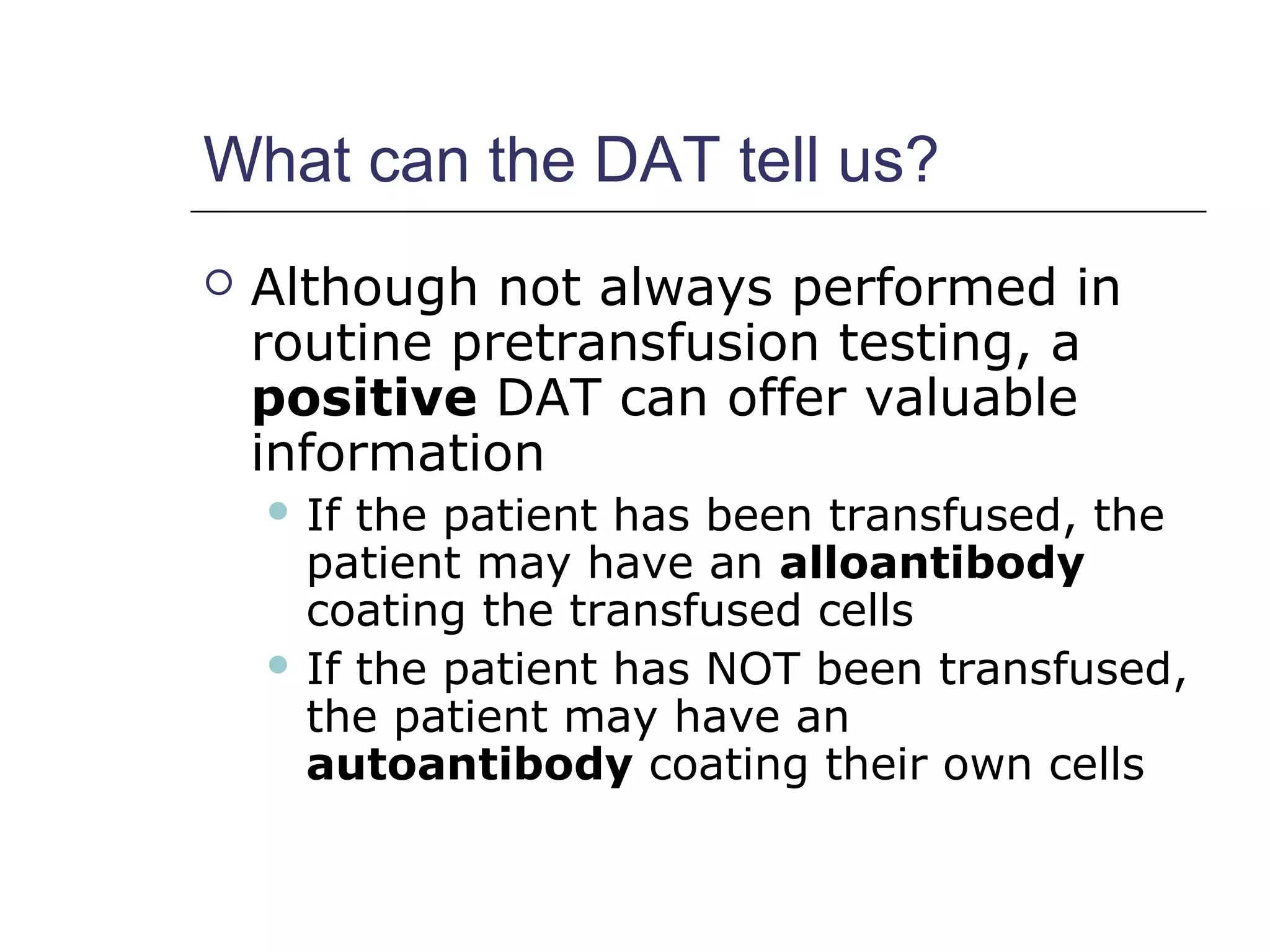
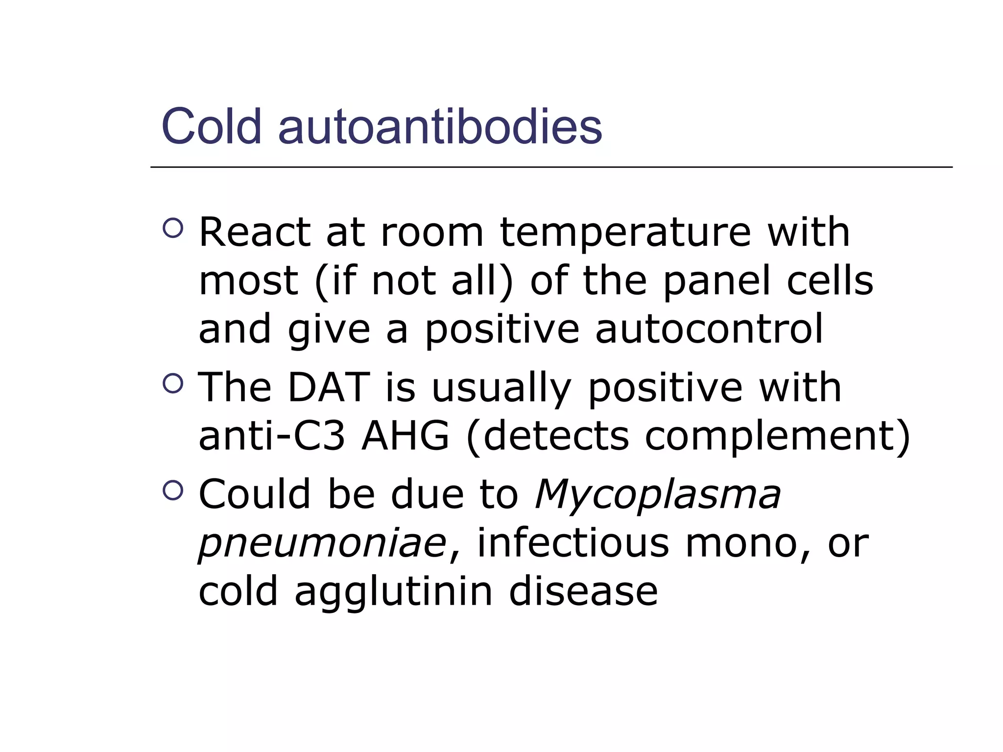
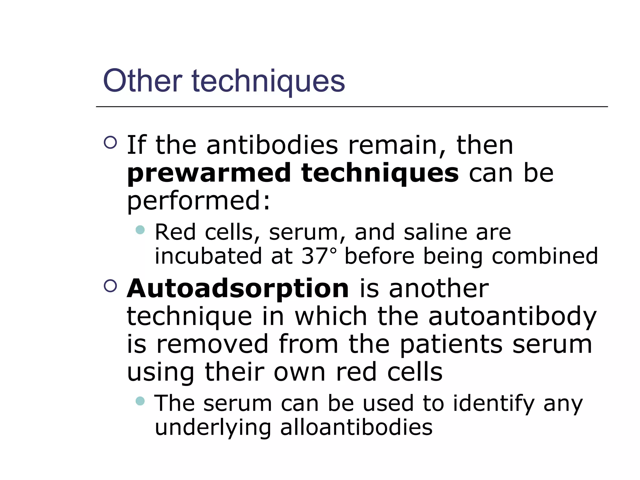
Antibody Identification
Renee Wilkins, PhD, MLS(ASCP)cm
CLS 325/435
School of Health Related Professions
University of Mississippi Medical Center
The Basics…..
 As you recall,
 Antibody Screens use 2 or 3 Screening
Cells to “detect” if antibodies are
present in the serum
 If antibodies are detected, they must
be identified…
present
Not present
Why do we need to identify?
 Antibody identification is needed for
transfusion purposes and is an
important component of
compatibility testing
 It will identify any unexpected
antibodies in the patient’s serum
 If a person with an antibody is
exposed to donor cells with the
corresponding antigen, serious side
effects can occur
Key Concepts
 In blood banking, we test “knowns” with
“unknowns”
 When detecting and/or identifying
antibodies, we test patient serum
(unknown) with reagent RBCs (known)
Known: Unknown:
Reagent RBCs + patient serum
Reagent antisera + patient RBCs
Reagent RBCs
 Screening Cells and Panel Cells are
the same with minor differences:
 Screening cells
 Antibody detection
 Sets of 2 or 3 vials
 Panel cells
 Antibody identification
 At least 10 vials per set
Antibody Panel vs. Screen
 An antibody panel is just an
extended version of an antibody
screen
 The screen only uses 2-3 cells:
Antibody Panel
 An antibody panel usually includes
at least 10 panel cells:
Panel
 Group O red blood cells
Panel
 Each of the panel cells has been
antigen typed (shown on antigram)
 + refers to the presence of the antigen
 0 refers to the absence of the antigen
Example: Panel Cell #10 has 9 antigens present: c, e, f, M, s, Leb
, k, Fya
, and Jka
Panel
 An autocontrol should also be run
with ALL panels
Autocontrol
Patient RBCs
+
Patient serum
Panel
 The same phases used in an
antibody screen are used in a panel
• IS
• 37°
• AHG
Antibody ID Testing
 A tube is labeled for each of the
panel cells plus one tube for AC:
AC
1 2 3 4 5 6 7 8 9 10 11
 1 drop of each panel cell
+
2 drops of the patients serum 
IS Phase
 Perform immediate spin (IS) and
grade agglutination; inspect for
hemolysis
 Record the results in the
appropriate space as shown:
2+
0
0
Last
tube
(LISS) 37°C Phase
 2 drops of LISS are added, mixed
and incubated for 10-15 minutes
 Centrifuge and check for
agglutination
 Record results
(LISS) 37°C Phase
2+
0
0
2+
0
0
2+
0
0
2+
0
0
0
0
IAT Phase (or AHG)
 Indirect Antiglobulin Test (IAT) –
we’re testing whether or not
possible antibodies in patient’s
serum will react with RBCs in vitro
 To do this we use the Anti-Human
Globulin reagent (AHG)
 Polyspecific
 Anti-IgG
 Anti-complement
AHG Phase
 Wash cells 3 times with saline
(manual or automated)
 Add 2 drops of AHG and gently mix
 Centrifuge
 Read
 Record reactions
AHG Phase
2+
0
0
2+
0
0
2+
0
0
2+
0
0
0
0
0
0
0
0
0
0
0
0
0
0
0
0
0
0
0
0
0
0
0
0 0 0
And don’t forget….
….add “check” cells to
any negative AHG !
IS LISS
37°
AHG CC
2+ 0 0 
0 0 0 
0 0 0 
2+ 0 0 
0 0 0 
0 0 0 
2+ 0 0 
0 0 0 
2+ 0 0 
0 0 0 
0 0 0 
All cells are
negative at
AHG, so
add
“Check”
Cells
You have agglutination…now what?
2+
0
0
2+
0
0
2+
0
0
2+
0
0
0
0
0
0
0
0
0
0
0
0
0
0
0
0
0
0
0
0
0
0
0
0 0 0












??
CC
Interpreting Antibody Panels
 There are a few basic steps to
follow when interpreting panels
1. “Ruling out” means crossing out
antigens that did not react
2. Circle the antigens that are not
crossed out
3. Consider antibody’s usual reactivity
4. Look for a matching pattern
An antibody will only react
with cells that have the
corresponding antigen;
antibodies will not react with
cells that do not have the
antigen
Always remember:
Here’s an example:
1. Ruling Out
2+
0
0
2+
0
0
2+
0
0
2+
0
0
0
0
0
0
0
0
0
0
0
0
0
0
0
0
0
0
0
0
0
0
0
0 0 0












Cross out antigens that show NO REACTION in any phase; do
NOT cross out heterozygous antigens that show dosage.
2. Circle antigens not crossed out
2+
0
0
2+
0
0
2+
0
0
2+
0
0
0
0
0
0
0
0
0
0
0
0
0
0
0
0
0
0
0
0
0
0
0
0 0 0












3. Consider antibody’s usual reactivity
2+
0
0
2+
0
0
2+
0
0
2+
0
0
0
0
0
0
0
0
0
0
0
0
0
0
0
0
0
0
0
0
0
0
0
0 0 0












Lea
is normally a Cold-Reacting antibody (IgM), so it makes
sense that we see the reaction in the IS phase of testing;
The E antigen will usually react at warmer temperatures
4. Look for a matching pattern
2+
0
0
2+
0
0
2+
0
0
2+
0
0
0
0
0
0
0
0
0
0
0
0
0
0
0
0
0
0
0
0
0
0
0
0 0 0












…Yes, there is a matching pattern!
E doesn’t match and
it’s a warmer rx Ab
Interpretation
anti-
Leaa
Guidelines
 Again, it’s important to look at:
 Autocontrol
 Negative - alloantibody
 Positive – autoantibody or DTR (i.e.,alloantibodies)
 Phases
 IS – cold (IgM)
 37° - cold (some have higher thermal range) or
warm reacting
 AHG – warm (IgG)…significant!!
 Reaction strength
 1 consistent strength – one antibody
 Different strengths – multiple antibodies or dosage
About reaction strengths……
 Strength of reaction may be due to
“dosage”
 If panel cells are homozygous, a strong
reaction may be seen
 If panel cells are heterozygous,
reaction may be weak or even non-
reactive
 Panel cells that are heterozygous
should not be crossed out because
antibody may be too weak to react
(see first example)
Guidelines (continued)
 Matching the pattern
 Single antibodies usually shows a
pattern that matches one of the
antigens (see previous panel example)
 Multiple antibodies are more difficult to
match because they often show mixed
reaction strengths
Rule of three
 The rule of three must be met to
confirm the presence of the antibody
 A p-value ≤ 0.05 must be observed
 This gives a 95% confidence interval
 How is it demonstrated?
 Patient serum MUST be:
 Positive with 3 cells with the antigen
 Negative with 3 cells without the antigen
Our previous example fulfills the
“rule of three”
2+
0
0
2+
0
0
2+
0
0
2+
0
0
0
0
0
0
0
0
0
0
0
0
0
0
0
0
0
0
0
0
0
0
0
0 0 0












3 Negative
cells
3 Positive
cells
Panel Cells 1, 4, and 7 are positive for the antigen and gave a reaction at immediate spin
Panel Cells 8, 10, and 11 are negative for the antigen and did not give a reaction at immediate spin
What if the “rule of three” is not fulfilled?
 If there are not enough cells in the
panel to fulfill the rule, then
additional cells from another panel
could be used
 Most labs carry different lot
numbers of panel cells
Phenotyping
 In addition to the rule of three,
antigen typing the patient red cells
can also confirm an antibody
 How is this done?
 Only perform this if the patient has NOT
been recently transfused (donor cells
could react)
 If reagent antisera (of the suspected
antibody) is added to the patient RBCs, a
negative reaction should result…Why?
Remember Landsteiner’s Rule
Individuals DO NOT make
allo-antibodies against
antigens they have
Multiple antibodies
 Multiple antibodies may be more of
a challenge than a single antibody
 Why?
 Reaction strengths can vary
 Matching the pattern is difficult
So what is a tech to do?
 Several procedures can be
performed to identify multiple
antibodies
 Selected Cells
 Neutralization
 Chemical treatment
 Proteolytic enzymes
 Sulfhydryl reagents
 ZZAP
Selected Cells
 Selected cells are chosen from other
panel or screening cells to confirm
or eliminate the antibody
 The cells are “selected” from other
panels because of their
characteristics
 The number of selected cells needed
depends on how may antibodies are
identified
Selected Cells
 Every cell should be positive for
each of the antibodies and negative
for the remaining antibodies
 For example:
 Let’s say you ran a panel and identified
3 different antibodies: anti-S, anti-Jka
,
and anti-P1
 Selected cells could help…
Selected Cells
Selected
cells
S Jka
P1 IS LISS
37°
AHG
#1 + 0 0 0 0 2+
#5 0 + 0 0 0 3+
#8 0 0 + 0 0 0
These results show that instead of 3 antibodies, there
are actually 2: anti-S and anti-Jka

Neutralization
 Some antibodies may be neutralized
as a way of confirmation
 Commercial “substances” bind to
the antibodies in the patient serum,
causing them to show no reaction
when tested with the corresponding
antigen (in panel)
Neutralization
 Manufacturer’s directions should be
followed and a dilutional control
should always be used
 The control contains saline and serum
(no substance) and should remain
positive
 A control shows that a loss of reactivity
is due to the neutralization and not to
the dilution of the antibody strength
when the substance is added
Neutralization
 Common substances
 P1 substance (sometimes derived from hydatid
cyst fluid)
 Lea
and Leb
substance (soluble antigen found in
plasma and saliva)
 I substance can be found in breast milk
 Sda
substance derived from human or guinea
pig urine
**you should be aware that many of these
substances neutralize COLD antibodies; Cold
antibodies can sometimes mask more clinically
significant antibodies (IgG), an important reason
to use neutralization techniques
Enzymes (proteolytic)
 Can be used to enhance or destroy
certain blood group antigens
 Several enzymes exist:
 Ficin (figs)
 Bromelin (pineapple)
 Papain (papaya)
 In addition, enzyme procedures
may be
 One-step
 Two-step
Enzymes
 Enzymes remove the sialic acid
from the RBC membrane, thus
“destroying” it and allowing other
antigens to be “enhanced”
 Antigens destroyed: M, N, S, s,
Duffy
 Antigens enhanced: Rh, Kidd,
Lewis, I, and P
Enzyme techniques
 One-stage
 Enzyme is added directly to the
serum/cell mixture
 Two-stage
 Panel cells are pre-treated with
enzyme, incubated and washed
 Patient serum is added to panel cells
and tested
Enzyme techniques
 If there is no agglutination after
treatment, then it is assumed the
enzymes destroyed the antigen
Enzyme
treament
Anti-K
Perfect match for anti-Fya
•Duffy antigens destroyed
•Kell antigens not affected
Enzyme treatment
Sulfhydryl Reagents
 Cleave the disulfide bonds of IgM
molecules and help differentiate
between IgM and IgG antibodies
 Good to use when you have both
IgG and IgM antibodies (warm/cold)
 Dithiothreitol (DTT) is a thiol and will
denature Kell antigens
 2-mercaptoethanol (2-ME)
ZZAP
 A combination of proteolytic
enzymes and DTT
 Denatures Kell, M, N, S, Duffy and
other less frequent blood group
antigens
 Does not denature the Kx antigen
 Good for adsorption techniques
 “frees” autoantibody off patient’s cell, so that
autoantibody can then be adsorbed onto another
RBC
Autoantibodies….
Warm & Cold Reacting
Autoantibodies
 Autoantibodies can be cold or
warm reacting
 A positive autocontrol or DAT may
indicate that an auto-antibody is
present
 Sometimes the autocontrol may be
positive, but the antibody screening
may be negative, meaning
something is coating the RBC
Getting a positive DAT
 We have focused a lot on the IAT
used in antibody screening and ID,
but what about the DAT?
 The direct antiglobulin test
(DAT) tests for the in vivo coating
of RBCs with antibody (in the body)
 AHG is added to washed patient red
cells to determine this
What can the DAT tell us?
 Although not always performed in
routine pretransfusion testing, a
positive DAT can offer valuable
information
 If the patient has been transfused, the
patient may have an alloantibody
coating the transfused cells
 If the patient has NOT been transfused,
the patient may have an
autoantibody coating their own cells
Identifying autoantibodies
 Auto-antibodies can sometimes
“mask” clinically significant allo-
antibodies, so it’s important to
differentiate between auto- and
allo-antibodies
Cold autoantibodies
 React at room temperature with
most (if not all) of the panel cells
and give a positive autocontrol
 The DAT is usually positive with
anti-C3 AHG (detects complement)
 Could be due to Mycoplasma
pneumoniae, infectious mono, or
cold agglutinin disease
Cold autoantibodies
 Mini-cold panels can be used to help
identify cold autoantibodies
 Since anti-I is a common
autoantibody, cord blood cells (no I
antigen) are usually included
Group O
individual with
cold autoanti-I
Group A
individual with
cold autoanti-IH
Anti-IH is reacting weakly with the cord
cells (some H antigen present)
Avoiding reactivity
 Cold autoantibodies can be a
nuisance at times. Here are a few
ways to avoid a reaction:
 Use anti-IgG AHG instead of
polyspecific. Most cold antibodies react
with polyspecific AHG and anti-C AHG
because they fix complement
 Skipping the IS phase avoids the
attachment of cold autoantibodies to
the red cells
 Use 22% BSA instead of LISS
Other techniques
 If the antibodies remain, then
prewarmed techniques can be
performed:
 Red cells, serum, and saline are
incubated at 37° before being combined
 Autoadsorption is another
technique in which the autoantibody
is removed from the patients serum
using their own red cells
 The serum can be used to identify any
underlying alloantibodies
Warm autoantibodies
 More common that cold
autoantibodies
 Positive DAT due to IgG antibodies
coating the red cell
 Again, the majority of panel or
screening cells will be positive
 The Rh system (e antigen) seems to
be the main target although others
occur
Warm autoantibodies
 Cause warm autoimmune hemolytic
anemia (WAIHA)…H&H
 How do you get a warm autoantibody?
 Idiopathic
 Known disorder (SLE, RA, leukemias, UC,
pregnancy, infectious diseases, etc)
 Medications
 Several techniques are used when
warm autoantibodies are suspected…
Elution (whenever DAT is positive)
Elution techniques “free”
antibodies from the sensitized
red cells so that the antibodies
can be identified
Y
Y
Y
Y
Sensitized
RBC
Positive DAT
Elution
Y
Y
Y
Y
Y
Y
Frees antibody Antibody ID
Elution
 The eluate is a term used for the
removed antibodies
 Testing the eluate is useful in
investigations of positive DATs
 HDN
 Transfusion reactions
 Autoimmune disease
 The red cells can also be used after
elution for RBC phenotyping if needed
 When tested with panel cells, the eluate
usually remains reactive with all cells if a
warm autoantibody is present
Elution Methods
 Acid elutions (glycine acid)
 Most common
 Lowers pH, causing antibody to
dissociate
 Organic solvents (ether, chloroform)
 Dissolve bilipid layer of RBC
 Heat (conformational change)
 Freeze-Thaw (lyses cells)
ABO
antibodies
Adsorption
 Adsorption procedures can be used
to investigate underlying
alloantibodies
 ZZAP or chloroquine
diphosphate can be used to
dissociate IgG antibodies from the
RBC (may take several repeats)
 After the patient RBCs are
incubated, the adsorbed serum is
tested with panel cells to ID the
alloantibody (if present)
Adsorption
 Two types:
 Autoadsorption
 No recent transfusion
 Autoantibodies are removed using patient
RBCs, so alloantibodies can be identified
 Allogenic (Differential) adsorption
 If recently transfused
 Uses other cells with the patients serum
Wash x3 after
incubation
Centrifuge after
incubating; and
transfer serum to 2nd
tube of treated cells;
incubate and
centrifuge again
2
tubes
Remove
serum and
test for
alloantibody
More reagents….
 Many of elution tests can damage
the antigens on the RBC
 Choroquine diphosphate (CDP)
and glycine acid EDTA reagents can
dissociate IgG from the RBC without
damaging the antigens
 Very useful if the RBC needs to be
antigen typed
Chloroquine diphosphate
 Quinilone derivative often used as
an antimalarial
 May not remove autoantibody
completely from DAT positive cells
 Partial removal may be enough to
antigen type the cells or to be used
for autoadsorption of warm
autoantibodies
THE END!!

Ab identificationreneewilkins

  • 1.
    Antibody Identification Renee Wilkins,PhD, MLS(ASCP)cm CLS 325/435 School of Health Related Professions University of Mississippi Medical Center
  • 2.
    The Basics…..  Asyou recall,  Antibody Screens use 2 or 3 Screening Cells to “detect” if antibodies are present in the serum  If antibodies are detected, they must be identified… present Not present
  • 3.
    Why do weneed to identify?  Antibody identification is needed for transfusion purposes and is an important component of compatibility testing  It will identify any unexpected antibodies in the patient’s serum  If a person with an antibody is exposed to donor cells with the corresponding antigen, serious side effects can occur
  • 4.
    Key Concepts  Inblood banking, we test “knowns” with “unknowns”  When detecting and/or identifying antibodies, we test patient serum (unknown) with reagent RBCs (known) Known: Unknown: Reagent RBCs + patient serum Reagent antisera + patient RBCs
  • 5.
    Reagent RBCs  ScreeningCells and Panel Cells are the same with minor differences:  Screening cells  Antibody detection  Sets of 2 or 3 vials  Panel cells  Antibody identification  At least 10 vials per set
  • 6.
    Antibody Panel vs.Screen  An antibody panel is just an extended version of an antibody screen  The screen only uses 2-3 cells:
  • 7.
    Antibody Panel  Anantibody panel usually includes at least 10 panel cells:
  • 8.
    Panel  Group Ored blood cells
  • 9.
    Panel  Each ofthe panel cells has been antigen typed (shown on antigram)  + refers to the presence of the antigen  0 refers to the absence of the antigen Example: Panel Cell #10 has 9 antigens present: c, e, f, M, s, Leb , k, Fya , and Jka
  • 10.
    Panel  An autocontrolshould also be run with ALL panels Autocontrol Patient RBCs + Patient serum
  • 11.
    Panel  The samephases used in an antibody screen are used in a panel • IS • 37° • AHG
  • 12.
    Antibody ID Testing A tube is labeled for each of the panel cells plus one tube for AC: AC 1 2 3 4 5 6 7 8 9 10 11  1 drop of each panel cell + 2 drops of the patients serum 
  • 13.
    IS Phase  Performimmediate spin (IS) and grade agglutination; inspect for hemolysis  Record the results in the appropriate space as shown: 2+ 0 0 Last tube
  • 14.
    (LISS) 37°C Phase 2 drops of LISS are added, mixed and incubated for 10-15 minutes  Centrifuge and check for agglutination  Record results
  • 17.
  • 18.
    IAT Phase (orAHG)  Indirect Antiglobulin Test (IAT) – we’re testing whether or not possible antibodies in patient’s serum will react with RBCs in vitro  To do this we use the Anti-Human Globulin reagent (AHG)  Polyspecific  Anti-IgG  Anti-complement
  • 19.
    AHG Phase  Washcells 3 times with saline (manual or automated)  Add 2 drops of AHG and gently mix  Centrifuge  Read  Record reactions
  • 20.
  • 21.
    And don’t forget…. ….add“check” cells to any negative AHG !
  • 22.
    IS LISS 37° AHG CC 2+0 0  0 0 0  0 0 0  2+ 0 0  0 0 0  0 0 0  2+ 0 0  0 0 0  2+ 0 0  0 0 0  0 0 0  All cells are negative at AHG, so add “Check” Cells
  • 23.
    You have agglutination…nowwhat? 2+ 0 0 2+ 0 0 2+ 0 0 2+ 0 0 0 0 0 0 0 0 0 0 0 0 0 0 0 0 0 0 0 0 0 0 0 0 0 0             ?? CC
  • 24.
    Interpreting Antibody Panels There are a few basic steps to follow when interpreting panels 1. “Ruling out” means crossing out antigens that did not react 2. Circle the antigens that are not crossed out 3. Consider antibody’s usual reactivity 4. Look for a matching pattern
  • 25.
    An antibody willonly react with cells that have the corresponding antigen; antibodies will not react with cells that do not have the antigen Always remember:
  • 26.
  • 27.
    1. Ruling Out 2+ 0 0 2+ 0 0 2+ 0 0 2+ 0 0 0 0 0 0 0 0 0 0 0 0 0 0 0 0 0 0 0 0 0 0 0 00 0             Cross out antigens that show NO REACTION in any phase; do NOT cross out heterozygous antigens that show dosage.
  • 28.
    2. Circle antigensnot crossed out 2+ 0 0 2+ 0 0 2+ 0 0 2+ 0 0 0 0 0 0 0 0 0 0 0 0 0 0 0 0 0 0 0 0 0 0 0 0 0 0            
  • 29.
    3. Consider antibody’susual reactivity 2+ 0 0 2+ 0 0 2+ 0 0 2+ 0 0 0 0 0 0 0 0 0 0 0 0 0 0 0 0 0 0 0 0 0 0 0 0 0 0             Lea is normally a Cold-Reacting antibody (IgM), so it makes sense that we see the reaction in the IS phase of testing; The E antigen will usually react at warmer temperatures
  • 30.
    4. Look fora matching pattern 2+ 0 0 2+ 0 0 2+ 0 0 2+ 0 0 0 0 0 0 0 0 0 0 0 0 0 0 0 0 0 0 0 0 0 0 0 0 0 0             …Yes, there is a matching pattern! E doesn’t match and it’s a warmer rx Ab
  • 31.
  • 32.
    Guidelines  Again, it’simportant to look at:  Autocontrol  Negative - alloantibody  Positive – autoantibody or DTR (i.e.,alloantibodies)  Phases  IS – cold (IgM)  37° - cold (some have higher thermal range) or warm reacting  AHG – warm (IgG)…significant!!  Reaction strength  1 consistent strength – one antibody  Different strengths – multiple antibodies or dosage
  • 33.
    About reaction strengths…… Strength of reaction may be due to “dosage”  If panel cells are homozygous, a strong reaction may be seen  If panel cells are heterozygous, reaction may be weak or even non- reactive  Panel cells that are heterozygous should not be crossed out because antibody may be too weak to react (see first example)
  • 34.
    Guidelines (continued)  Matchingthe pattern  Single antibodies usually shows a pattern that matches one of the antigens (see previous panel example)  Multiple antibodies are more difficult to match because they often show mixed reaction strengths
  • 35.
    Rule of three The rule of three must be met to confirm the presence of the antibody  A p-value ≤ 0.05 must be observed  This gives a 95% confidence interval  How is it demonstrated?  Patient serum MUST be:  Positive with 3 cells with the antigen  Negative with 3 cells without the antigen
  • 36.
    Our previous examplefulfills the “rule of three” 2+ 0 0 2+ 0 0 2+ 0 0 2+ 0 0 0 0 0 0 0 0 0 0 0 0 0 0 0 0 0 0 0 0 0 0 0 0 0 0             3 Negative cells 3 Positive cells Panel Cells 1, 4, and 7 are positive for the antigen and gave a reaction at immediate spin Panel Cells 8, 10, and 11 are negative for the antigen and did not give a reaction at immediate spin
  • 37.
    What if the“rule of three” is not fulfilled?  If there are not enough cells in the panel to fulfill the rule, then additional cells from another panel could be used  Most labs carry different lot numbers of panel cells
  • 38.
    Phenotyping  In additionto the rule of three, antigen typing the patient red cells can also confirm an antibody  How is this done?  Only perform this if the patient has NOT been recently transfused (donor cells could react)  If reagent antisera (of the suspected antibody) is added to the patient RBCs, a negative reaction should result…Why?
  • 39.
    Remember Landsteiner’s Rule IndividualsDO NOT make allo-antibodies against antigens they have
  • 40.
    Multiple antibodies  Multipleantibodies may be more of a challenge than a single antibody  Why?  Reaction strengths can vary  Matching the pattern is difficult
  • 41.
    So what isa tech to do?  Several procedures can be performed to identify multiple antibodies  Selected Cells  Neutralization  Chemical treatment  Proteolytic enzymes  Sulfhydryl reagents  ZZAP
  • 42.
    Selected Cells  Selectedcells are chosen from other panel or screening cells to confirm or eliminate the antibody  The cells are “selected” from other panels because of their characteristics  The number of selected cells needed depends on how may antibodies are identified
  • 43.
    Selected Cells  Everycell should be positive for each of the antibodies and negative for the remaining antibodies  For example:  Let’s say you ran a panel and identified 3 different antibodies: anti-S, anti-Jka , and anti-P1  Selected cells could help…
  • 44.
    Selected Cells Selected cells S Jka P1IS LISS 37° AHG #1 + 0 0 0 0 2+ #5 0 + 0 0 0 3+ #8 0 0 + 0 0 0 These results show that instead of 3 antibodies, there are actually 2: anti-S and anti-Jka 
  • 45.
    Neutralization  Some antibodiesmay be neutralized as a way of confirmation  Commercial “substances” bind to the antibodies in the patient serum, causing them to show no reaction when tested with the corresponding antigen (in panel)
  • 46.
    Neutralization  Manufacturer’s directionsshould be followed and a dilutional control should always be used  The control contains saline and serum (no substance) and should remain positive  A control shows that a loss of reactivity is due to the neutralization and not to the dilution of the antibody strength when the substance is added
  • 47.
    Neutralization  Common substances P1 substance (sometimes derived from hydatid cyst fluid)  Lea and Leb substance (soluble antigen found in plasma and saliva)  I substance can be found in breast milk  Sda substance derived from human or guinea pig urine **you should be aware that many of these substances neutralize COLD antibodies; Cold antibodies can sometimes mask more clinically significant antibodies (IgG), an important reason to use neutralization techniques
  • 48.
    Enzymes (proteolytic)  Canbe used to enhance or destroy certain blood group antigens  Several enzymes exist:  Ficin (figs)  Bromelin (pineapple)  Papain (papaya)  In addition, enzyme procedures may be  One-step  Two-step
  • 49.
    Enzymes  Enzymes removethe sialic acid from the RBC membrane, thus “destroying” it and allowing other antigens to be “enhanced”  Antigens destroyed: M, N, S, s, Duffy  Antigens enhanced: Rh, Kidd, Lewis, I, and P
  • 50.
    Enzyme techniques  One-stage Enzyme is added directly to the serum/cell mixture  Two-stage  Panel cells are pre-treated with enzyme, incubated and washed  Patient serum is added to panel cells and tested
  • 51.
    Enzyme techniques  Ifthere is no agglutination after treatment, then it is assumed the enzymes destroyed the antigen
  • 52.
    Enzyme treament Anti-K Perfect match foranti-Fya •Duffy antigens destroyed •Kell antigens not affected Enzyme treatment
  • 53.
    Sulfhydryl Reagents  Cleavethe disulfide bonds of IgM molecules and help differentiate between IgM and IgG antibodies  Good to use when you have both IgG and IgM antibodies (warm/cold)  Dithiothreitol (DTT) is a thiol and will denature Kell antigens  2-mercaptoethanol (2-ME)
  • 54.
    ZZAP  A combinationof proteolytic enzymes and DTT  Denatures Kell, M, N, S, Duffy and other less frequent blood group antigens  Does not denature the Kx antigen  Good for adsorption techniques  “frees” autoantibody off patient’s cell, so that autoantibody can then be adsorbed onto another RBC
  • 55.
  • 56.
    Autoantibodies  Autoantibodies canbe cold or warm reacting  A positive autocontrol or DAT may indicate that an auto-antibody is present  Sometimes the autocontrol may be positive, but the antibody screening may be negative, meaning something is coating the RBC
  • 57.
    Getting a positiveDAT  We have focused a lot on the IAT used in antibody screening and ID, but what about the DAT?  The direct antiglobulin test (DAT) tests for the in vivo coating of RBCs with antibody (in the body)  AHG is added to washed patient red cells to determine this
  • 58.
    What can theDAT tell us?  Although not always performed in routine pretransfusion testing, a positive DAT can offer valuable information  If the patient has been transfused, the patient may have an alloantibody coating the transfused cells  If the patient has NOT been transfused, the patient may have an autoantibody coating their own cells
  • 59.
    Identifying autoantibodies  Auto-antibodiescan sometimes “mask” clinically significant allo- antibodies, so it’s important to differentiate between auto- and allo-antibodies
  • 60.
    Cold autoantibodies  Reactat room temperature with most (if not all) of the panel cells and give a positive autocontrol  The DAT is usually positive with anti-C3 AHG (detects complement)  Could be due to Mycoplasma pneumoniae, infectious mono, or cold agglutinin disease
  • 61.
    Cold autoantibodies  Mini-coldpanels can be used to help identify cold autoantibodies  Since anti-I is a common autoantibody, cord blood cells (no I antigen) are usually included Group O individual with cold autoanti-I Group A individual with cold autoanti-IH Anti-IH is reacting weakly with the cord cells (some H antigen present)
  • 62.
    Avoiding reactivity  Coldautoantibodies can be a nuisance at times. Here are a few ways to avoid a reaction:  Use anti-IgG AHG instead of polyspecific. Most cold antibodies react with polyspecific AHG and anti-C AHG because they fix complement  Skipping the IS phase avoids the attachment of cold autoantibodies to the red cells  Use 22% BSA instead of LISS
  • 63.
    Other techniques  Ifthe antibodies remain, then prewarmed techniques can be performed:  Red cells, serum, and saline are incubated at 37° before being combined  Autoadsorption is another technique in which the autoantibody is removed from the patients serum using their own red cells  The serum can be used to identify any underlying alloantibodies
  • 64.
    Warm autoantibodies  Morecommon that cold autoantibodies  Positive DAT due to IgG antibodies coating the red cell  Again, the majority of panel or screening cells will be positive  The Rh system (e antigen) seems to be the main target although others occur
  • 65.
    Warm autoantibodies  Causewarm autoimmune hemolytic anemia (WAIHA)…H&H  How do you get a warm autoantibody?  Idiopathic  Known disorder (SLE, RA, leukemias, UC, pregnancy, infectious diseases, etc)  Medications  Several techniques are used when warm autoantibodies are suspected…
  • 66.
    Elution (whenever DATis positive) Elution techniques “free” antibodies from the sensitized red cells so that the antibodies can be identified Y Y Y Y Sensitized RBC Positive DAT Elution Y Y Y Y Y Y Frees antibody Antibody ID
  • 67.
    Elution  The eluateis a term used for the removed antibodies  Testing the eluate is useful in investigations of positive DATs  HDN  Transfusion reactions  Autoimmune disease  The red cells can also be used after elution for RBC phenotyping if needed  When tested with panel cells, the eluate usually remains reactive with all cells if a warm autoantibody is present
  • 68.
    Elution Methods  Acidelutions (glycine acid)  Most common  Lowers pH, causing antibody to dissociate  Organic solvents (ether, chloroform)  Dissolve bilipid layer of RBC  Heat (conformational change)  Freeze-Thaw (lyses cells) ABO antibodies
  • 69.
    Adsorption  Adsorption procedurescan be used to investigate underlying alloantibodies  ZZAP or chloroquine diphosphate can be used to dissociate IgG antibodies from the RBC (may take several repeats)  After the patient RBCs are incubated, the adsorbed serum is tested with panel cells to ID the alloantibody (if present)
  • 70.
    Adsorption  Two types: Autoadsorption  No recent transfusion  Autoantibodies are removed using patient RBCs, so alloantibodies can be identified  Allogenic (Differential) adsorption  If recently transfused  Uses other cells with the patients serum
  • 71.
    Wash x3 after incubation Centrifugeafter incubating; and transfer serum to 2nd tube of treated cells; incubate and centrifuge again 2 tubes Remove serum and test for alloantibody
  • 72.
    More reagents….  Manyof elution tests can damage the antigens on the RBC  Choroquine diphosphate (CDP) and glycine acid EDTA reagents can dissociate IgG from the RBC without damaging the antigens  Very useful if the RBC needs to be antigen typed
  • 73.
    Chloroquine diphosphate  Quinilonederivative often used as an antimalarial  May not remove autoantibody completely from DAT positive cells  Partial removal may be enough to antigen type the cells or to be used for autoadsorption of warm autoantibodies
  • 74.

Editor's Notes

  • #33 DTR – delayed transfusion reaction (donor cells are sensitized with patient’s antibody)
  • #45 These are panel cells that are being used, I just didn’t include the rest of the antigram because it is not necessary.