Downloaded 194 times
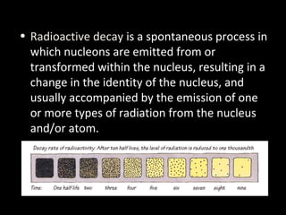


![Suffering from thyroid cancer, Oleg, 54, and Dima, 13,
receive care at a thyroid hospital in Minsk, where
surgery is performed daily.
As a liquidator, Oleg was exposed to extreme levels of
radiation. This was his third thyroid operation.
Dima’s mother claims that Chernobyl’s nuclear fallout is
responsible for her son’s cancer,
but Belarusian officials are often instructed to downplay
the severity of the radiation. [Minsk, Belarus 2005]](https://image.slidesharecdn.com/nuclearradiation-160323093157/85/Nuclear-radiation-58-320.jpg)


Okay, let's solve this step-by-step: * Original amount: 64 g * Amount after t hours: 2 g * Half-life: 1/2 hours * To find t, we set up the half-life equation: N = N0 * (1/2)^(t/T1/2) * Plug in the values: 2 = 64 * (1/2)^(t/0.5) * Take the log of both sides: log(2) = log(64) - (t/0.5)log(2) * Solve for t: t = 1 hour Therefore, the time for the amount to reduce to 2


![谷歌留痕技术 [ 𝙩𝙤𝙥 𝟮𝟯𝟯. 𝙘 𝙤𝙢 ]](https://cdn.slidesharecdn.com/ss_thumbnails/top233-260130174328-3833018c-thumbnail.jpg?width=640&height=640&fit=bounds)


