Downloaded 461 times
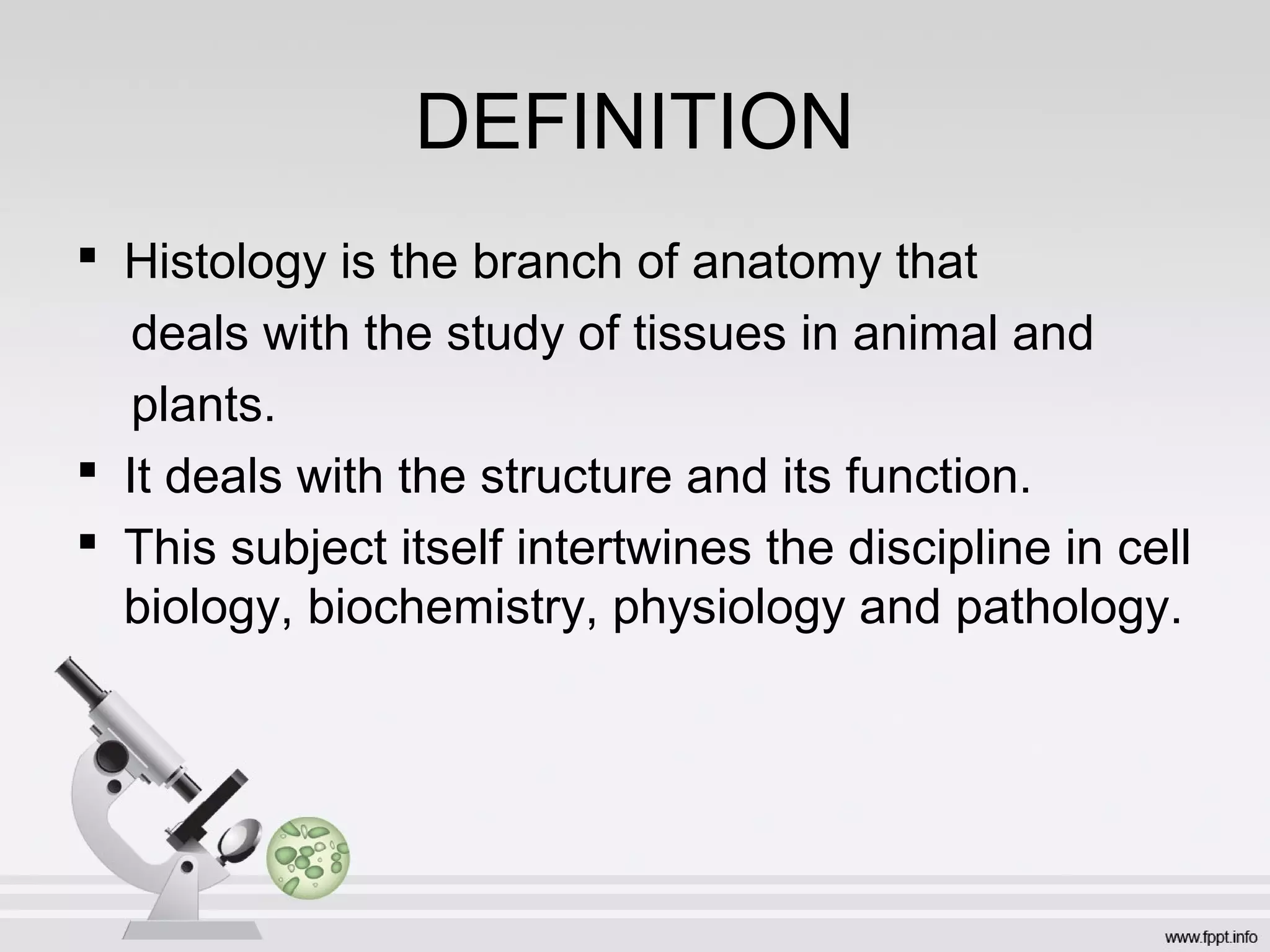
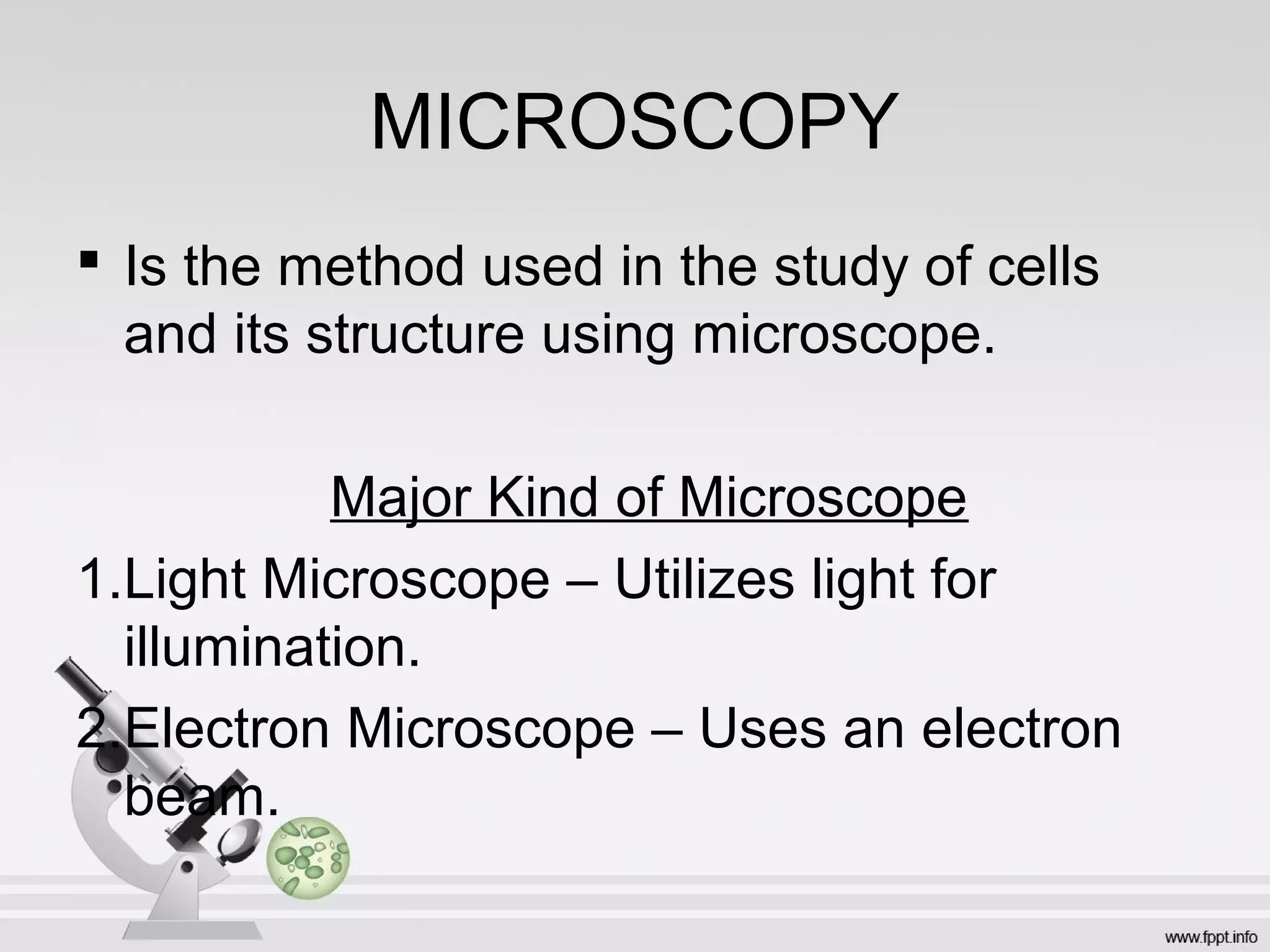
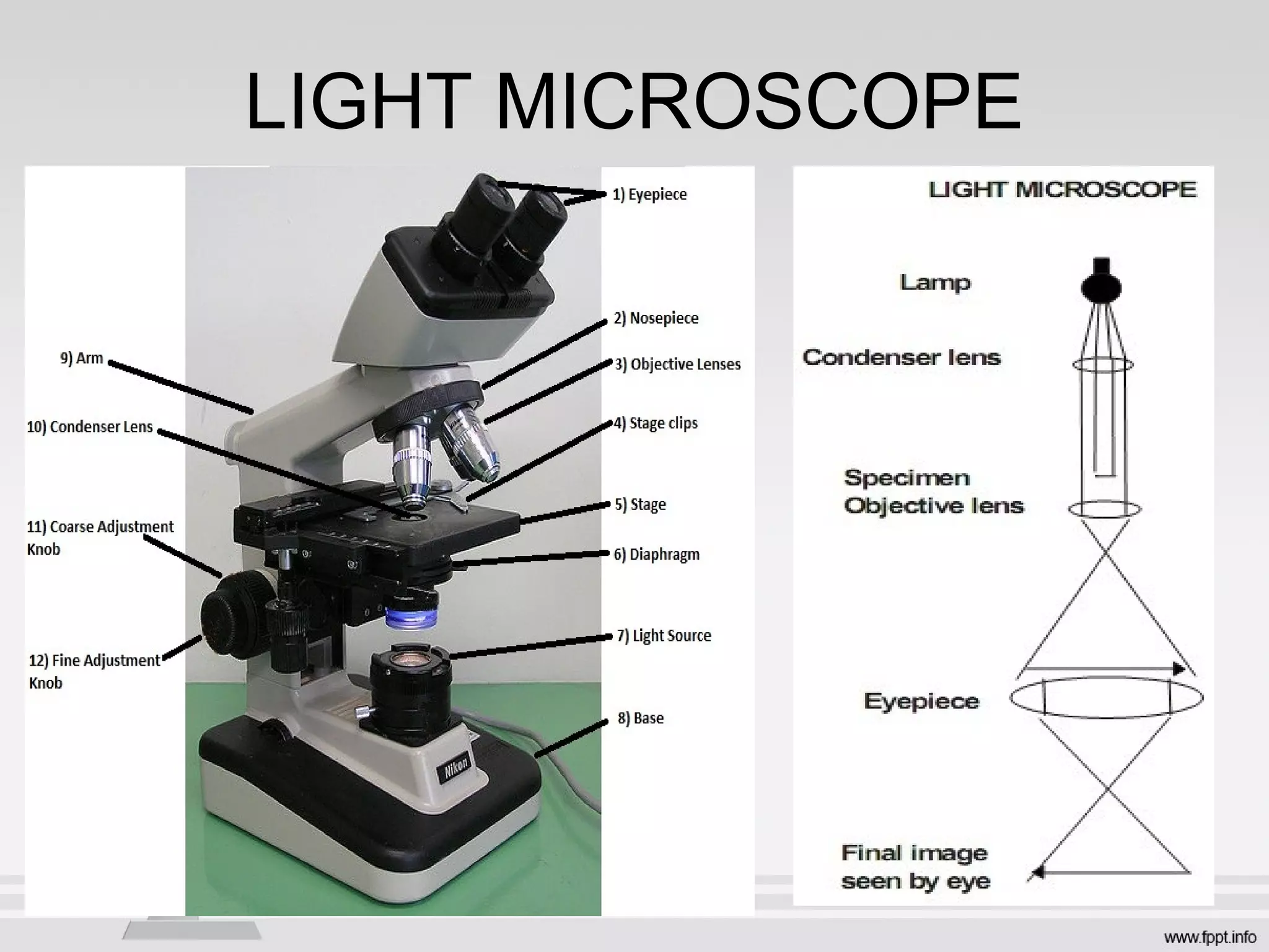
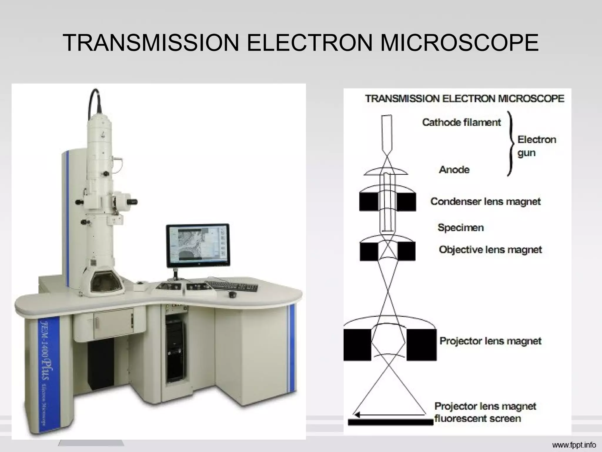
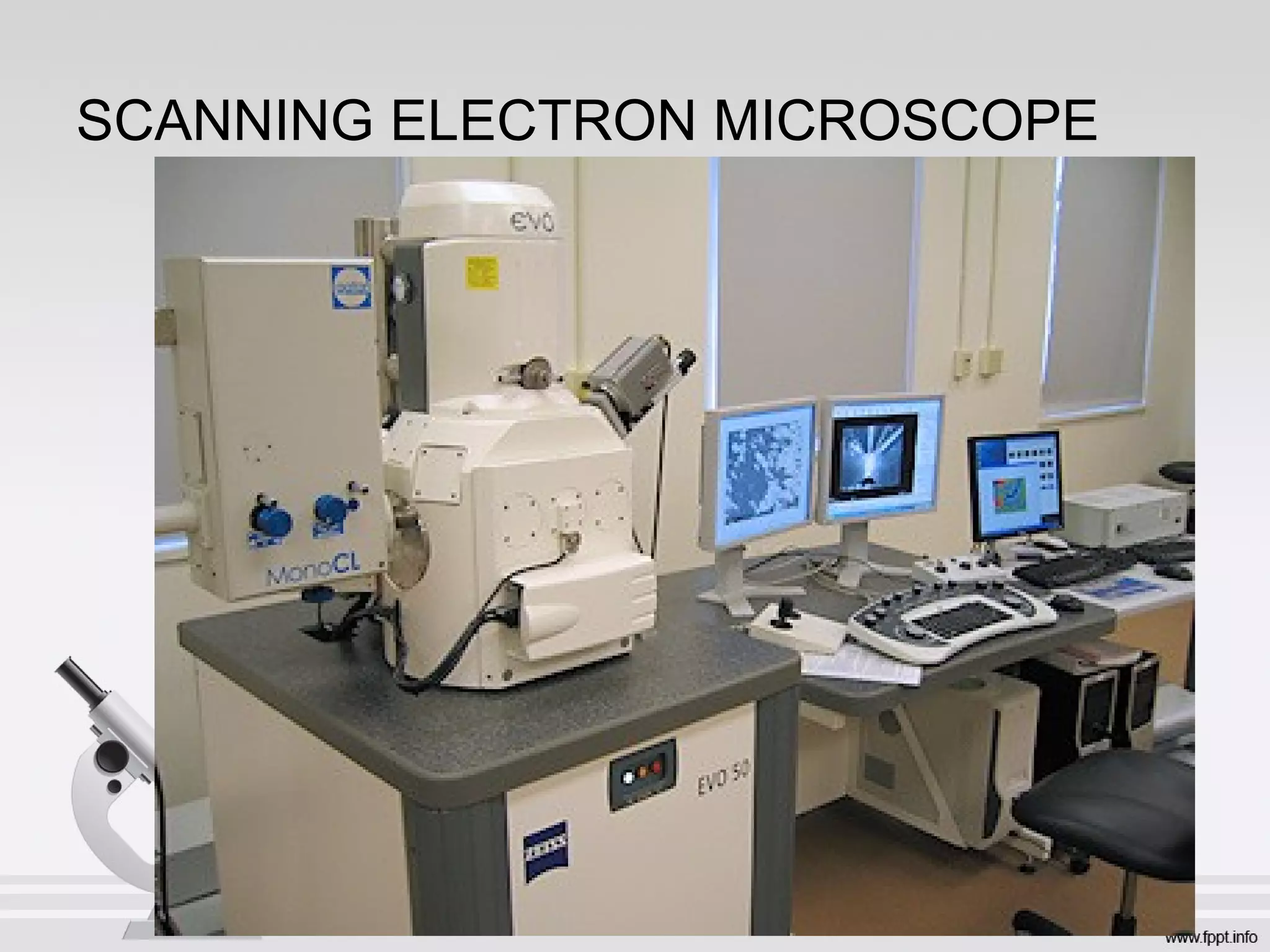
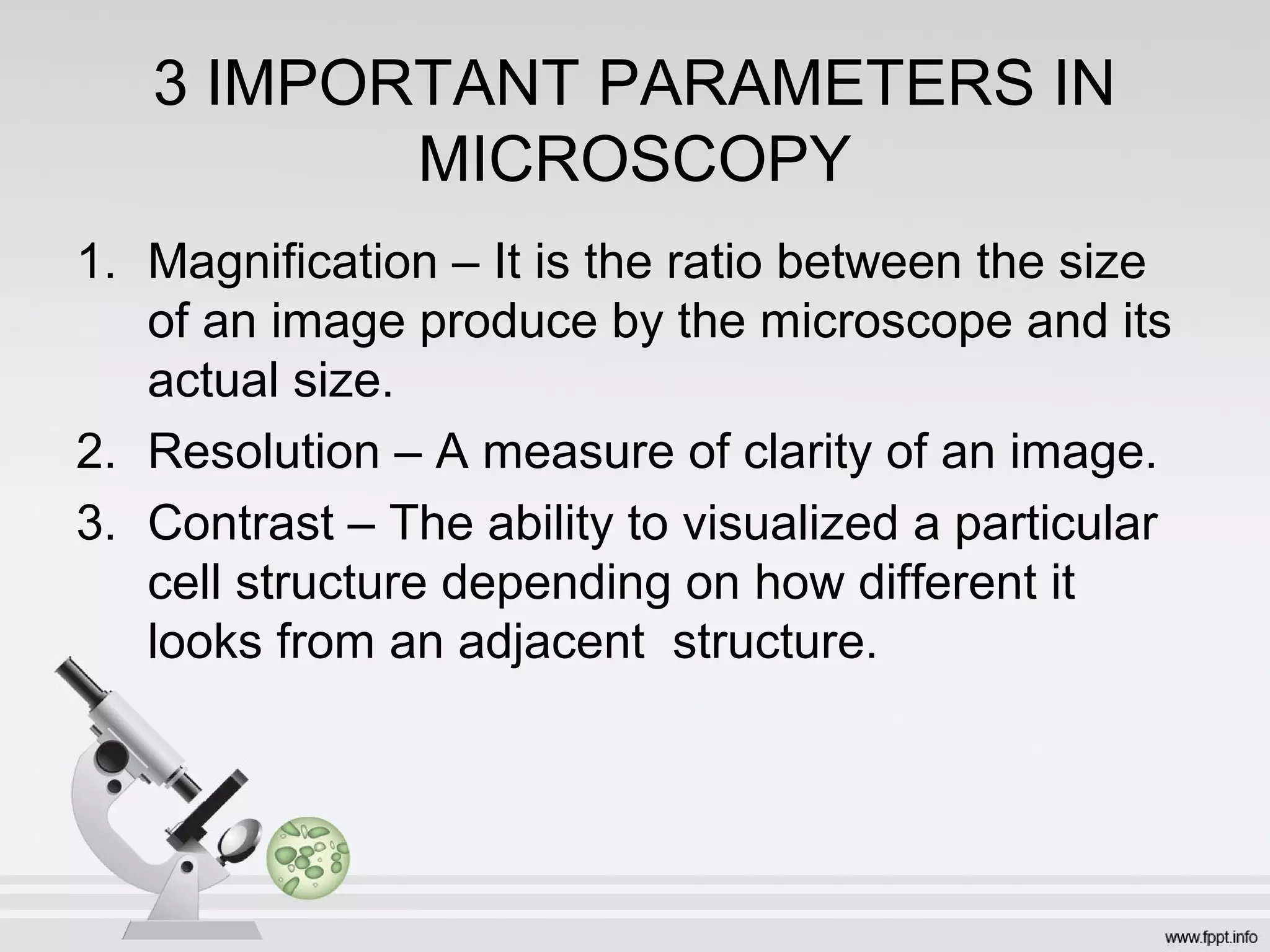
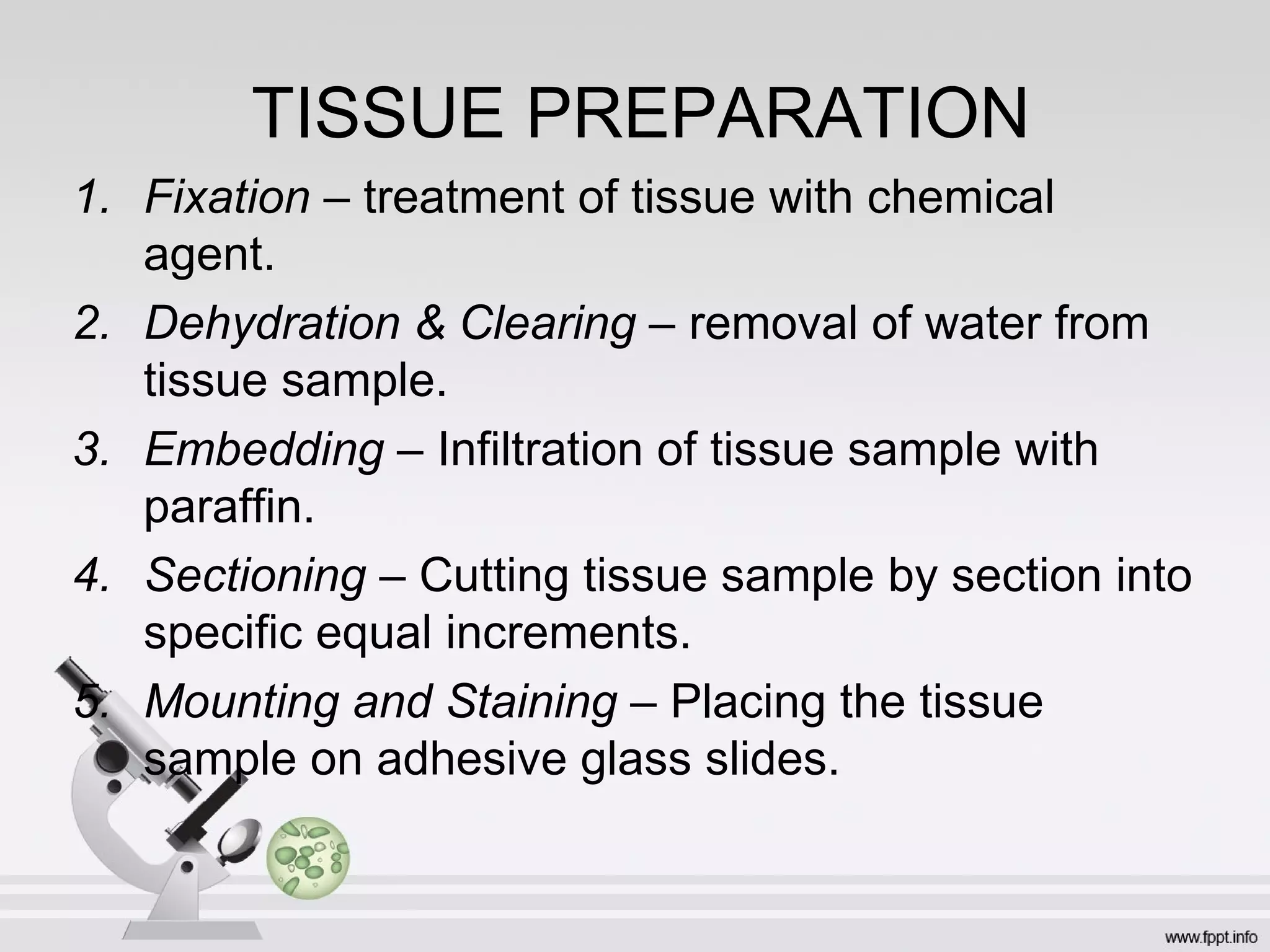
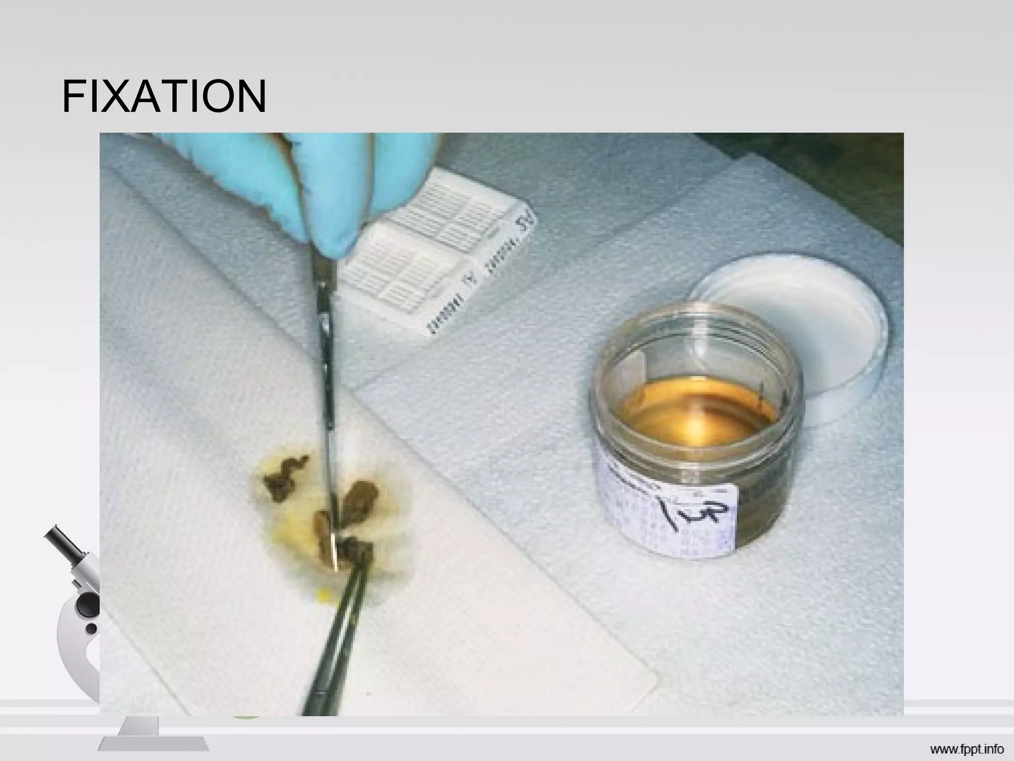
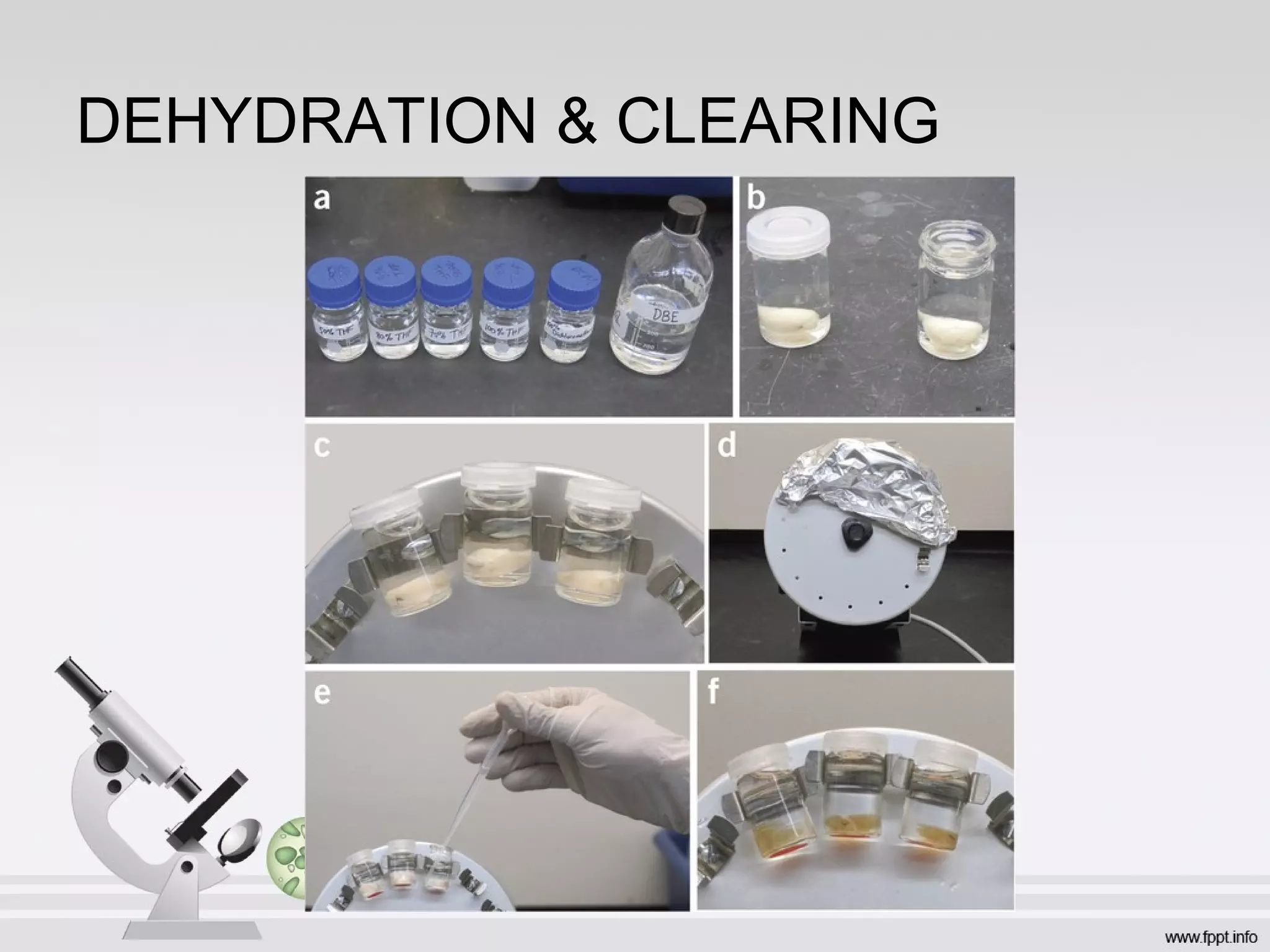
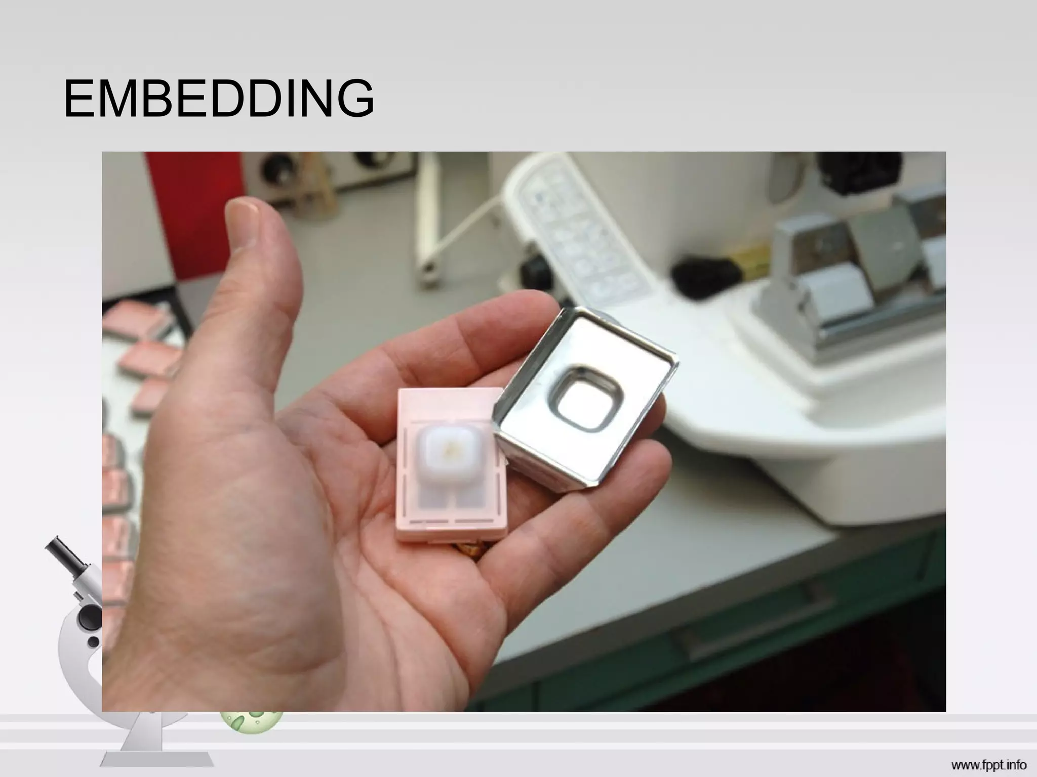
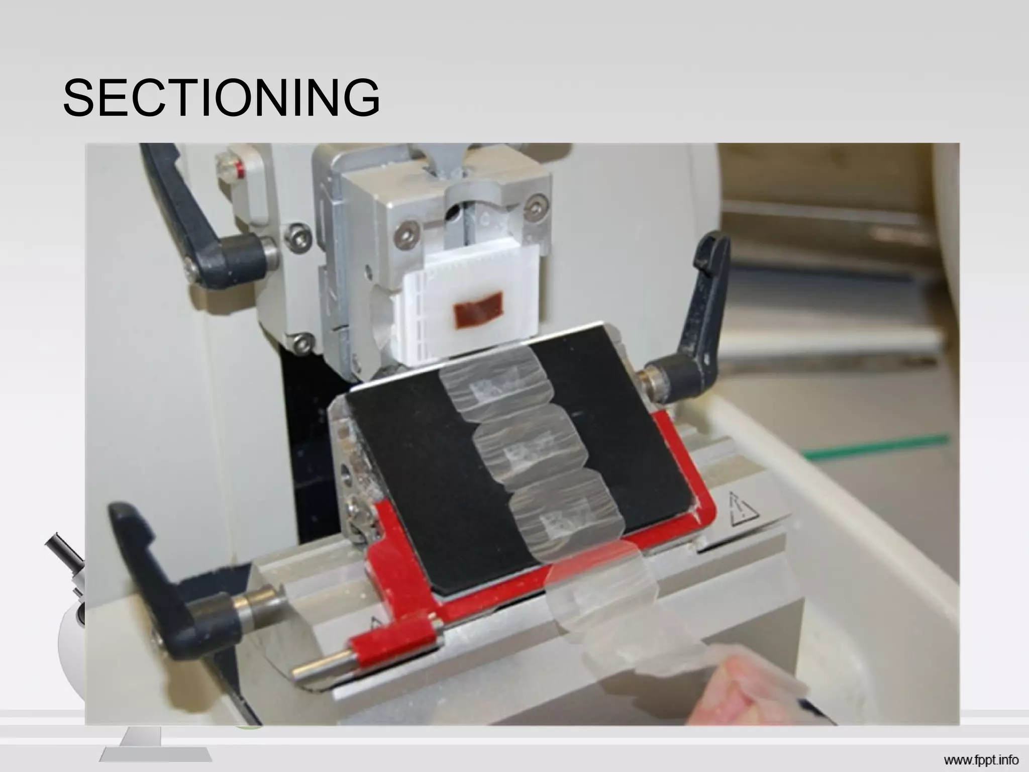
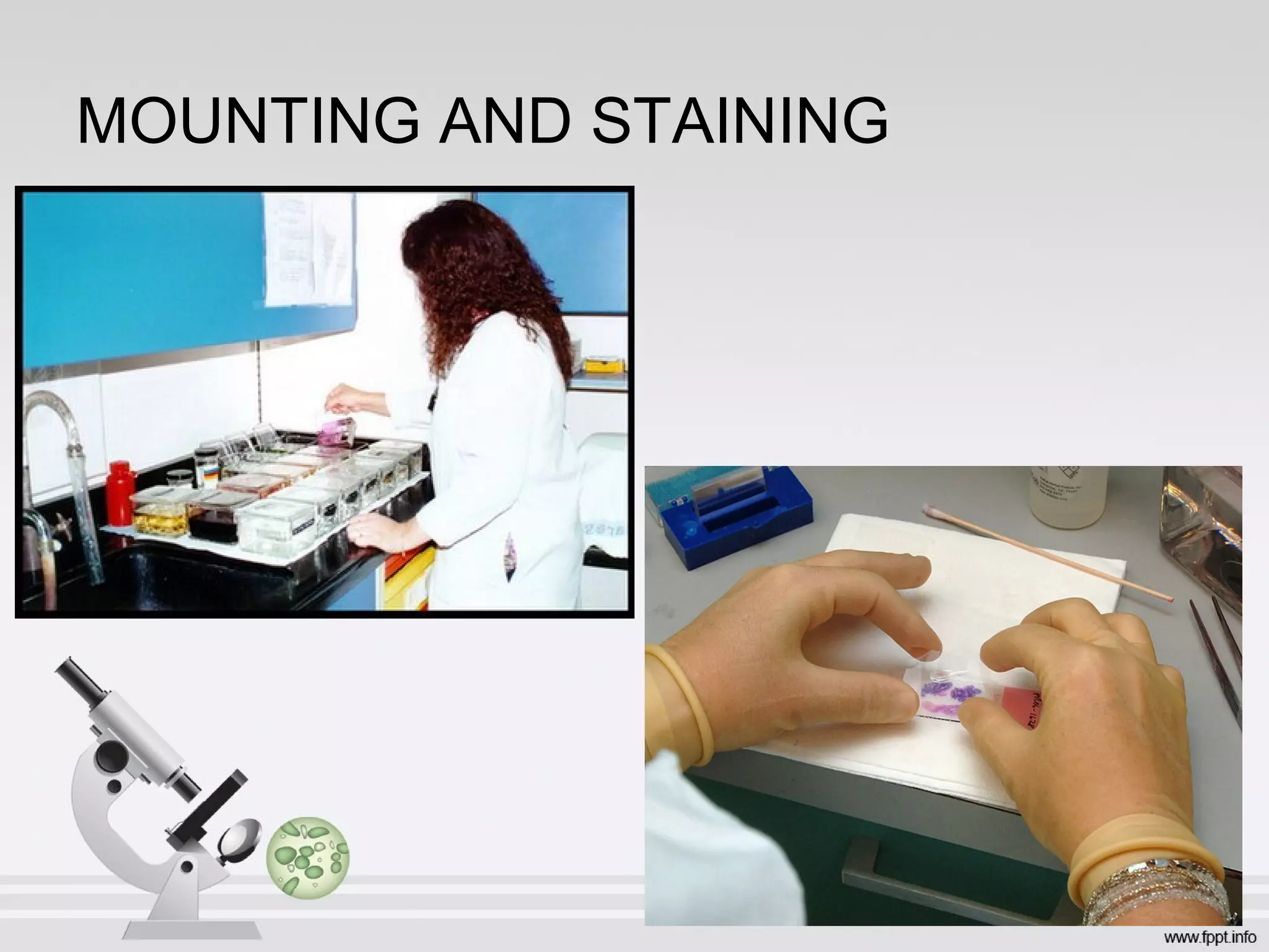

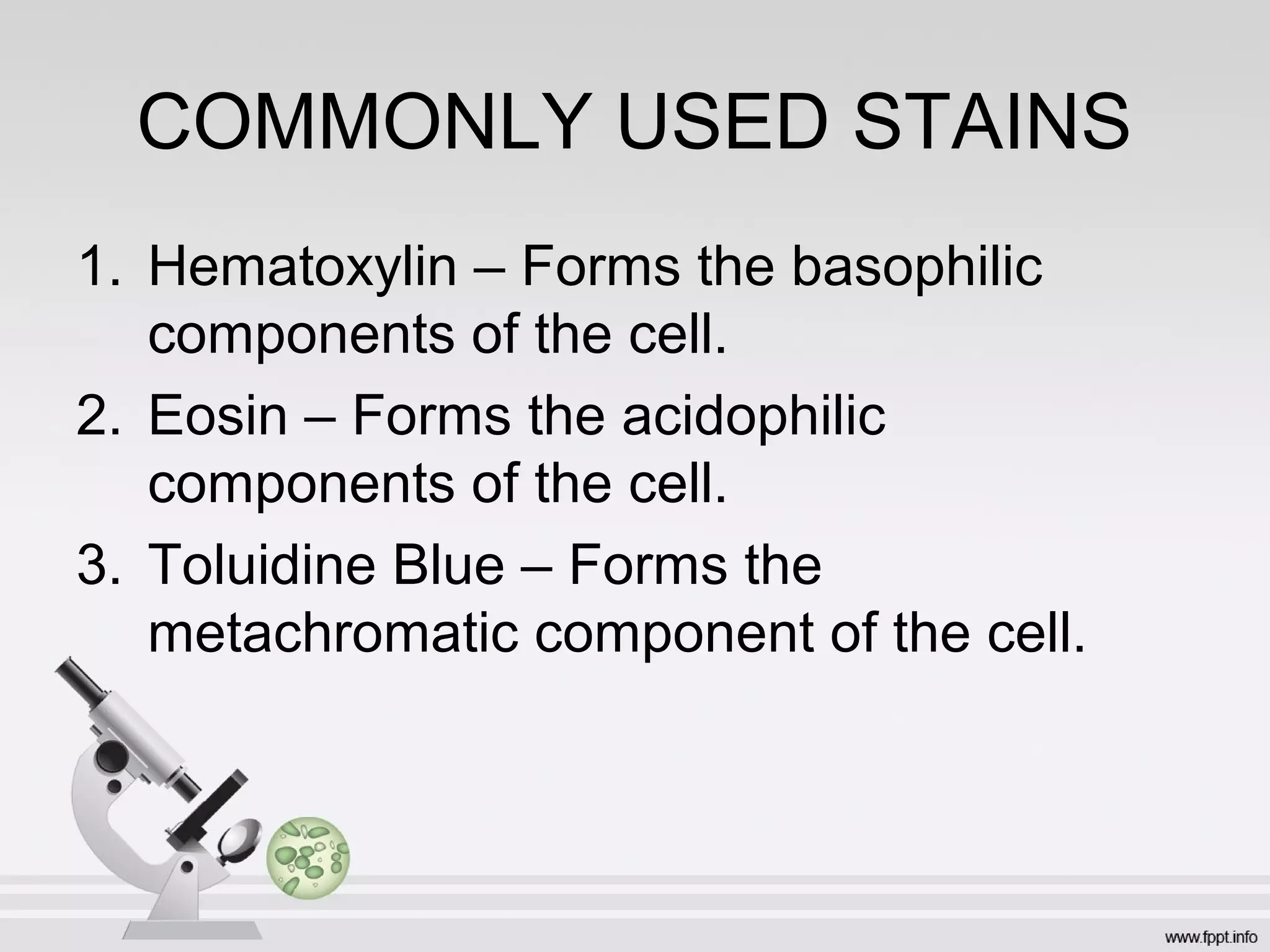
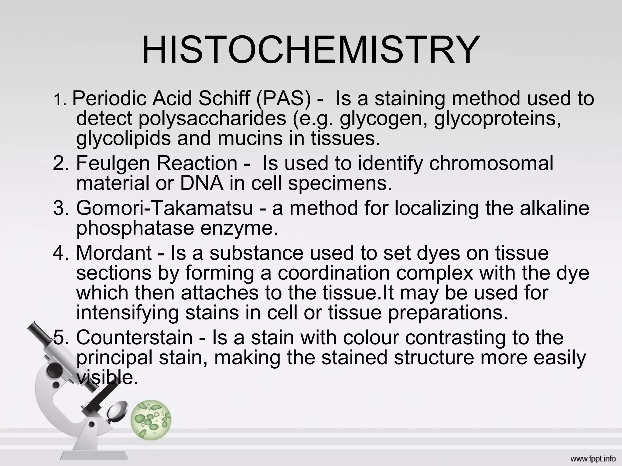
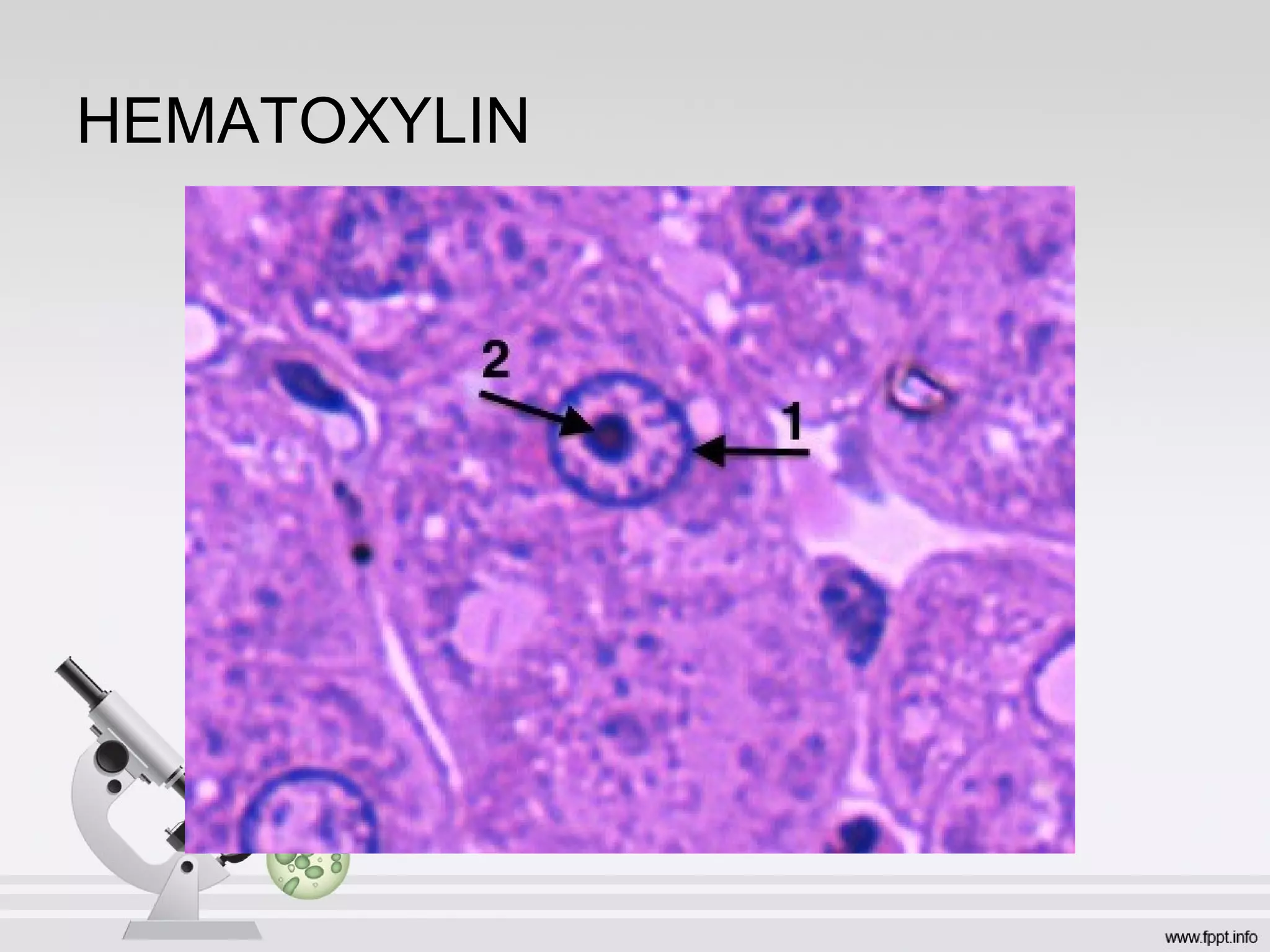
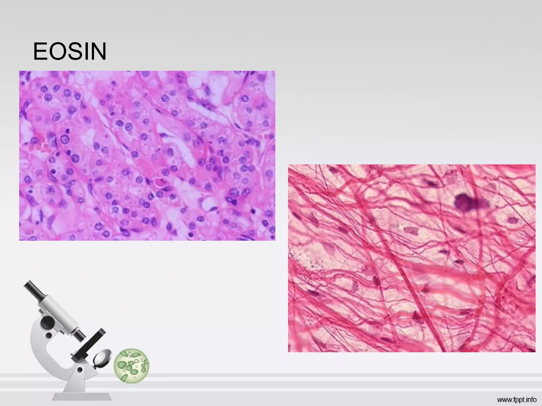
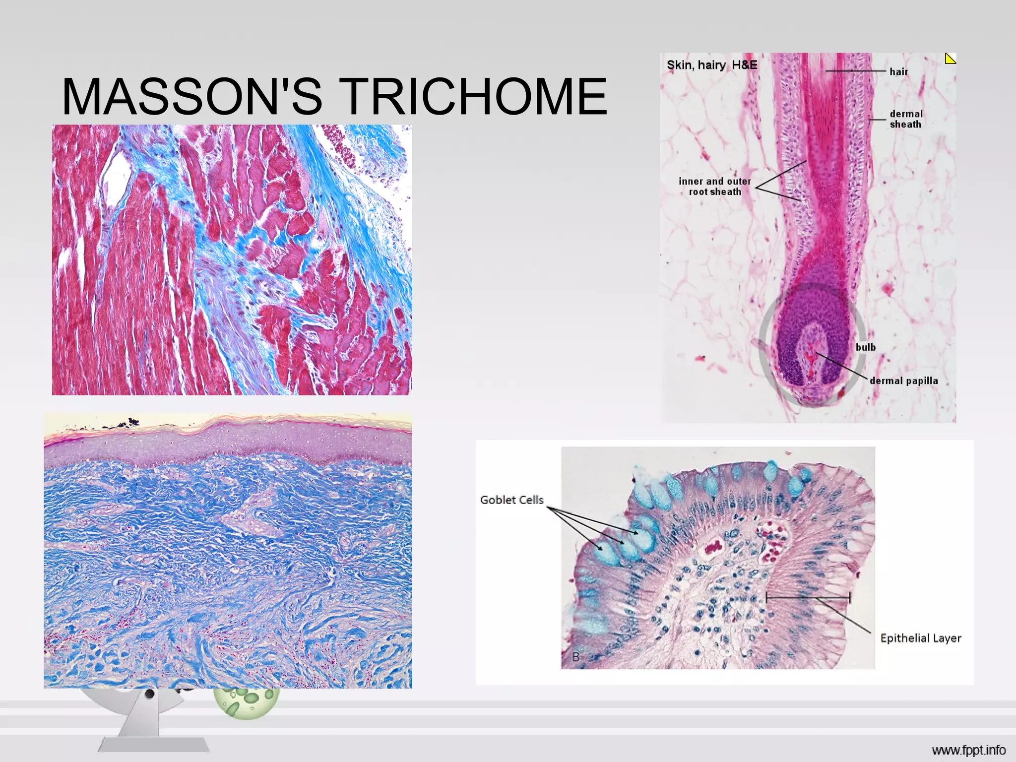
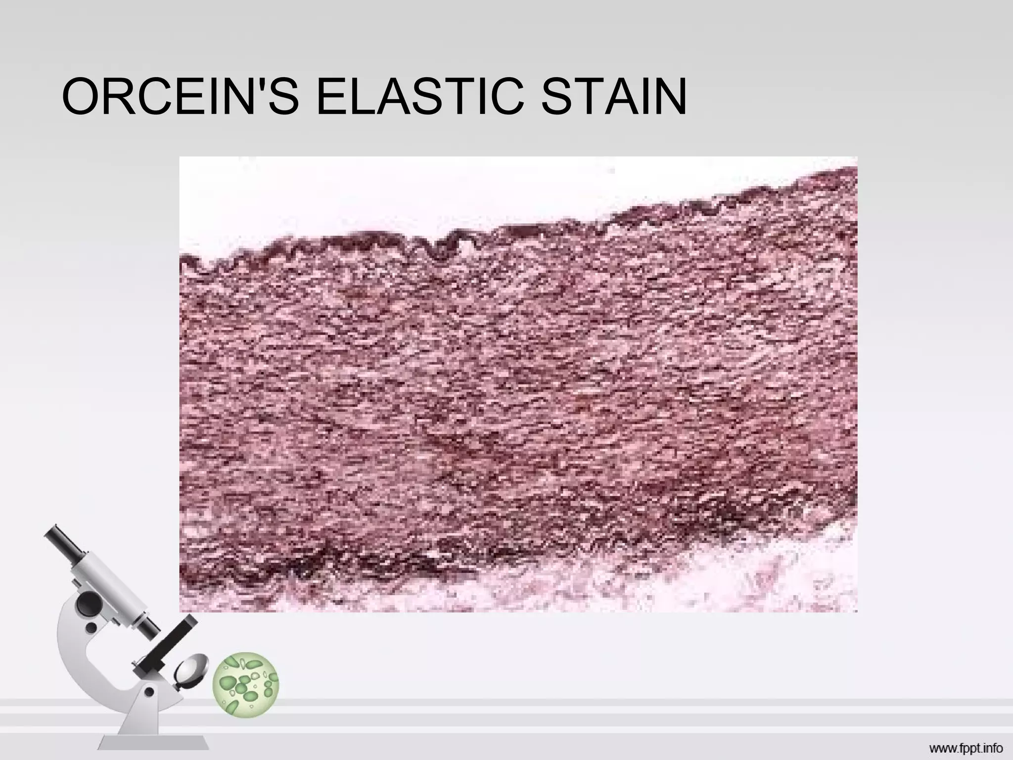
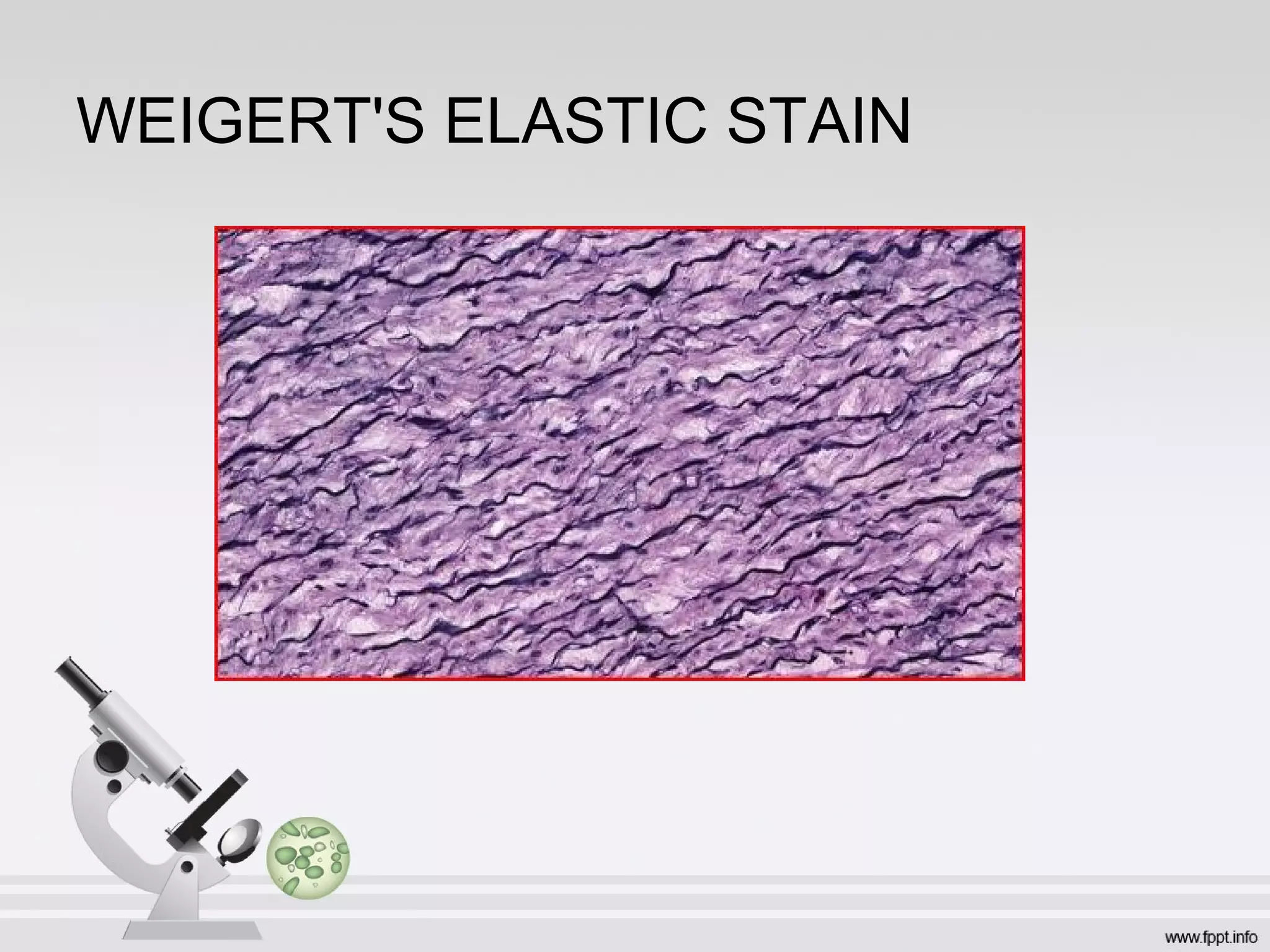
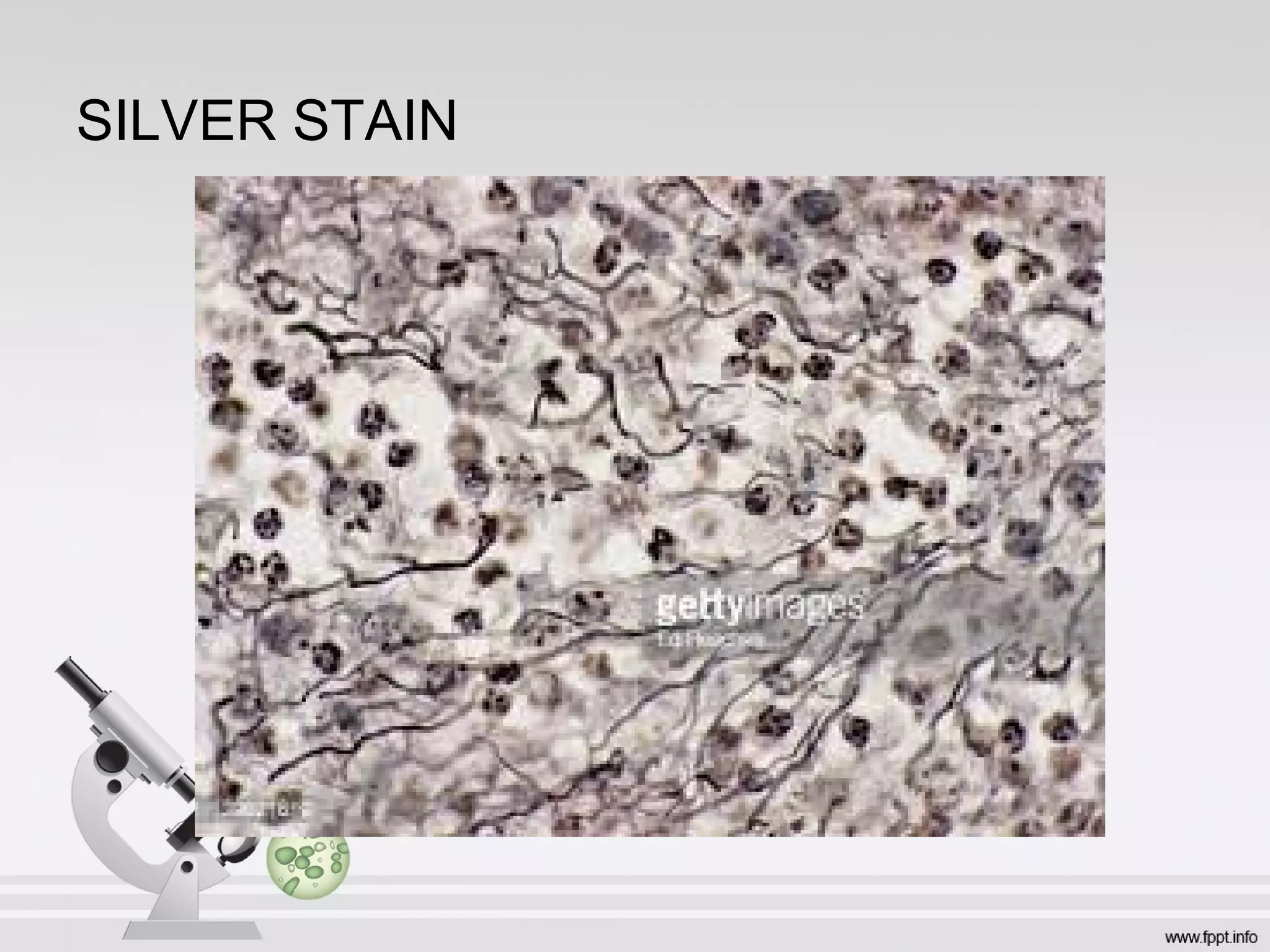
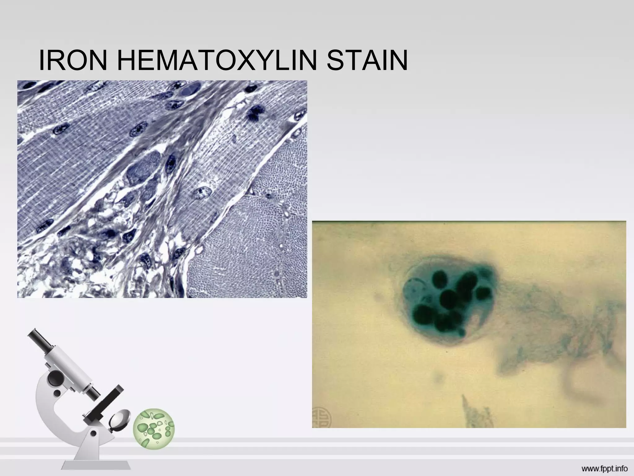
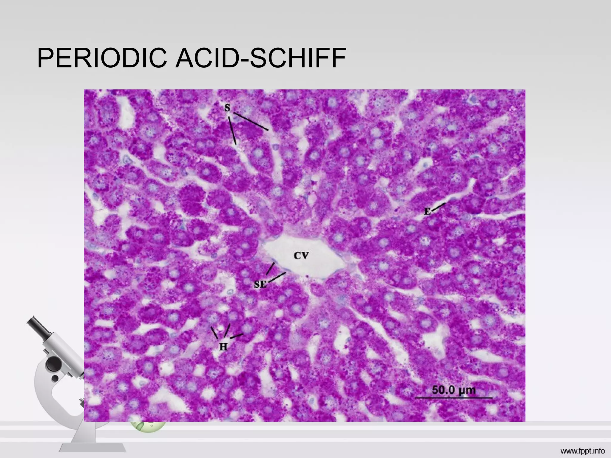
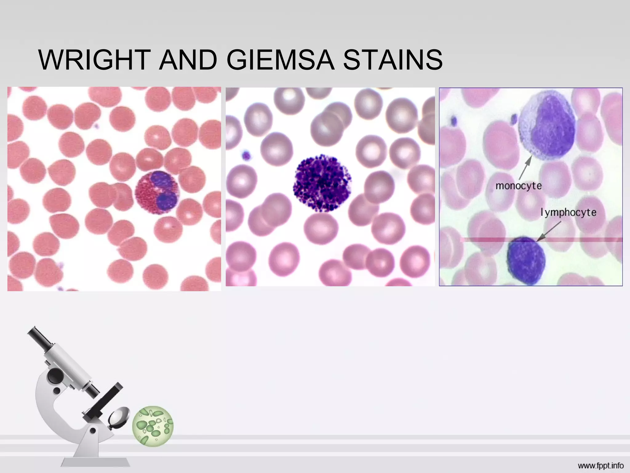

Histology is the study of tissues at a microscopic level. It involves preparing tissue samples using processes like fixation, dehydration, embedding, sectioning, and staining. Different types of microscopes like light, transmission electron, and scanning electron microscopes are used to examine cells and structures at varying levels of magnification, resolution, and contrast. Common staining techniques include hematoxylin and eosin, periodic acid-schiff, and trichrome stains which allow visualization of different cellular components. Histochemistry and immunocytochemistry further aid in localization of macromolecules within tissues.


