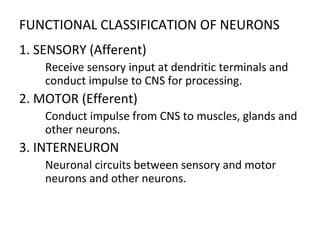
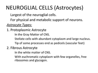
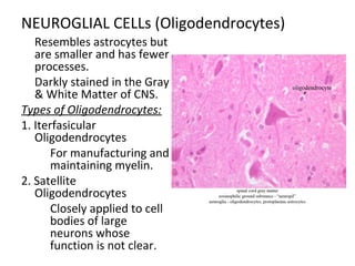
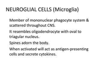
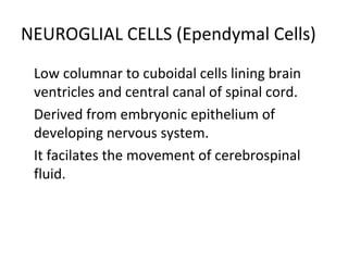
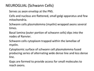
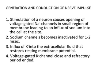
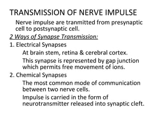
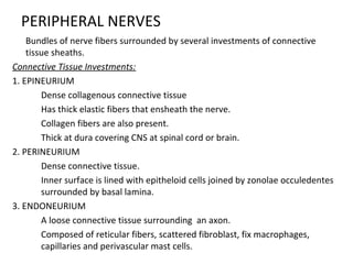
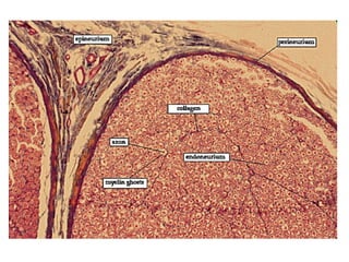
NERVOUS TISSUE
Prof. Redem C. Deligero, MATMRS, MSES
SOMA OF A NEURONS

For the reception and transmission
of nerve impulses to and from the
CNS.
Component of the Soma/Perikaryon:
1. Nucleus

Large, spherical to ovoid &
centrally located.
2. Cytoplasm

Abundant RER with many
cisternae in parallel arrays.

With scattered polyribsomes.

Prominent Golgi Complex &
numerous mitochondria

There is only one centriole
present.
SOMA OF NEURONS
3. Inclusions

Nonliving substance that encompass the neuronal
cell bodies.
A. Melanin Granules

Found in regions of CNS mostly in the substancia
nigra, locus ceruleus & symphathetic ganglia of PNS.
B. Lipofuscin

Irregularly shaped, yellowish brown pigment
granules.

Prevalent in neuronal cytoplasm of older adult.
C. Lipid Droplets

Maybe observed as a result of faulty metabolism or
energy reserves.
DENDRITES OF NEURONS

A ramification of a single short trunk into
smaller and smaller branches tapering at the
end like branches of trees.

Dendritic base contain the usual complement
organelle except Golgi Complexes.

Spines are found on the surface of some
dendrites.
AXON OF NEURONS

Transmit impulses to other neurons or
effector cells. (e.g. muscles or glands)

Arises from axon hillock of the cell body
extending in long distance.

Its thickness is related to conduction velocity.

On the axolemma of of the initial segment
lacks RER & ribosomes but with abundant
microtubules and neurofilaments.

Spike trigger zone is also present in the initial
segment.
AXON OF NEURONS

The axoplasm has short profiles of SER, long thin
mitochondria & numerous microtubules.

RER and polyribosomes are absent.

Somes axons of the CNS and PNS are myelinated.

Axonal Transport of materials between soma and axon
terminals is crucial to trophic relationship.(e.g. neurons
and muscle or glands)

Organelles, vesicles, actin, myosine, clathrin and
enzymes for neurotransmitter synthesis at axon
terminals.

Protien building blocks of neurofilament, sub units of
microtubules, soluble ezymes and materials taken by
endocytosis.
MORPHOLOGICALCLASSIFICATION OF NEURONS
1. BIPOLAR

Has 2 processes from the soma, a single denrite and single
axon.
 Located at vestibular and cochlear ganglia, & in olfactory
epithelium of nasal cavity.
2. UNIPOLAR

Only one process that branch in peripheral and central
branches.
 Develop from embryonic bipolar neurons.

Present in dorsal root ganglia and some cranial nerve ganglia.
3. MULTIPOLAR

Most common with various arrangement of multiple
dendrites from soma and single axon.

Present throughout the nervous system and most are motor
neurons.
FUNCTIONAL CLASSIFICATION OF NEURONS
1. SENSORY (Afferent)

Receive sensory input at dendritic terminals and
conduct impulse to CNS for processing.
2. MOTOR (Efferent)

Conduct impulse from CNS to muscles, glands and
other neurons.
3. INTERNEURON

Neuronal circuits between sensory and motor
neurons and other neurons.
NEUROGLIAL CELLS (Astrocytes)

Largest of the neuroglial cells.

For physical and metabolic support of neurons.
Astrocyte Types:
1. Protoplasmic Astrocyte

In the Gray Matter of CNS.

Stellate cells with abundant cytoplasm and large nucleus.

Tip of some processes end as pedicels (vascular feet)
2. Fibrous Astrocyte

In the white matter of CNS.

With euchromatic cytoplasm with few organelles, free
ribosomes and glycogen.
NEUROGLIAL CELLs (Oligodendrocytes)

Resembles astrocytes but
are smaller and has fewer
processes.

Darkly stained in the Gray
& White Matter of CNS.
Types of Oligodendrocytes:
1. Iterfasicular
Oligodendrocytes

For manufacturing and
maintaining myelin.
2. Satellite
Oligodendrocytes

Closely applied to cell
bodies of large
neurons whose
function is not clear.
NEUROGLIAL CELLS (Microglia)

Member of mononuclear phagocyte system &
scattered throughout CNS.

It resembles oligodendrocyte with oval to
triagular nucleus.

Spines adorn the body.

When activated will act as antigen-presenting
cells and secrete cytokines.
NEUROGLIAL CELLS (Ependymal Cells)

Low columnar to cuboidal cells lining brain
ventricles and central canal of spinal cord.

Derived from embryonic epithelium of
developing nervous system.

It facilates the movement of cerebrospinal
fluid.
NEUROGLIAL (Schwann Cells)

Serves as axon envelop at the PNS.

Cells and nucleus are flattened, small golgi apparatus and few
mitochondria.

Schwann cells plasmalemma (myelin) wrapped axons several
times.

Basal lamina (outer portion of schwann cells) dips into the
nodes of Ranvier.

Schwann cells cytoplasm trapped within the lamellae of
myelin.

Cytoplasmic surface of schwann cell plasmalemma fused
producing series of alternating wide dense line and less dense
line.

Gaps are formed to provide access for small molecules to
reach axons.
GENERATION AND CONDUCTION OF NERVE IMPULSE
1. Stimulation of a neuron causes opening of
voltage gated Na+
channels in small region of
membrane leading to an influx of sodium into
the cell at the site.
2. Sodium channels becomes inactivated for 1-2
msec.
3. Influx of K+
into the extracellular fluid that
restores resting membrane potential.
4. Voltage-gated K+
channel close and refractory
period ended.
TRANSMISSION OF NERVE IMPULSE

Nerve impulse are tranmitted from presynaptic
cell to postsynaptic cell.
2 Ways of Synapse Transmission:
1. Electrical Synapses

At brain stem, retina & cerebral cortex.

This synapse is represented by gap junction
which permits free movement of ions.
2. Chemical Synapses

The most common mode of communication
between two nerve cells.

Impulse is carried in the form of
neurotransmitter released into synaptic cleft.
SYNAPTIC CONTACTS
1. AXODENDRITIC SYNAPSE - Axon & Dendrite
2. AXOSOMATIC SYNAPSE - Axon and Soma
3. AXOAXONIC SYNAPSE - 2 axons
4. DENDRODENDRITIC SYNAPSE - 2 dendrites
SYNAPTIC MORPHOLOGY

Synapse may form bulbous expansion at the
terminal end.

Some synapse form from swelling along the
axons.

Presynaptic membrane has mitochondria, few
elements of SER and abundance of synaptic
vessels.

Cell adhesion molecules are presents both at
presynaptic and postsynaptic aspects of
synapse.
DIFFERENT CELL ADHESION MOLECULES
1. SYNAPSIN I

Small proteins that assists in the clustering of
synaptic vesicle.
2. SYNAPSIN II

Also known as (rab3a) controls association of the
vesicles with actin microfilaments.

Synaptotagamin and Synaptophysin are additional
synaptovesicle proteins.

SNARE (SNAP receptor) influence fusion of
synaptic vesicle with presynaptic membrane.
PERIPHERAL NERVES

Bundles of nerve fibers surrounded by several investments of connective
tissue sheaths.
Connective Tissue Investments:
1. EPINEURIUM

Dense collagenous connective tissue

Has thick elastic fibers that ensheath the nerve.

Collagen fibers are also present.

Thick at dura covering CNS at spinal cord or brain.
2. PERINEURIUM

Dense connective tissue.

Inner surface is lined with epitheloid cells joined by zonolae occuledentes
surrounded by basal lamina.
3. ENDONEURIUM

A loose connective tissue surrounding an axon.

Composed of reticular fibers, scattered fibroblast, fix macrophages,
capillaries and perivascular mast cells.
Nervous tissue

Nervous tissue

  • 1.
    NERVOUS TISSUE Prof. RedemC. Deligero, MATMRS, MSES
  • 2.
    SOMA OF ANEURONS  For the reception and transmission of nerve impulses to and from the CNS. Component of the Soma/Perikaryon: 1. Nucleus  Large, spherical to ovoid & centrally located. 2. Cytoplasm  Abundant RER with many cisternae in parallel arrays.  With scattered polyribsomes.  Prominent Golgi Complex & numerous mitochondria  There is only one centriole present.
  • 3.
    SOMA OF NEURONS 3.Inclusions  Nonliving substance that encompass the neuronal cell bodies. A. Melanin Granules  Found in regions of CNS mostly in the substancia nigra, locus ceruleus & symphathetic ganglia of PNS. B. Lipofuscin  Irregularly shaped, yellowish brown pigment granules.  Prevalent in neuronal cytoplasm of older adult. C. Lipid Droplets  Maybe observed as a result of faulty metabolism or energy reserves.
  • 4.
    DENDRITES OF NEURONS  Aramification of a single short trunk into smaller and smaller branches tapering at the end like branches of trees.  Dendritic base contain the usual complement organelle except Golgi Complexes.  Spines are found on the surface of some dendrites.
  • 5.
    AXON OF NEURONS  Transmitimpulses to other neurons or effector cells. (e.g. muscles or glands)  Arises from axon hillock of the cell body extending in long distance.  Its thickness is related to conduction velocity.  On the axolemma of of the initial segment lacks RER & ribosomes but with abundant microtubules and neurofilaments.  Spike trigger zone is also present in the initial segment.
  • 6.
    AXON OF NEURONS  Theaxoplasm has short profiles of SER, long thin mitochondria & numerous microtubules.  RER and polyribosomes are absent.  Somes axons of the CNS and PNS are myelinated.  Axonal Transport of materials between soma and axon terminals is crucial to trophic relationship.(e.g. neurons and muscle or glands)  Organelles, vesicles, actin, myosine, clathrin and enzymes for neurotransmitter synthesis at axon terminals.  Protien building blocks of neurofilament, sub units of microtubules, soluble ezymes and materials taken by endocytosis.
  • 7.
    MORPHOLOGICALCLASSIFICATION OF NEURONS 1.BIPOLAR  Has 2 processes from the soma, a single denrite and single axon.  Located at vestibular and cochlear ganglia, & in olfactory epithelium of nasal cavity. 2. UNIPOLAR  Only one process that branch in peripheral and central branches.  Develop from embryonic bipolar neurons.  Present in dorsal root ganglia and some cranial nerve ganglia. 3. MULTIPOLAR  Most common with various arrangement of multiple dendrites from soma and single axon.  Present throughout the nervous system and most are motor neurons.
  • 9.
    FUNCTIONAL CLASSIFICATION OFNEURONS 1. SENSORY (Afferent)  Receive sensory input at dendritic terminals and conduct impulse to CNS for processing. 2. MOTOR (Efferent)  Conduct impulse from CNS to muscles, glands and other neurons. 3. INTERNEURON  Neuronal circuits between sensory and motor neurons and other neurons.
  • 10.
    NEUROGLIAL CELLS (Astrocytes)  Largestof the neuroglial cells.  For physical and metabolic support of neurons. Astrocyte Types: 1. Protoplasmic Astrocyte  In the Gray Matter of CNS.  Stellate cells with abundant cytoplasm and large nucleus.  Tip of some processes end as pedicels (vascular feet) 2. Fibrous Astrocyte  In the white matter of CNS.  With euchromatic cytoplasm with few organelles, free ribosomes and glycogen.
  • 11.
    NEUROGLIAL CELLs (Oligodendrocytes)  Resemblesastrocytes but are smaller and has fewer processes.  Darkly stained in the Gray & White Matter of CNS. Types of Oligodendrocytes: 1. Iterfasicular Oligodendrocytes  For manufacturing and maintaining myelin. 2. Satellite Oligodendrocytes  Closely applied to cell bodies of large neurons whose function is not clear.
  • 12.
    NEUROGLIAL CELLS (Microglia)  Memberof mononuclear phagocyte system & scattered throughout CNS.  It resembles oligodendrocyte with oval to triagular nucleus.  Spines adorn the body.  When activated will act as antigen-presenting cells and secrete cytokines.
  • 13.
    NEUROGLIAL CELLS (EpendymalCells)  Low columnar to cuboidal cells lining brain ventricles and central canal of spinal cord.  Derived from embryonic epithelium of developing nervous system.  It facilates the movement of cerebrospinal fluid.
  • 14.
    NEUROGLIAL (Schwann Cells)  Servesas axon envelop at the PNS.  Cells and nucleus are flattened, small golgi apparatus and few mitochondria.  Schwann cells plasmalemma (myelin) wrapped axons several times.  Basal lamina (outer portion of schwann cells) dips into the nodes of Ranvier.  Schwann cells cytoplasm trapped within the lamellae of myelin.  Cytoplasmic surface of schwann cell plasmalemma fused producing series of alternating wide dense line and less dense line.  Gaps are formed to provide access for small molecules to reach axons.
  • 16.
    GENERATION AND CONDUCTIONOF NERVE IMPULSE 1. Stimulation of a neuron causes opening of voltage gated Na+ channels in small region of membrane leading to an influx of sodium into the cell at the site. 2. Sodium channels becomes inactivated for 1-2 msec. 3. Influx of K+ into the extracellular fluid that restores resting membrane potential. 4. Voltage-gated K+ channel close and refractory period ended.
  • 17.
    TRANSMISSION OF NERVEIMPULSE  Nerve impulse are tranmitted from presynaptic cell to postsynaptic cell. 2 Ways of Synapse Transmission: 1. Electrical Synapses  At brain stem, retina & cerebral cortex.  This synapse is represented by gap junction which permits free movement of ions. 2. Chemical Synapses  The most common mode of communication between two nerve cells.  Impulse is carried in the form of neurotransmitter released into synaptic cleft.
  • 18.
    SYNAPTIC CONTACTS 1. AXODENDRITICSYNAPSE - Axon & Dendrite 2. AXOSOMATIC SYNAPSE - Axon and Soma 3. AXOAXONIC SYNAPSE - 2 axons 4. DENDRODENDRITIC SYNAPSE - 2 dendrites
  • 19.
    SYNAPTIC MORPHOLOGY  Synapse mayform bulbous expansion at the terminal end.  Some synapse form from swelling along the axons.  Presynaptic membrane has mitochondria, few elements of SER and abundance of synaptic vessels.  Cell adhesion molecules are presents both at presynaptic and postsynaptic aspects of synapse.
  • 20.
    DIFFERENT CELL ADHESIONMOLECULES 1. SYNAPSIN I  Small proteins that assists in the clustering of synaptic vesicle. 2. SYNAPSIN II  Also known as (rab3a) controls association of the vesicles with actin microfilaments.  Synaptotagamin and Synaptophysin are additional synaptovesicle proteins.  SNARE (SNAP receptor) influence fusion of synaptic vesicle with presynaptic membrane.
  • 21.
    PERIPHERAL NERVES  Bundles ofnerve fibers surrounded by several investments of connective tissue sheaths. Connective Tissue Investments: 1. EPINEURIUM  Dense collagenous connective tissue  Has thick elastic fibers that ensheath the nerve.  Collagen fibers are also present.  Thick at dura covering CNS at spinal cord or brain. 2. PERINEURIUM  Dense connective tissue.  Inner surface is lined with epitheloid cells joined by zonolae occuledentes surrounded by basal lamina. 3. ENDONEURIUM  A loose connective tissue surrounding an axon.  Composed of reticular fibers, scattered fibroblast, fix macrophages, capillaries and perivascular mast cells.