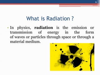
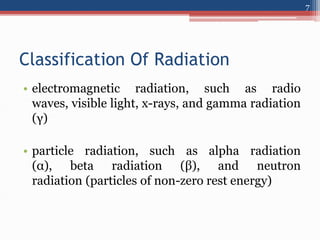
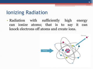
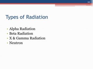
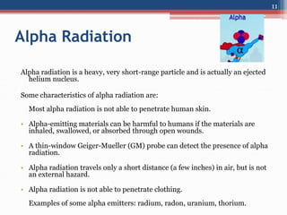
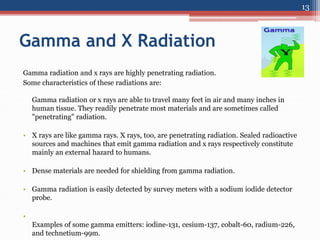
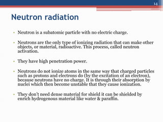
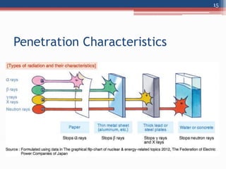
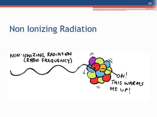
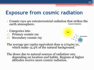
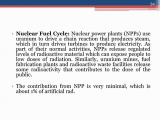
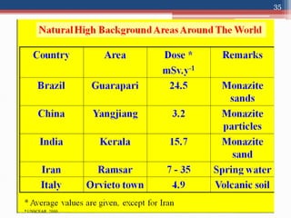
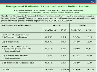
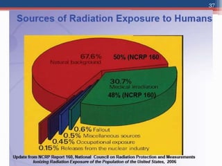
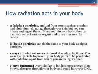
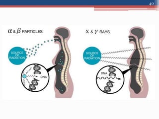
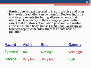
Radiation Hazards Evaluation
&
its Control
Dinesh Kr Baghel
PG Dip RP, MBAHAM, M.Sc. (Phy.)
Medical Physicist
Department of Medical Physics &
Radiotherapy
Delhi State Cancer Institute (DSCI)
Delhi, India.
1
Contents -
• Introduction
• Radiation
• Hazards of radiation
• Evaluation
• & Safety
2
Introduction
• Every day you are effected by
radiation whether from the natural or from
man-made radiation. It’s very important
to know what radiation is and how it
works to harm life. You also need to know
what you can do to limit your exposure
to radiation.
• The radiation hazards evaluation is very important
for justifying and ensuring satisfactory protection.
• Without such evaluation it may scare them who fear
radiation even if the levels are in safe limits & may
cause unwanted exposure to those who are careless
even if the exposures are really high.
3
• The extensive investigation have been made to
determine what constitutes a harmful dose of
ionizing radiation.
• The aim of the presentation is to give a
clear understanding of hazard, their
evaluation and control.
4
RADIATION HAZARDS EVALUATION
5
What is Radiation ?
• In physics, radiation is the emission or
transmission of energy in the form
of waves or particles through space or through a
material medium.
6
Classification Of Radiation
• electromagnetic radiation, such as radio
waves, visible light, x-rays, and gamma radiation
(γ)
• particle radiation, such as alpha radiation
(α), beta radiation (β), and neutron
radiation (particles of non-zero rest energy)
7
• Radiation is often categorized as either
ionizing or non-ionizing
depending on the energy of the radiated particles.
8
Ionizing Radiation
• Radiation with sufficiently high energy
can ionize atoms; that is to say it can
knock electrons off atoms and create ions.
9
Types of Radiation
• Alpha Radiation
• Beta Radiation
• X & Gamma Radiation
• Neutron
10
Alpha Radiation
Alpha radiation is a heavy, very short-range particle and is actually an ejected
helium nucleus.
Some characteristics of alpha radiation are:
Most alpha radiation is not able to penetrate human skin.
• Alpha-emitting materials can be harmful to humans if the materials are
inhaled, swallowed, or absorbed through open wounds.
• A thin-window Geiger-Mueller (GM) probe can detect the presence of alpha
radiation.
• Alpha radiation travels only a short distance (a few inches) in air, but is not
an external hazard.
• Alpha radiation is not able to penetrate clothing.
Examples of some alpha emitters: radium, radon, uranium, thorium.
11
Beta Radiation
Beta radiation is a light, short-range particle and is actually an ejected electron.
Some characteristics of beta radiation are:
Beta radiation may travel several feet in air and is moderately penetrating.
• Beta radiation can penetrate human skin to the "germinal layer," where new skin
cells are produced. If high levels of beta-emitting contaminants are allowed to remain
on the skin for a prolonged period of time, they may cause skin injury.
• Beta-emitting contaminants may be harmful if deposited internally.
• Most beta emitters can be detected with a survey instrument and a thin-window GM
probe . Some beta emitters, however, produce very low-energy, poorly penetrating
radiation that may be difficult or impossible to detect. Examples of these difficult-to-
detect beta emitters are hydrogen-3 (tritium), carbon-14, and sulfur-35.
• Examples of some pure beta emitters: strontium-90, carbon-14, tritium, and sulfur-
35.
12
Gamma and X Radiation
Gamma radiation and x rays are highly penetrating radiation.
Some characteristics of these radiations are:
Gamma radiation or x rays are able to travel many feet in air and many inches in
human tissue. They readily penetrate most materials and are sometimes called
"penetrating" radiation.
• X rays are like gamma rays. X rays, too, are penetrating radiation. Sealed radioactive
sources and machines that emit gamma radiation and x rays respectively constitute
mainly an external hazard to humans.
• Dense materials are needed for shielding from gamma radiation.
• Gamma radiation is easily detected by survey meters with a sodium iodide detector
probe.
•
Examples of some gamma emitters: iodine-131, cesium-137, cobalt-60, radium-226,
and technetium-99m.
13
Neutron radiation
• Neutron is a subatomic particle with no electric charge.
• Neutrons are the only type of ionizing radiation that can make other
objects, or material, radioactive. This process, called neutron
activation.
• They have high penetration power.
• Neutrons do not ionize atoms in the same way that charged particles
such as protons and electrons do (by the excitation of an electron),
because neutrons have no charge. It is through their absorption by
nuclei which then become unstable that they cause ionization.
• They don’t need dense material for shield it can be shielded by
enrich hydrogenous material like water & paraffin.
14
Penetration Characteristics
15
Non Ionizing Radiation
16
• Non-ionizing radiation is emitted from things
like cell phones, microwave ovens, cordless
telephones, satellite, radio towers, and stars like
our sun. This type of radiation is potentially
dangerous, however in most cases it is low
energy radiation and is generally harmless (cell
phones, wireless transmitters).
17
Can Radiation be felt physically
?
18
•Yes, if u have super natural power
19
•No, Radiation can not be feel by any
natural senses like- eyes, senstaion, taste, smell
etc.
20
• The only way to recognize its presence is from
readings on radiation monitoring machines.
21
Sources of Radiation
• People are constantly exposed to small amounts
of ionizing radiation from the environment as
they carry out their normal daily activities; this
is known as natural background radiation.
• We are also exposed through some medical
treatments and through activities involving
radioactive material.
22
Natural background radiation
• People are constantly exposed to small amounts
of ionizing radiation from the environment as
they carry out their normal daily activities; this
is known as natural background radiation.
• Radiation has always been present and is all
around us. Life has evolved in a world
containing significant levels of ionizing
radiation. Our bodies are adapted to it.
23
• The United Nations Scientific Committee on the
Effects of Atomic Radiation (UNSCEAR) &
(AERB) identifies four major sources of public
exposure to natural radiation:
• cosmic radiation
• terrestrial radiation
• inhalation
• ingestion
24
Exposure from cosmic radiation
• Cosmic rays are extraterrestrial radiation that strikes the
earth atmosphere.
• Categories into
1) Primary cosmic ray
2) Secondary cosmic ray
The average per capita equivalent dose is 270μSv/yr,
which make 15.4% of the natural background.
The doses due to natural sources of radiation vary
depending on location and habits. Regions at higher
altitudes receive more cosmic radiation.
25
Exposure from terrestrial radiation
• The composition of the earth's crust is a major
source of natural radiation. The main contributors
are natural deposits of uranium, potassium and
thorium which, in the process of natural decay, will
release small amounts of ionizing radiation.
Uranium and thorium are “ubiquitous”, meaning
they are found essentially everywhere. Traces of
these minerals are also found in building materials
so exposure to natural radiation can occur from
indoors as well as outdoors.
• It account for about 16.5% of natural background.
26
Exposure through inhalation
• Most of the variation in exposure to natural radiation results from
inhalation of radioactive gases that are produced by radioactive
minerals found in soil and bedrock. Radon is an odourless and
colourless radioactive gas that is produced by the decay of uranium.
Thoron is a radioactive gas produced by the thorium. Radon and
thoron levels vary considerably by location depending on the
composition of soil and bedrock. Once released into the air, these
gases will normally dilute to harmless levels in the atmosphere but
sometimes they become trapped and accumulate inside buildings
and are inhaled by occupants. Radon gas poses a health risk not
only to uranium miners, but also to homeowners if it is left to collect
in the home. On average, it is the largest source of natural radiation
exposure.
• Radon account equvlnt dose of 2mSv/yr to bronchial epithelium.
• It account for about 55% of natural background.
27
Exposure through ingestion
• Trace amounts of radioactive minerals are
naturally found in the contents of food and
drinking water. For instance, vegetables are
typically cultivated in soil and ground water
which contains radioactive minerals. Once
ingested, these minerals result in internal
exposure to natural radiation.
28
• Naturally occurring radioactive isotopes, such as
potassium-40 and carbon-14, have the same
chemical and biological properties as their non-
radioactive isotopes. These radioactive and non-
radioactive elements are used in building and
maintaining our bodies. Natural radioisotopes
continually expose us to radiation.
• It accounts an average equivalent dose of 400
μSv/yr.
29
Radioactive isotopes in the body (70
kg adult)
Isotopes Amount of radioactivity in Bq
Uranium 2.3
Thorium 0.21
Potassium-40 4000
Carbon-14 3700
Radium-266 1.1
30
The human body also contains several radioactive isotopes. The
table below contains a list of some of the isotopes naturally found
in the body.
Artificial sources of radiation
• Atmospheric testing: The atmospheric testing of atomic
weapons from the end of the Second World War until as late
as 1980 released radioactive material, called fallout, into the
air. As the fallout settled to the ground, it was incorporated
into the environment. Much of the fallout had short half-lives
and no longer exists, but some continues to decay to this day.
People and the environment receive smaller and smaller doses
from the fallout every year.
• Consist of C-14 (70%) & other radionuclide including H-3,
Mn-54, Cs-136,137, etc.
• It result in an equivalent eff. Dose of <10 μSv. It contributes
2% of the manmade rad. Exposure.
31
• Medical sources: Radiation has many uses in medicine.
The most well known use is X-ray machines, which use
radiation to find broken bones and diagnose disease. Another
example is nuclear medicine, which uses radioactive isotopes
to diagnose and treat diseases such as cancer. Another
example in Radiation therapy to treat cancer.
• The majority of the exposure is from medical X-ray which
contain 58% of the artificial radiation. Next contributor is NM
which is 21% so both produces an annual avg. eff. Dose equ.
Of 540 μSv /yr.
• It accounts for about 79% of artificial radiation.
32
• Industrial sources:Radiation has a variety of
industrial uses that range from nuclear gauges used
to build roads to density gauges that measure the
flow of material through pipes in factories. It is also
used for smoke detectors, some glow-in-the dark
exit signs, and to estimate reserves in oil fields.
Radiation is also used for sterilization which is done
by using large, heavily shielded irradiators.
33
• Nuclear Fuel Cycle: Nuclear power plants (NPPs) use
uranium to drive a chain reaction that produces steam,
which in turn drives turbines to produce electricity. As
part of their normal activities, NPPs release regulated
levels of radioactive material which can expose people to
low doses of radiation. Similarly, uranium mines, fuel
fabrication plants and radioactive waste facilities release
some radioactivity that contributes to the dose of the
public.
• The contribution from NPP is very minimal, which is
about 1% of artificial rad.
34
35
36
37
RADIATION HAZARDS EVALUATION
38
How radiation acts in your body
• α (alpha) particles, emitted from atoms such as uranium
and plutonium, do not go through your skin but you can
inhale and ingest them. If they get into your body, they can
irradiate cells of various organs and cause illnesses like
cancer.
• β (beta) particles can do the same to your body as alpha
particles.
• x-rays are what we are accustomed at medical facilities. You
wear lead jackets to prevent your body parts to be penetrated
with radiation apart from where you are being scanned.
• γ-rays (gamma) , very similar to but has more energy than
x-rays, also goes through your body and could hurt your DNA.
39
40
• Each dose you get exposed to is cumulative and even
low levels of radiation can be harmful. Nuclear industry
and its proponents (including all governments that
utilize nuclear energy in their energy program) often
assert that low doses of radiation produce no harmful
effects to human body, but as US National Academy of
Sciences report concludes, there is no safe dose of
radiation.
Hazard Alpha Beta Gamma
External Nil Not high Very high
Internal Very high Very high High
41
• This diagram shows human organs and radioactive materials that are likely
to accumulate in each organ.
42
Why Should You Be Concerned About Radiation?
• Radiation (especially ionizing) is harmful & potentially fatal to all life
on earth and we should limit our exposure to it when possible. Some
radiation exposure is unavoidable and cannot be removed such as our
exposure to background radiation from our sun.
• However, most of the exposure humans receive from harmful ionizing
radiation is not from background radiation sources but rather from man-
made sources.
• Much of the fallout from the nuclear testing done in the 40’s, 50’s and 60’s
is still active and either circulating the globe as airborne contamination or
deposited throughout the world in soils and vegetation to be taken up by
biological life.
• Radiation is much worse than all other forms of environmental
contamination. Radioactive substances not only irradiate their
environment and harm life in the process but worse they stay radioactive
for hundreds to thousands of years and continue to irradiate all throughout
their entire lives.
• Radioactive contamination is also infinitely more difficult to decontaminate
and remove
43
What radiation can do
• When Ionizing radiation interact with human body they
induced some biological effects
• They deposits energy in tissues (<10
-10
sec) via Excitation,
ionization & heating in turn produces electron.
• These electrons interact with atoms & molecule leading to
chemical & molecular changes.
• These changes may appear as biological changes such as
chromosome breakage, cell death, oncogenic transformation
acute radiation sicknenss.
• These effects appear after a period of time (latent period) may
vary from minutes to years.
44
Radiation interactions that produce biologic
effects are classified as :
• Direct Interaction
• In-Direct Interaction
45
In Direct interaction
• The radiation ionize or excite the molecules such
as DNA, RNA & protein directly.
• It involves rupture of cell membrane & break of
chromosome structure, resulting in DNA strands
break.
• The fragments of chromosomes produced in a
direct interaction, can join together to form
abnormal chromosome structure.
46
In indirect interaction
• Radiation interacts with medium and produce
radicals which in turn interact with target
molecules.
• Radicals is an atom, molecule, or ion that has
unpaired valence electrons.
• For E.g.- Human body composed of 70-80%
water and the major (interaction) is indirect
action.
47
• The absorption of radiation by a water molecule
(radiolysis) results in ion pair (H2O+, H2O-) which
are unstable and form free radicals( FR).
• These FR are extremely reactive chemical species
and perform variety of reaction.
• For E.g. these FR interfere with cell function and
may inactive cellular mechanism or break DNA
bonds.
48
Direct & indirect interaction of radiation with cell
ultimately result in
• Cell modification
• Cell transformation
• Chromosomal Aberration
• & Cell death
49
Effects of Radiation
The harmful effects of radiation in human body
are classified as
• Somatic effects
• Genetic effects
50
Somatic Effects
• The radiation effect arises due to the damage of
the somatic cell are called somatic effects.
• The magnitude of the somatic effects vary with
nature of exposure (whole body 0r partial body).
51
Early Somatic effects
(whole body irradiation)
• Effects may appear immediately after exposure.
• Early effects due to an acute exposure (large exp.
Over a short time)
• Amount of radiation damage depends on the
rate at which the radiation delivered.
52
Dose Range Effects
Less than 0.1Gy No detectable
Above 0.1Gy Chromosomal Abb. Detection
Above 0.5Gy Above + transient reduction in blood count
Above 1.0Gy Above + Radiation sickness like appetite, NVD,
Above 3.0Gy Severity of above + damaging blood forming organs.
3.0 – 5.0Gy Severity of above + Anemia, bone marrow synd, LD50/60.
8.0-10.0Gy Severity of above + Severity GIS weight loss
Above 10Gy All above + depression, CNSD.
53
Early Somatic effects
(Acute whole body irradiation)
Early somatic effects
(Partial body irradiation)
• A whole body exposure to a dose of 4Gy can be
lethal but partial will not be the life threatening.
• However it can produce severe local effect
54
55
Early somatic effects
(Partial body irradiation)
Dose Region Effect
0.15Gy Testes Temporary sterility
3.5-6.0Gy Testes Permanent Sterility
1.5-2.0Gy Ovaries Temporary sterility
2.5-6.0Gy Ovaries Permanent Sterility
3Gy Hair Epilation
5Gy Eye, Skin Cataract, skin reddening
10-20Gy Skin Bur n tissue.
Genetic Effects
• Rad. Effects produced in the successive
generation of the exposed individual are called
genetic effects.
• It is caused by radiation induced damage to the
genes or chromosomes in the ova or
spermatozoa.
56
• Deterministic Effect (Non Stochastic)
• Stochastic Effect
57
Deterministic Effect
• Is one which increases in severity with
increasing absorbed dose in affected individuals.
• It may appear at highest doses (>0.5Gy).
• It have threshold level.
58
Stochastic Effect
• Is one in which the probability of
occurrence increases with increasing
dose rather than severity.
• It has no threshold limit
• The chances of occurrence increases witch dose and
independent of sex and age.
• Cancer and genetic effects are examples for
stochastic.
59
RADIATION HAZARDS EVALUATION
60
Cardinal Principle
• All medical exposures must be subject to the
principles of justification and optimization of
radiological protection, which are common to all
practices dealing with potential exposures of
humans to ionizing radiation.
• Justification of medical exposures requires that all
medical imaging exposures must show a sufficient
net benefit when balanced against possible
detriment that the examination might cause.
61
• For patients undergoing medical diagnosis or
treatment, there are different levels of justification
• The practice involving exposure to radiation must be
justified in principle through the endorsement of
relevant professional societies,
• Also, each procedure should be subject to a further,
case by case, justification by both the referring
clinician who is responsible for the management of
the patient and the radiologist who selects the most
appropriate imaging examination to answer the
referrer’s question.
62
Justification
• Justification of medical exposures is the
responsibility of both the radiological medical
practitioner and the referring medical
practitioner.
• A medical exposure is justified if it provides a
benefit to the patient in terms of relevant
diagnostic information and a potential
therapeutic result that exceeds the detriment
caused by the examination.
63
Justification of medical exposures
should be made on three levels
Level Description
1 Use of radiation for diagnosis in
medicine is generally accepted.
2 Use of radiation in a specific
procedure for a specific objective is
justified, for
example, mammography to follow up
after breast cancer.
3 Use of radiation for an individual
patient should be justified prior to the
examination.
Here, the specific reasons for the
exposure and the explicit conditions of
the patient
should be considered.
64
Optimization
• Working as a medical physicist with
responsibility for optimization of radiation
procedures, it is necessary to use a strategy to
perform the optimization work in an efficient
way.
• But at last Technologist are the main switch who
are responsible for delivering radiation to the
patient.
65
ALARA
• ALARA is an acronym used in radiation safety
for “As Low As Reasonably Achievable.”
• The ALARA radiation safety principle is based
on the minimization of radiation doses and
limiting the release of radioactive materials into
the environment by employing all “reasonable
methods.”
66
Good Practice
• Positioning of the patient
• Limiting the radiation field
• Proper Execution of plan
• Proper Attachment of bolus , wedge etc.
accessory.
67
Dose limits by AERB, Govt. of
India,2001
• Workers
1) Cumulative dose over a block of five year shall
not exceed 100mSv.
2) Eff. Dose in any calender year during a five year
block shall not exceed 30 mSv.
3) In case of women worker of reproductive age,
once pregnancy has been established , the dose
limit to fetus shall be of 1mSv.
68
Trainee
1) 6mSv.
Public
1) Not exceed 1 mSv
69
Safety Methods
• The four principal methods by which radiation
exposures to persons can be minimized are :
• Time
• Distance
• Shielding &
• Contamination Control
• Training & Awareness
70
Time
• The total dose received by a radiation worker is
directly proportional to the total time spent in
handling the radiation source.
• Lesser the time spent near the radiation source,
lesser will be the radiation dose.
• As the time spent in the radiation field increase, the
radiation dose received also increases.
71
• E.g. an operator is handling 10mci of I-131 source
with 30cm tongs. Within how much time the
technician will receive the weekly permissible
equivalent dose ?
(assume Exp. Rate Const. = 2.18 R-cm
2
/mci-hr for I-
131)
Problem?............
72
• Exposure level at 30cm from 10 mci of I-131
source = 2.18 x 10 mci /(30)
2
= 0.024 R/hr
= 24 mR/hr
Weekly permissible exposure
= 2000mR/50 Weeks= 40mR
Allowed time of work
= 40mR/24mR/hr = 100mint.
Solution............
73
Distance
• Radiation Intensity (exposure rate) from a point
source decrease with distance, due to divergence
of the beam.
• It is governed by the inverse square law (ISL)
• Which states that the exposure rate from a point
source of radiation is inversely proportional to
the square of the distance.
74
75
distance
dose-rate
Dose-rate  1/(distance)2
76
• If the exposure rate is X1 at distance D1, then the
exposure rate X2 at another distance D2 is given by
–
X2 = X1 (D1/D2)2
• Doubling the distance from the source decrease the
beam intensity by a factor of 4.
• E.g. If the exposure rate is 100 mR/hr at 1 m, then it
will be ____________ at 2m.
77
• 25mR/hr at 2 m.
• Larger the distance, lesser will be the radiation
dose.
• This relationship is only valid for point source,
whose dimension are very small compared to
distance under consideration.
• Thus the relationship is not valid near (<1m) a
patient injected with radioisotopes , since the
exposure rate decreases less rapidly than inverse
square law.
78
• E.g. – What would be the radiation level at 10cm
from a 5mci source of C0-60?
(Assume Exp. Rate Const. – 13R-cm2/mci-hr for
co-60)
Problem?............
79
Exposure level at 1 cm from 5 mci co-60 =
13x5=65R/hr
X1= 65R/hr, D1=1cm, D2= 10cm, X2=?
X2 = X1 (D1/D2)2
= 65R/hr x (1cm)2
(10 cm)2
= 0.65 R/hr
= 650 mR/hr
Solution............
80
Stay as far away as possible when
radiation is ON, use extension tubing,
remote injector, longer instruments, etc.
Practical rule
81
Shielding
• When maximum distance and minimum time do
not ensure an acceptably low radiation dose,
then adequate shielding must be provided so
that the radiation beam will be sufficiently
attenuated.
• The material that attenuate the radiation
exponentially is called a shield & shield will
reduce exposure to patients, staff and the public.
82
• When photon passes through an absorber of
thickness x, both absorption and scattering takes
place.
• As a result, the transmitted beam will have less
number of photons.
• This can be represented by the relation
I(t) = I(0) exp(-µt)
Where I= is the number of transmitted photons
Io= is the number of incident photons
e = base of natural logarithm
µ = Linear attenuation coefficient
x = Absorber thickness
83
84
Half Value Layer
• The required thickness of material that attenuate
radiation beam or reduces by 50% of the primary
beam, called HVL or HVT.
• The reduction factor offered by one HVT is 2 and by
2HVT is 2x2 or 22 .
• Hence the reduction factor offered by n HVT of the
shielding material is 2n.
• Relation between Linear Attenuation Coefficient
and HVT
µ = 0.693/HVT
85
• If dose-rate at a position is 2 mSv/h, it can be
reduced to 1 mSv/h by placing an HVT between
the source and the measuring position.
86
• The dose-rate at a location is 800 mGy/h. For
reducing it to 2 mGy/h, the shielding required is
▫ 400 HVLs
▫ 2 TVLs + 2 HVLs
▫ 4 HVLs + 2 TVLs
▫ 1 TVLs + 1 HVLs
Problem?............
87
Relation Between HVT & % Transmission
No. Of HVTs % Transmission
0 100
1 50
2 25
3 12.5
4 6.25
5 3.12
88
Tenth Value Layer
• There is another interesting tern called TVL or
TVT.
• Which gives the thickness of material that
attenuates the radiation beam by 90%.
• The reduction factor offered by one TVT is 10
and by 2 TVT is 10x10 or 102 and 10n for n TVT.
89
Relationship between TVT & HVT
TVT = 2.303/ µ
µ = 0.693/HVT
TVT = 2.303/(0.693/HVT)
TVT= 3.32 HVT
90
• For a given material, both HVT & TVT depend
upon the energy of the incident radiation and
shielding material.
Source Energy
(Mev)
Concret
e
(HVT)
Concret
e
(TVT)
Lead
(HVT)
Lead
(TVT)
Co-60 1.17, 1.33 62 218 12 41
Cs-137 0.662 48 175 6.5 22
Ir-192 0.14,1.06 43 152 6 16
91
Ref. IAEA R.No. 47
Energy
(MV)
HVL Values in terms of
Lead
(cm)
TVL Values in terms
of Lead
(cm)
C0 1.2 4
4 MV 1.59 53
6 MV 1.65 55
10 MV 1.68 56
15 MV 1.71 57
18 MV 1.65 56
92
Practical Exercise : HVL Measurement
detector
collimator
Different
absorbers
source
93
X Cm
94
% Transmission
Thickness
?
Contamination Control
• Radioactive contamination is the deposition
of, or presence of radioactive substances on
surfaces or within solids, liquids or gases
(including the human body), where their
presence is unintended or undesirable
(from IAEA definition)
• contamination presents a hazard because of
the radioactive decay of the contaminants, which
emit harmful ionizing radiation
95
• Radioactive contamination is typically the result
of a spill or accident during the production, or
use of, radionuclide (radioisotopes)
96
Training & Awareness
• Educated Staff or worker
• Must provide periodic training
97
References
• RTT Lecture notes, BARC
• IAEA handbook of Diagnostic Radiology for
students & Teachers.
• IAEA safety report series no. 47
• NCRP Report no. 160
• UNSCEAR Report
• AERB SC, India
98
Thanks
99
Deenesh.kr05@gmail.com

Radiation safety and hazards, Control

  • 1.
    Radiation Hazards Evaluation & itsControl Dinesh Kr Baghel PG Dip RP, MBAHAM, M.Sc. (Phy.) Medical Physicist Department of Medical Physics & Radiotherapy Delhi State Cancer Institute (DSCI) Delhi, India. 1
  • 2.
    Contents - • Introduction •Radiation • Hazards of radiation • Evaluation • & Safety 2
  • 3.
    Introduction • Every dayyou are effected by radiation whether from the natural or from man-made radiation. It’s very important to know what radiation is and how it works to harm life. You also need to know what you can do to limit your exposure to radiation. • The radiation hazards evaluation is very important for justifying and ensuring satisfactory protection. • Without such evaluation it may scare them who fear radiation even if the levels are in safe limits & may cause unwanted exposure to those who are careless even if the exposures are really high. 3
  • 4.
    • The extensiveinvestigation have been made to determine what constitutes a harmful dose of ionizing radiation. • The aim of the presentation is to give a clear understanding of hazard, their evaluation and control. 4
  • 5.
  • 6.
    What is Radiation? • In physics, radiation is the emission or transmission of energy in the form of waves or particles through space or through a material medium. 6
  • 7.
    Classification Of Radiation •electromagnetic radiation, such as radio waves, visible light, x-rays, and gamma radiation (γ) • particle radiation, such as alpha radiation (α), beta radiation (β), and neutron radiation (particles of non-zero rest energy) 7
  • 8.
    • Radiation isoften categorized as either ionizing or non-ionizing depending on the energy of the radiated particles. 8
  • 9.
    Ionizing Radiation • Radiationwith sufficiently high energy can ionize atoms; that is to say it can knock electrons off atoms and create ions. 9
  • 10.
    Types of Radiation •Alpha Radiation • Beta Radiation • X & Gamma Radiation • Neutron 10
  • 11.
    Alpha Radiation Alpha radiationis a heavy, very short-range particle and is actually an ejected helium nucleus. Some characteristics of alpha radiation are: Most alpha radiation is not able to penetrate human skin. • Alpha-emitting materials can be harmful to humans if the materials are inhaled, swallowed, or absorbed through open wounds. • A thin-window Geiger-Mueller (GM) probe can detect the presence of alpha radiation. • Alpha radiation travels only a short distance (a few inches) in air, but is not an external hazard. • Alpha radiation is not able to penetrate clothing. Examples of some alpha emitters: radium, radon, uranium, thorium. 11
  • 12.
    Beta Radiation Beta radiationis a light, short-range particle and is actually an ejected electron. Some characteristics of beta radiation are: Beta radiation may travel several feet in air and is moderately penetrating. • Beta radiation can penetrate human skin to the "germinal layer," where new skin cells are produced. If high levels of beta-emitting contaminants are allowed to remain on the skin for a prolonged period of time, they may cause skin injury. • Beta-emitting contaminants may be harmful if deposited internally. • Most beta emitters can be detected with a survey instrument and a thin-window GM probe . Some beta emitters, however, produce very low-energy, poorly penetrating radiation that may be difficult or impossible to detect. Examples of these difficult-to- detect beta emitters are hydrogen-3 (tritium), carbon-14, and sulfur-35. • Examples of some pure beta emitters: strontium-90, carbon-14, tritium, and sulfur- 35. 12
  • 13.
    Gamma and XRadiation Gamma radiation and x rays are highly penetrating radiation. Some characteristics of these radiations are: Gamma radiation or x rays are able to travel many feet in air and many inches in human tissue. They readily penetrate most materials and are sometimes called "penetrating" radiation. • X rays are like gamma rays. X rays, too, are penetrating radiation. Sealed radioactive sources and machines that emit gamma radiation and x rays respectively constitute mainly an external hazard to humans. • Dense materials are needed for shielding from gamma radiation. • Gamma radiation is easily detected by survey meters with a sodium iodide detector probe. • Examples of some gamma emitters: iodine-131, cesium-137, cobalt-60, radium-226, and technetium-99m. 13
  • 14.
    Neutron radiation • Neutronis a subatomic particle with no electric charge. • Neutrons are the only type of ionizing radiation that can make other objects, or material, radioactive. This process, called neutron activation. • They have high penetration power. • Neutrons do not ionize atoms in the same way that charged particles such as protons and electrons do (by the excitation of an electron), because neutrons have no charge. It is through their absorption by nuclei which then become unstable that they cause ionization. • They don’t need dense material for shield it can be shielded by enrich hydrogenous material like water & paraffin. 14
  • 15.
  • 16.
  • 17.
    • Non-ionizing radiationis emitted from things like cell phones, microwave ovens, cordless telephones, satellite, radio towers, and stars like our sun. This type of radiation is potentially dangerous, however in most cases it is low energy radiation and is generally harmless (cell phones, wireless transmitters). 17
  • 18.
    Can Radiation befelt physically ? 18
  • 19.
    •Yes, if uhave super natural power 19
  • 20.
    •No, Radiation cannot be feel by any natural senses like- eyes, senstaion, taste, smell etc. 20
  • 21.
    • The onlyway to recognize its presence is from readings on radiation monitoring machines. 21
  • 22.
    Sources of Radiation •People are constantly exposed to small amounts of ionizing radiation from the environment as they carry out their normal daily activities; this is known as natural background radiation. • We are also exposed through some medical treatments and through activities involving radioactive material. 22
  • 23.
    Natural background radiation •People are constantly exposed to small amounts of ionizing radiation from the environment as they carry out their normal daily activities; this is known as natural background radiation. • Radiation has always been present and is all around us. Life has evolved in a world containing significant levels of ionizing radiation. Our bodies are adapted to it. 23
  • 24.
    • The UnitedNations Scientific Committee on the Effects of Atomic Radiation (UNSCEAR) & (AERB) identifies four major sources of public exposure to natural radiation: • cosmic radiation • terrestrial radiation • inhalation • ingestion 24
  • 25.
    Exposure from cosmicradiation • Cosmic rays are extraterrestrial radiation that strikes the earth atmosphere. • Categories into 1) Primary cosmic ray 2) Secondary cosmic ray The average per capita equivalent dose is 270μSv/yr, which make 15.4% of the natural background. The doses due to natural sources of radiation vary depending on location and habits. Regions at higher altitudes receive more cosmic radiation. 25
  • 26.
    Exposure from terrestrialradiation • The composition of the earth's crust is a major source of natural radiation. The main contributors are natural deposits of uranium, potassium and thorium which, in the process of natural decay, will release small amounts of ionizing radiation. Uranium and thorium are “ubiquitous”, meaning they are found essentially everywhere. Traces of these minerals are also found in building materials so exposure to natural radiation can occur from indoors as well as outdoors. • It account for about 16.5% of natural background. 26
  • 27.
    Exposure through inhalation •Most of the variation in exposure to natural radiation results from inhalation of radioactive gases that are produced by radioactive minerals found in soil and bedrock. Radon is an odourless and colourless radioactive gas that is produced by the decay of uranium. Thoron is a radioactive gas produced by the thorium. Radon and thoron levels vary considerably by location depending on the composition of soil and bedrock. Once released into the air, these gases will normally dilute to harmless levels in the atmosphere but sometimes they become trapped and accumulate inside buildings and are inhaled by occupants. Radon gas poses a health risk not only to uranium miners, but also to homeowners if it is left to collect in the home. On average, it is the largest source of natural radiation exposure. • Radon account equvlnt dose of 2mSv/yr to bronchial epithelium. • It account for about 55% of natural background. 27
  • 28.
    Exposure through ingestion •Trace amounts of radioactive minerals are naturally found in the contents of food and drinking water. For instance, vegetables are typically cultivated in soil and ground water which contains radioactive minerals. Once ingested, these minerals result in internal exposure to natural radiation. 28
  • 29.
    • Naturally occurringradioactive isotopes, such as potassium-40 and carbon-14, have the same chemical and biological properties as their non- radioactive isotopes. These radioactive and non- radioactive elements are used in building and maintaining our bodies. Natural radioisotopes continually expose us to radiation. • It accounts an average equivalent dose of 400 μSv/yr. 29
  • 30.
    Radioactive isotopes inthe body (70 kg adult) Isotopes Amount of radioactivity in Bq Uranium 2.3 Thorium 0.21 Potassium-40 4000 Carbon-14 3700 Radium-266 1.1 30 The human body also contains several radioactive isotopes. The table below contains a list of some of the isotopes naturally found in the body.
  • 31.
    Artificial sources ofradiation • Atmospheric testing: The atmospheric testing of atomic weapons from the end of the Second World War until as late as 1980 released radioactive material, called fallout, into the air. As the fallout settled to the ground, it was incorporated into the environment. Much of the fallout had short half-lives and no longer exists, but some continues to decay to this day. People and the environment receive smaller and smaller doses from the fallout every year. • Consist of C-14 (70%) & other radionuclide including H-3, Mn-54, Cs-136,137, etc. • It result in an equivalent eff. Dose of <10 μSv. It contributes 2% of the manmade rad. Exposure. 31
  • 32.
    • Medical sources:Radiation has many uses in medicine. The most well known use is X-ray machines, which use radiation to find broken bones and diagnose disease. Another example is nuclear medicine, which uses radioactive isotopes to diagnose and treat diseases such as cancer. Another example in Radiation therapy to treat cancer. • The majority of the exposure is from medical X-ray which contain 58% of the artificial radiation. Next contributor is NM which is 21% so both produces an annual avg. eff. Dose equ. Of 540 μSv /yr. • It accounts for about 79% of artificial radiation. 32
  • 33.
    • Industrial sources:Radiationhas a variety of industrial uses that range from nuclear gauges used to build roads to density gauges that measure the flow of material through pipes in factories. It is also used for smoke detectors, some glow-in-the dark exit signs, and to estimate reserves in oil fields. Radiation is also used for sterilization which is done by using large, heavily shielded irradiators. 33
  • 34.
    • Nuclear FuelCycle: Nuclear power plants (NPPs) use uranium to drive a chain reaction that produces steam, which in turn drives turbines to produce electricity. As part of their normal activities, NPPs release regulated levels of radioactive material which can expose people to low doses of radiation. Similarly, uranium mines, fuel fabrication plants and radioactive waste facilities release some radioactivity that contributes to the dose of the public. • The contribution from NPP is very minimal, which is about 1% of artificial rad. 34
  • 35.
  • 36.
  • 37.
  • 38.
  • 39.
    How radiation actsin your body • α (alpha) particles, emitted from atoms such as uranium and plutonium, do not go through your skin but you can inhale and ingest them. If they get into your body, they can irradiate cells of various organs and cause illnesses like cancer. • β (beta) particles can do the same to your body as alpha particles. • x-rays are what we are accustomed at medical facilities. You wear lead jackets to prevent your body parts to be penetrated with radiation apart from where you are being scanned. • γ-rays (gamma) , very similar to but has more energy than x-rays, also goes through your body and could hurt your DNA. 39
  • 40.
  • 41.
    • Each doseyou get exposed to is cumulative and even low levels of radiation can be harmful. Nuclear industry and its proponents (including all governments that utilize nuclear energy in their energy program) often assert that low doses of radiation produce no harmful effects to human body, but as US National Academy of Sciences report concludes, there is no safe dose of radiation. Hazard Alpha Beta Gamma External Nil Not high Very high Internal Very high Very high High 41
  • 42.
    • This diagramshows human organs and radioactive materials that are likely to accumulate in each organ. 42
  • 43.
    Why Should YouBe Concerned About Radiation? • Radiation (especially ionizing) is harmful & potentially fatal to all life on earth and we should limit our exposure to it when possible. Some radiation exposure is unavoidable and cannot be removed such as our exposure to background radiation from our sun. • However, most of the exposure humans receive from harmful ionizing radiation is not from background radiation sources but rather from man- made sources. • Much of the fallout from the nuclear testing done in the 40’s, 50’s and 60’s is still active and either circulating the globe as airborne contamination or deposited throughout the world in soils and vegetation to be taken up by biological life. • Radiation is much worse than all other forms of environmental contamination. Radioactive substances not only irradiate their environment and harm life in the process but worse they stay radioactive for hundreds to thousands of years and continue to irradiate all throughout their entire lives. • Radioactive contamination is also infinitely more difficult to decontaminate and remove 43
  • 44.
    What radiation cando • When Ionizing radiation interact with human body they induced some biological effects • They deposits energy in tissues (<10 -10 sec) via Excitation, ionization & heating in turn produces electron. • These electrons interact with atoms & molecule leading to chemical & molecular changes. • These changes may appear as biological changes such as chromosome breakage, cell death, oncogenic transformation acute radiation sicknenss. • These effects appear after a period of time (latent period) may vary from minutes to years. 44
  • 45.
    Radiation interactions thatproduce biologic effects are classified as : • Direct Interaction • In-Direct Interaction 45
  • 46.
    In Direct interaction •The radiation ionize or excite the molecules such as DNA, RNA & protein directly. • It involves rupture of cell membrane & break of chromosome structure, resulting in DNA strands break. • The fragments of chromosomes produced in a direct interaction, can join together to form abnormal chromosome structure. 46
  • 47.
    In indirect interaction •Radiation interacts with medium and produce radicals which in turn interact with target molecules. • Radicals is an atom, molecule, or ion that has unpaired valence electrons. • For E.g.- Human body composed of 70-80% water and the major (interaction) is indirect action. 47
  • 48.
    • The absorptionof radiation by a water molecule (radiolysis) results in ion pair (H2O+, H2O-) which are unstable and form free radicals( FR). • These FR are extremely reactive chemical species and perform variety of reaction. • For E.g. these FR interfere with cell function and may inactive cellular mechanism or break DNA bonds. 48
  • 49.
    Direct & indirectinteraction of radiation with cell ultimately result in • Cell modification • Cell transformation • Chromosomal Aberration • & Cell death 49
  • 50.
    Effects of Radiation Theharmful effects of radiation in human body are classified as • Somatic effects • Genetic effects 50
  • 51.
    Somatic Effects • Theradiation effect arises due to the damage of the somatic cell are called somatic effects. • The magnitude of the somatic effects vary with nature of exposure (whole body 0r partial body). 51
  • 52.
    Early Somatic effects (wholebody irradiation) • Effects may appear immediately after exposure. • Early effects due to an acute exposure (large exp. Over a short time) • Amount of radiation damage depends on the rate at which the radiation delivered. 52
  • 53.
    Dose Range Effects Lessthan 0.1Gy No detectable Above 0.1Gy Chromosomal Abb. Detection Above 0.5Gy Above + transient reduction in blood count Above 1.0Gy Above + Radiation sickness like appetite, NVD, Above 3.0Gy Severity of above + damaging blood forming organs. 3.0 – 5.0Gy Severity of above + Anemia, bone marrow synd, LD50/60. 8.0-10.0Gy Severity of above + Severity GIS weight loss Above 10Gy All above + depression, CNSD. 53 Early Somatic effects (Acute whole body irradiation)
  • 54.
    Early somatic effects (Partialbody irradiation) • A whole body exposure to a dose of 4Gy can be lethal but partial will not be the life threatening. • However it can produce severe local effect 54
  • 55.
    55 Early somatic effects (Partialbody irradiation) Dose Region Effect 0.15Gy Testes Temporary sterility 3.5-6.0Gy Testes Permanent Sterility 1.5-2.0Gy Ovaries Temporary sterility 2.5-6.0Gy Ovaries Permanent Sterility 3Gy Hair Epilation 5Gy Eye, Skin Cataract, skin reddening 10-20Gy Skin Bur n tissue.
  • 56.
    Genetic Effects • Rad.Effects produced in the successive generation of the exposed individual are called genetic effects. • It is caused by radiation induced damage to the genes or chromosomes in the ova or spermatozoa. 56
  • 57.
    • Deterministic Effect(Non Stochastic) • Stochastic Effect 57
  • 58.
    Deterministic Effect • Isone which increases in severity with increasing absorbed dose in affected individuals. • It may appear at highest doses (>0.5Gy). • It have threshold level. 58
  • 59.
    Stochastic Effect • Isone in which the probability of occurrence increases with increasing dose rather than severity. • It has no threshold limit • The chances of occurrence increases witch dose and independent of sex and age. • Cancer and genetic effects are examples for stochastic. 59
  • 60.
  • 61.
    Cardinal Principle • Allmedical exposures must be subject to the principles of justification and optimization of radiological protection, which are common to all practices dealing with potential exposures of humans to ionizing radiation. • Justification of medical exposures requires that all medical imaging exposures must show a sufficient net benefit when balanced against possible detriment that the examination might cause. 61
  • 62.
    • For patientsundergoing medical diagnosis or treatment, there are different levels of justification • The practice involving exposure to radiation must be justified in principle through the endorsement of relevant professional societies, • Also, each procedure should be subject to a further, case by case, justification by both the referring clinician who is responsible for the management of the patient and the radiologist who selects the most appropriate imaging examination to answer the referrer’s question. 62
  • 63.
    Justification • Justification ofmedical exposures is the responsibility of both the radiological medical practitioner and the referring medical practitioner. • A medical exposure is justified if it provides a benefit to the patient in terms of relevant diagnostic information and a potential therapeutic result that exceeds the detriment caused by the examination. 63
  • 64.
    Justification of medicalexposures should be made on three levels Level Description 1 Use of radiation for diagnosis in medicine is generally accepted. 2 Use of radiation in a specific procedure for a specific objective is justified, for example, mammography to follow up after breast cancer. 3 Use of radiation for an individual patient should be justified prior to the examination. Here, the specific reasons for the exposure and the explicit conditions of the patient should be considered. 64
  • 65.
    Optimization • Working asa medical physicist with responsibility for optimization of radiation procedures, it is necessary to use a strategy to perform the optimization work in an efficient way. • But at last Technologist are the main switch who are responsible for delivering radiation to the patient. 65
  • 66.
    ALARA • ALARA isan acronym used in radiation safety for “As Low As Reasonably Achievable.” • The ALARA radiation safety principle is based on the minimization of radiation doses and limiting the release of radioactive materials into the environment by employing all “reasonable methods.” 66
  • 67.
    Good Practice • Positioningof the patient • Limiting the radiation field • Proper Execution of plan • Proper Attachment of bolus , wedge etc. accessory. 67
  • 68.
    Dose limits byAERB, Govt. of India,2001 • Workers 1) Cumulative dose over a block of five year shall not exceed 100mSv. 2) Eff. Dose in any calender year during a five year block shall not exceed 30 mSv. 3) In case of women worker of reproductive age, once pregnancy has been established , the dose limit to fetus shall be of 1mSv. 68
  • 69.
  • 70.
    Safety Methods • Thefour principal methods by which radiation exposures to persons can be minimized are : • Time • Distance • Shielding & • Contamination Control • Training & Awareness 70
  • 71.
    Time • The totaldose received by a radiation worker is directly proportional to the total time spent in handling the radiation source. • Lesser the time spent near the radiation source, lesser will be the radiation dose. • As the time spent in the radiation field increase, the radiation dose received also increases. 71
  • 72.
    • E.g. anoperator is handling 10mci of I-131 source with 30cm tongs. Within how much time the technician will receive the weekly permissible equivalent dose ? (assume Exp. Rate Const. = 2.18 R-cm 2 /mci-hr for I- 131) Problem?............ 72
  • 73.
    • Exposure levelat 30cm from 10 mci of I-131 source = 2.18 x 10 mci /(30) 2 = 0.024 R/hr = 24 mR/hr Weekly permissible exposure = 2000mR/50 Weeks= 40mR Allowed time of work = 40mR/24mR/hr = 100mint. Solution............ 73
  • 74.
    Distance • Radiation Intensity(exposure rate) from a point source decrease with distance, due to divergence of the beam. • It is governed by the inverse square law (ISL) • Which states that the exposure rate from a point source of radiation is inversely proportional to the square of the distance. 74
  • 75.
  • 76.
  • 77.
    • If theexposure rate is X1 at distance D1, then the exposure rate X2 at another distance D2 is given by – X2 = X1 (D1/D2)2 • Doubling the distance from the source decrease the beam intensity by a factor of 4. • E.g. If the exposure rate is 100 mR/hr at 1 m, then it will be ____________ at 2m. 77
  • 78.
    • 25mR/hr at2 m. • Larger the distance, lesser will be the radiation dose. • This relationship is only valid for point source, whose dimension are very small compared to distance under consideration. • Thus the relationship is not valid near (<1m) a patient injected with radioisotopes , since the exposure rate decreases less rapidly than inverse square law. 78
  • 79.
    • E.g. –What would be the radiation level at 10cm from a 5mci source of C0-60? (Assume Exp. Rate Const. – 13R-cm2/mci-hr for co-60) Problem?............ 79
  • 80.
    Exposure level at1 cm from 5 mci co-60 = 13x5=65R/hr X1= 65R/hr, D1=1cm, D2= 10cm, X2=? X2 = X1 (D1/D2)2 = 65R/hr x (1cm)2 (10 cm)2 = 0.65 R/hr = 650 mR/hr Solution............ 80
  • 81.
    Stay as faraway as possible when radiation is ON, use extension tubing, remote injector, longer instruments, etc. Practical rule 81
  • 82.
    Shielding • When maximumdistance and minimum time do not ensure an acceptably low radiation dose, then adequate shielding must be provided so that the radiation beam will be sufficiently attenuated. • The material that attenuate the radiation exponentially is called a shield & shield will reduce exposure to patients, staff and the public. 82
  • 83.
    • When photonpasses through an absorber of thickness x, both absorption and scattering takes place. • As a result, the transmitted beam will have less number of photons. • This can be represented by the relation I(t) = I(0) exp(-µt) Where I= is the number of transmitted photons Io= is the number of incident photons e = base of natural logarithm µ = Linear attenuation coefficient x = Absorber thickness 83
  • 84.
  • 85.
    Half Value Layer •The required thickness of material that attenuate radiation beam or reduces by 50% of the primary beam, called HVL or HVT. • The reduction factor offered by one HVT is 2 and by 2HVT is 2x2 or 22 . • Hence the reduction factor offered by n HVT of the shielding material is 2n. • Relation between Linear Attenuation Coefficient and HVT µ = 0.693/HVT 85
  • 86.
    • If dose-rateat a position is 2 mSv/h, it can be reduced to 1 mSv/h by placing an HVT between the source and the measuring position. 86
  • 87.
    • The dose-rateat a location is 800 mGy/h. For reducing it to 2 mGy/h, the shielding required is ▫ 400 HVLs ▫ 2 TVLs + 2 HVLs ▫ 4 HVLs + 2 TVLs ▫ 1 TVLs + 1 HVLs Problem?............ 87
  • 88.
    Relation Between HVT& % Transmission No. Of HVTs % Transmission 0 100 1 50 2 25 3 12.5 4 6.25 5 3.12 88
  • 89.
    Tenth Value Layer •There is another interesting tern called TVL or TVT. • Which gives the thickness of material that attenuates the radiation beam by 90%. • The reduction factor offered by one TVT is 10 and by 2 TVT is 10x10 or 102 and 10n for n TVT. 89
  • 90.
    Relationship between TVT& HVT TVT = 2.303/ µ µ = 0.693/HVT TVT = 2.303/(0.693/HVT) TVT= 3.32 HVT 90
  • 91.
    • For agiven material, both HVT & TVT depend upon the energy of the incident radiation and shielding material. Source Energy (Mev) Concret e (HVT) Concret e (TVT) Lead (HVT) Lead (TVT) Co-60 1.17, 1.33 62 218 12 41 Cs-137 0.662 48 175 6.5 22 Ir-192 0.14,1.06 43 152 6 16 91
  • 92.
    Ref. IAEA R.No.47 Energy (MV) HVL Values in terms of Lead (cm) TVL Values in terms of Lead (cm) C0 1.2 4 4 MV 1.59 53 6 MV 1.65 55 10 MV 1.68 56 15 MV 1.71 57 18 MV 1.65 56 92
  • 93.
    Practical Exercise :HVL Measurement detector collimator Different absorbers source 93 X Cm
  • 94.
  • 95.
    Contamination Control • Radioactivecontamination is the deposition of, or presence of radioactive substances on surfaces or within solids, liquids or gases (including the human body), where their presence is unintended or undesirable (from IAEA definition) • contamination presents a hazard because of the radioactive decay of the contaminants, which emit harmful ionizing radiation 95
  • 96.
    • Radioactive contaminationis typically the result of a spill or accident during the production, or use of, radionuclide (radioisotopes) 96
  • 97.
    Training & Awareness •Educated Staff or worker • Must provide periodic training 97
  • 98.
    References • RTT Lecturenotes, BARC • IAEA handbook of Diagnostic Radiology for students & Teachers. • IAEA safety report series no. 47 • NCRP Report no. 160 • UNSCEAR Report • AERB SC, India 98
  • 99.