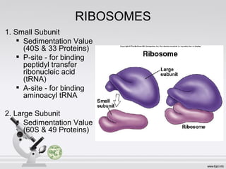
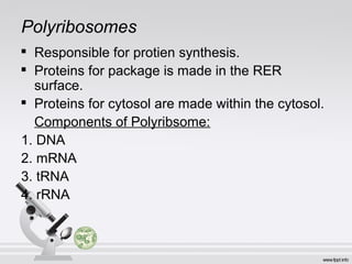
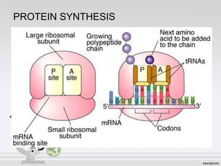
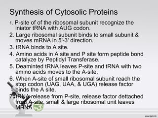
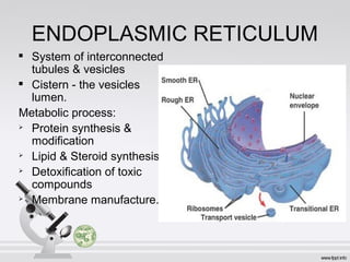
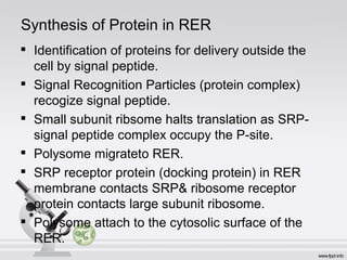
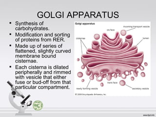
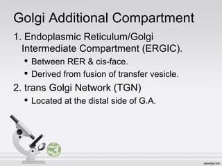
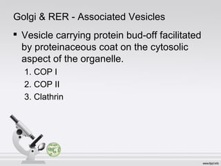
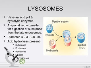
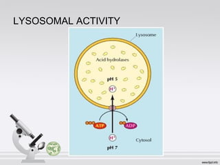
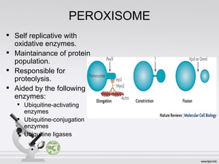
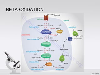
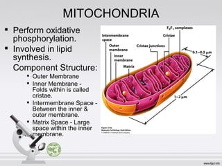
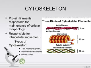
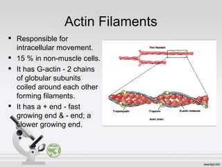
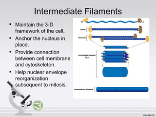
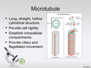
CELL STRUCTURE
Prof. Redem C. Deligero, MATMRS;MSES
RIBOSOMES
1. Small Subunit

Sedimentation Value
(40S & 33 Proteins)

P-site - for binding
peptidyl transfer
ribonucleic acid
(tRNA)

A-site - for binding
aminoacyl tRNA
2. Large Subunit

Sedimentation Value
(60S & 49 Proteins)
Polyribosomes

Responsible for protien synthesis.

Proteins for package is made in the RER
surface.

Proteins for cytosol are made within the cytosol.
Components of Polyribsome:
1. DNA
2. mRNA
3. tRNA
4. rRNA
PROTEIN SYNTHESIS
Synthesis of Cytosolic Proteins
1. P-site of of the ribosomal subunit recognize the
iniator tRNA with AUG codon.
2. Large ribosomal subunit binds to small subunit &
moves mRNA in 5'-3' direction.
3. tRNA binds to A site.
4. Amino acids in A site and P site form peptide bond
catalyze by Peptidyl Transferase.
5. Deaminted tRNA leaves P-site and tRNA with two
amino acids moves to the A-site.
6. When A-site of small ribosomal subunit reach the
stop codon (UAG, UAA, & UGA) release factor
binds the A site.
7. tRNA release from P-site, release factor dettached
from A-site, small & large ribosomal unit leaves
MRNA
ENDOPLASMIC RETICULUM

System of interconnected
tubules & vesicles

Cistern - the vesicles
lumen.
Metabolic process:

Protein synthesis &
modification

Lipid & Steroid synthesis

Detoxification of toxic
compounds

Membrane manufacture.
Synthesis of Protein in RER

Identification of proteins for delivery outside the
cell by signal peptide.

Signal Recognition Particles (protein complex)
recogize signal peptide.

Small subunit ribsome halts translation as SRP-
signal peptide complex occupy the P-site.

Polysome migrateto RER.

SRP receptor protein (docking protein) in RER
membrane contacts SRP& ribosome receptor
protein contacts large subunit ribosome.

Polysome attach to the cytosolic surface of the
RER.
GOLGI APPARATUS

Synthesis of
carbohydrates.

Modification and sorting
of proteins from RER.

Made up of series of
flattened, slightly curved
membrane bound
cisternae.

Each cisterna is dilated
peripherally and rimmed
with vesicle that either
fuse or bud-off from that
particular compartment.
Golgi Additional Compartment
1. Endoplasmic Reticulum/Golgi
Intermediate Compartment (ERGIC).

Between RER & cis-face.

Derived from fusion of transfer vesicle.
2. trans Golgi Network (TGN)

Located at the distal side of G.A.
Golgi & RER - Associated Vesicles

Vesicle carrying protein bud-off facilitated
by proteinaceous coat on the cytosolic
aspect of the organelle.
1. COP I
2. COP II
3. Clathrin
LYSOSOMES

Have an acid pH &
hydrolytic enzymes.

A specialized organelle
for digestion of substance
from the late endosomes.

Diameter is 0.3 - 0.8 µm.

Acid hydrolyses present:

Sulfatases

Proteases

Nucleases

Lipases

Glycosideses
LYSOSOMAL ACTIVITY
ENDOSOMES

Also involved in ingestion,
sequestering &
degradation of substance
internalized from
extracellular space.

Has two compartments:

Early endosomes - at the
cell periphery

Late endosomes - deeper
into cytoplasm.
PEROXISOME

Self replicative with
oxidative enzymes.

Maintainance of protein
population.

Responsible for
proteolysis.

Aided by the following
enzymes:

Ubiquitine-activating
enzymes

Ubiquitine-conjugation
enzymes

Ubiquitine ligases
BETA-OXIDATION
MITOCHONDRIA

Perform oxidative
phosphorylation.

Involved in lipid
synthesis.
Component Structure:

Outer Membrane

Inner Membrane -
Folds within is called
cristae.

Intermembrane Space -
Between the inner &
outer membrane.

Matrix Space - Large
space within the inner
membrane.
CYTOSKELETON

Protein filaments
responsible for
maintenance of cellular
morphology.

Responsible for
intracellular movement.
Types of
Cytoskeleton:

Thin Filaments (Actin)

Intermediate Filaments

Microtubules
Actin Filaments

Responsible for
intracellular movement.

15 % in non-muscle cells.

It has G-actin - 2 chains
of globular subunits
coiled around each other
forming filaments.

It has a + end - fast
growing end & - end; a
slower growing end.
Intermediate Filaments

Maintain the 3-D
framework of the cell.

Anchor the nucleus in
place.

Provide connection
between cell membrane
and cytoskeleton.

Help nuclear envelope
reorganization
subsequent to mitosis.
Microtubule

Long, straight, hollow
cylindrical structure.

Provide cell rigidity.

Establish intracellular
compartments.

Provide ciliary and
flagellated movement.

Cell Structure

  • 1.
    CELL STRUCTURE Prof. RedemC. Deligero, MATMRS;MSES
  • 2.
    RIBOSOMES 1. Small Subunit  SedimentationValue (40S & 33 Proteins)  P-site - for binding peptidyl transfer ribonucleic acid (tRNA)  A-site - for binding aminoacyl tRNA 2. Large Subunit  Sedimentation Value (60S & 49 Proteins)
  • 3.
    Polyribosomes  Responsible for protiensynthesis.  Proteins for package is made in the RER surface.  Proteins for cytosol are made within the cytosol. Components of Polyribsome: 1. DNA 2. mRNA 3. tRNA 4. rRNA
  • 4.
  • 5.
    Synthesis of CytosolicProteins 1. P-site of of the ribosomal subunit recognize the iniator tRNA with AUG codon. 2. Large ribosomal subunit binds to small subunit & moves mRNA in 5'-3' direction. 3. tRNA binds to A site. 4. Amino acids in A site and P site form peptide bond catalyze by Peptidyl Transferase. 5. Deaminted tRNA leaves P-site and tRNA with two amino acids moves to the A-site. 6. When A-site of small ribosomal subunit reach the stop codon (UAG, UAA, & UGA) release factor binds the A site. 7. tRNA release from P-site, release factor dettached from A-site, small & large ribosomal unit leaves MRNA
  • 6.
    ENDOPLASMIC RETICULUM  System ofinterconnected tubules & vesicles  Cistern - the vesicles lumen. Metabolic process:  Protein synthesis & modification  Lipid & Steroid synthesis  Detoxification of toxic compounds  Membrane manufacture.
  • 7.
    Synthesis of Proteinin RER  Identification of proteins for delivery outside the cell by signal peptide.  Signal Recognition Particles (protein complex) recogize signal peptide.  Small subunit ribsome halts translation as SRP- signal peptide complex occupy the P-site.  Polysome migrateto RER.  SRP receptor protein (docking protein) in RER membrane contacts SRP& ribosome receptor protein contacts large subunit ribosome.  Polysome attach to the cytosolic surface of the RER.
  • 8.
    GOLGI APPARATUS  Synthesis of carbohydrates.  Modificationand sorting of proteins from RER.  Made up of series of flattened, slightly curved membrane bound cisternae.  Each cisterna is dilated peripherally and rimmed with vesicle that either fuse or bud-off from that particular compartment.
  • 9.
    Golgi Additional Compartment 1.Endoplasmic Reticulum/Golgi Intermediate Compartment (ERGIC).  Between RER & cis-face.  Derived from fusion of transfer vesicle. 2. trans Golgi Network (TGN)  Located at the distal side of G.A.
  • 10.
    Golgi & RER- Associated Vesicles  Vesicle carrying protein bud-off facilitated by proteinaceous coat on the cytosolic aspect of the organelle. 1. COP I 2. COP II 3. Clathrin
  • 11.
    LYSOSOMES  Have an acidpH & hydrolytic enzymes.  A specialized organelle for digestion of substance from the late endosomes.  Diameter is 0.3 - 0.8 µm.  Acid hydrolyses present:  Sulfatases  Proteases  Nucleases  Lipases  Glycosideses
  • 12.
  • 13.
    ENDOSOMES  Also involved iningestion, sequestering & degradation of substance internalized from extracellular space.  Has two compartments:  Early endosomes - at the cell periphery  Late endosomes - deeper into cytoplasm.
  • 14.
    PEROXISOME  Self replicative with oxidativeenzymes.  Maintainance of protein population.  Responsible for proteolysis.  Aided by the following enzymes:  Ubiquitine-activating enzymes  Ubiquitine-conjugation enzymes  Ubiquitine ligases
  • 15.
  • 16.
    MITOCHONDRIA  Perform oxidative phosphorylation.  Involved inlipid synthesis. Component Structure:  Outer Membrane  Inner Membrane - Folds within is called cristae.  Intermembrane Space - Between the inner & outer membrane.  Matrix Space - Large space within the inner membrane.
  • 17.
    CYTOSKELETON  Protein filaments responsible for maintenanceof cellular morphology.  Responsible for intracellular movement. Types of Cytoskeleton:  Thin Filaments (Actin)  Intermediate Filaments  Microtubules
  • 18.
    Actin Filaments  Responsible for intracellularmovement.  15 % in non-muscle cells.  It has G-actin - 2 chains of globular subunits coiled around each other forming filaments.  It has a + end - fast growing end & - end; a slower growing end.
  • 19.
    Intermediate Filaments  Maintain the3-D framework of the cell.  Anchor the nucleus in place.  Provide connection between cell membrane and cytoskeleton.  Help nuclear envelope reorganization subsequent to mitosis.
  • 20.
    Microtubule  Long, straight, hollow cylindricalstructure.  Provide cell rigidity.  Establish intracellular compartments.  Provide ciliary and flagellated movement.