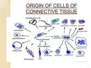
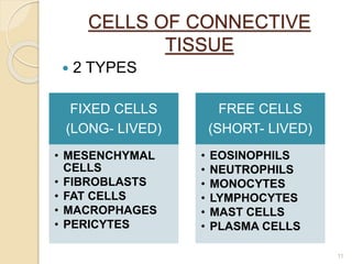
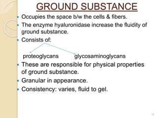
GOOD MORNING
1
CONNECTIVE
TISSUE
CHECKED BY: PRESENTED BY:
DR. NEHA MAHAJAN DR. VIRSHALI GUPTA
PG.1ST YEAR
2
CONTENTS
 INTRODUCTION
 DEFINITION
 ORIGIN
 FEATURES -
a. MATRIX
b. FIBRES
c. CELLS
 CLASSIFICATION
 FUNCTIONS
 DISORDERS OF CONNECTIVE TISSUE
 CONCLUSION
 REFRENCES
3
WHAT IS CONNECTIVE
TISSUE??
 Connective tissue is the group of tissues
that fills the interstices b/w more
specialised elements & serves to hold
them together & support them.
BY- ‘’GP PAL 3rd edition’’
 Defined as that group of tissues
predominantly composed of intercellular
matrix , secreted mainly by its cells
which are therefore , usually widely
spread.
BY – ‘’HAMS 9th edition’’ 4
ORIGIN OF CELLS OF
CONNECTIVE TISSUE
5
FEATURES OF CONNECTIVE
TISSUE
 CONSISTS OF 2 BASIC COMPONENTS
CELLS MATRIX
FIBERS GROUND
SUBSTANCE
6
FIBERS
COLLAGEN
FIBERS
RETICULAR
FIBERS
ELASTIC
FIBERS
OXYTALAN
FIBRES
7
FIBERS OF CONNECTIVE
TISSUE
COLLAGEN
FIBERS
RETICULAR
FIBERS
ELASTIC
FIBERS
OXYTALAN
FIBERS
DIAMETE
R
2-10nm 0.5 -2”m 0.1-2”m 0.2-1.5”m
LOCATION Walls of blood
vessels
Developing
matrices of
dentin & pulp
Lungs &
large
arterioles
PDL, gums,
dental pulp
COURSE Run in bundles
which may
branch and
anastomose
with
neighbouring
bundles.
Mesh like
pattern or
network.
Run singly &
branch to
form network.
3D
meshwork
extends from
cementum to
peripheral
blood
vessels.
OTHER
NAME
White fibers Argyrophilic
fibers
Yellow fibers -------
8
SYNTHESIS OF COLLAGEN
FIBERS
9
MAIN CHARACTERISTICS OF DIFFERENT
COLLAGEN TYPES
COLLAGEN
TYPE
TISSUE
DISTRIBUTION
SITE OF
SYNTHESIS
MAIN
FUNCTION
1 DERMIS, BONE,
TENDON, DENTIN,
SCLERA, PDL,
LAMINA PROPRIA
FIBROBLAST,
OSTEOBLAST,
ODONTOBLAST,
CHONDROBLAST
RESISTANCE TO
TENSION
11 HYALNE AND
ELASTIC
CARTILAGES
CHONDROBLAST RESISTANCE TO
INTERMEDIATE
PRESSURE
111 SMOOTH MUSCLE,
SPLEEN, LIVER,
KIDNEY , LUNG, PDL,
LAMINA PROPRIA
SMOOTH MUSCLE
CELLS,
FIBROBLAST,
RETICULAR
CELLS
STRUCTURAL
MAINTAINANCE IN
EXPANSIBLE
ORGANS
1V EPITHELIAL AND
ENDOTHELIAL BASAL
LAMINAE AND
BASEMEMENT
MEMBRANES
ENDOTHELIAL
CELLS,
EPITHELIAL
CELLS, MUSCLE
CELLS
SUPPORT AND
FILTRATION
V MUSCLE BASAL ----- ----- 10
CELLS OF CONNECTIVE
TISSUE
 2 TYPES
FIXED CELLS
(LONG- LIVED)
‱ MESENCHYMAL
CELLS
‱ FIBROBLASTS
‱ FAT CELLS
‱ MACROPHAGES
‱ PERICYTES
FREE CELLS
(SHORT- LIVED)
‱ EOSINOPHILS
‱ NEUTROPHILS
‱ MONOCYTES
‱ LYMPHOCYTES
‱ MAST CELLS
‱ PLASMA CELLS
11
CELL TYPE MAIN PRODUCT OR
ACTIVITY
MAIN FUNCTION
FIBROBLASTS,
CHONDROBLASTS,
OSTEOBLASTS,
ODONTOBLASTS
PRODUCTION OF FIBERS AND
GROUND SUBSTANCE
STRUCTURAL
PLASMA CELL PRODUCTION OF
ANTIBODIES
IMMUNOLOGIC
LYMPHOCYTE PRODUCTION OF IMMUNE
CELLS
IMMUNOLOGIC
EOSINOPHILIC
LEUKOCYTE
PHAGOCYTOSIS OF
ANTIGEN-ANTIBODY
COMPLEX
IMMUNOLOGIC
MACROPHAGE,
NEUTROPHIL
PHAGOCYTOSIS OF FOREIGN
SUBSTANCES, BACTERIA
DEFENSE
MAST CELL, BASOPHIL LIBERATION OF
PHARMACOLOGICALLY
ACTIVE SUBSTANCES
(HISTAMINE)
DEFENSE
ADIPOSE CELL STORAGE OF NEUTRAL FATS ENERGY RESERVOIR,
HEAT PRODUCTION
12
DIAGRAM SHOWING CELLS AND
FIBRES OF CONNECTIVE FIBERS
13
GROUND SUBSTANCE
 Occupies the space b/w the cells & fibers.
 The enzyme hyaluronidase increase the fluidity of
ground substance.
 Consists of:
proteoglycans glycosaminoglycans
 These are responsible for physical properties
of ground substance.
 Granular in appearance.
 Consistency: varies, fluid to gel.
14
CLASSIFICATION OF CONNECTIVE
TISSUE
CONNECTIVE TISSUE PROPER
A. EMBRYONAL CT
i.Mesenchymal
ii. Mucous
B. REGULAR CT
i. Loose CT
ii. Dense CT
C. SPECIAL FORMS
i . Elastic
ii. Adipose
iii. Reticular
SPECIALISED CONNECTIVE
TISSUE
A. FLUID CT
i. Blood
B. RIGID
i. Mineralized
ii. Non-
mineralized
15
16
CONNECTIVE TISSUE
PROPER
17
EMBRYONIC CONNECTIVE
TISSUE
 It consist of two varieties 1.mesenchymal and 2.
mucous .
 Present for most of the parts during embryonic and
foetal development.
 When present post- natally , associated with healing
of injured tissue or tumorous lesion.
 Made up of small cells with slender branching
processes that join to form a fine network.
MESENCHYMAL
CELLS
18
CARTILAGE
 Type of connective tissue composed of special cells
known as chondrocytes along with collagen or
yellow elastic fibers.
 Types –
Hyaline
Fibrous
Elastic
19
BONE
COMPACT BONE SPONGY BONE
OTHER NAME CORTICAL BONE TRABECULAE BONE
DENSITY COMPACT POROUS
LOCATION OUTER BONE INNER BONE
FUNCTIONAL UNIT OSTEON TRABECULAE
20
 DENTIN
 3 TYPES-
PRIMARY
SECONDARY
TERTIARY
 CEMENTUM –
 2 TYPES
ACELLULAR
CELLULAR
21
FUNCTIONS OF CONNECTIVE TISSUE
 SUPPORT- To epithelium e.g.: lamina propria.
 STRENGTH- Provides tensile strength to
mechanical stress e.g.: dermis of skin , ligament.
 STORAGE- Fat cells store fat while ground
substance store water , ions and inorganic
materials.
 TRANSPORT- Water, ions and inorganic materials
are transported from blood to various tissues of
body through connective tissue matrix.
 PACKING- They fill spaces e.g.: loose connective
tissue and adipose tissue.
 REPAIR- Helps in wound healing
 DEFENSE- Cells helps in defense action e.g.:
plasma cells, macrophages, lymphocytes,
monocytes, eosinophils.
22
GINGIVAL CONNECTIVE
TISSUE
 The gingival connective tissue provides strength to
gums and make the gums to cement root and alveolar
bone.
 The dense gingival connective tissue is referred to as a
lamina propria.
 Consists of 2 layers:
Papillary layer reticular layer
23
HOW EPITHELIUM CONNECTS WITH
CONNECTIVE TISSUE ??
24STRUCTURE OF BASEMENT MEMBRANE
DISORDERS OF CONNECTIVE TISSUE
 MARFAN’S SYNDROME –
25
SCURVY
 Deficiency of vitamin C
 Characteristic features:
 Bleeding gums, loosening of teeth with grade 1 – 2
mobility , gingivitis n sometimes bluish
pigmentation.
26
EHLERS- DANLOS SYNDROME
 Skin hyperextensibility.
 Joint hypermobility.
 Tissue fragility.
SCLERODEMA
 Difficulty in opening mouth due to involvement of
perioral skin.
PEMPHIGUS
 Blisters in mouth which erupts later.
 Blisters are painful, but don’t itch.
 Difficulty in swallowing.
27
28
29
SPONTANEOUS
PNEUMOTHORAX
30
CONCLUSION
 Basic tissue of the body.
 Includes fat, cartilage, bone and blood.
 Provides support, fills the spaces between
organs, protecting organs and aiding in the
transport of materials around the body.
31
REFERENCES
 Textbook of histology GP Pal 3rd edition.
 Textbook of human histology 3rd edition by Inderbir
Singh.
 Tencate’s oral histology.
 Ham’s histology 9th edition by David.
 Textbook of oral histology & embryology (orban’s) 12th
edition by GS Kumar.
 Textbook of oral histology by D.Vincent Provenza 2nd
edition.
32
33

connective tissue.

  • 1.
  • 2.
    CONNECTIVE TISSUE CHECKED BY: PRESENTEDBY: DR. NEHA MAHAJAN DR. VIRSHALI GUPTA PG.1ST YEAR 2
  • 3.
    CONTENTS  INTRODUCTION  DEFINITION ORIGIN  FEATURES - a. MATRIX b. FIBRES c. CELLS  CLASSIFICATION  FUNCTIONS  DISORDERS OF CONNECTIVE TISSUE  CONCLUSION  REFRENCES 3
  • 4.
    WHAT IS CONNECTIVE TISSUE?? Connective tissue is the group of tissues that fills the interstices b/w more specialised elements & serves to hold them together & support them. BY- ‘’GP PAL 3rd edition’’  Defined as that group of tissues predominantly composed of intercellular matrix , secreted mainly by its cells which are therefore , usually widely spread. BY – ‘’HAMS 9th edition’’ 4
  • 5.
    ORIGIN OF CELLSOF CONNECTIVE TISSUE 5
  • 6.
    FEATURES OF CONNECTIVE TISSUE CONSISTS OF 2 BASIC COMPONENTS CELLS MATRIX FIBERS GROUND SUBSTANCE 6
  • 7.
  • 8.
    FIBERS OF CONNECTIVE TISSUE COLLAGEN FIBERS RETICULAR FIBERS ELASTIC FIBERS OXYTALAN FIBERS DIAMETE R 2-10nm0.5 -2”m 0.1-2”m 0.2-1.5”m LOCATION Walls of blood vessels Developing matrices of dentin & pulp Lungs & large arterioles PDL, gums, dental pulp COURSE Run in bundles which may branch and anastomose with neighbouring bundles. Mesh like pattern or network. Run singly & branch to form network. 3D meshwork extends from cementum to peripheral blood vessels. OTHER NAME White fibers Argyrophilic fibers Yellow fibers ------- 8
  • 9.
  • 10.
    MAIN CHARACTERISTICS OFDIFFERENT COLLAGEN TYPES COLLAGEN TYPE TISSUE DISTRIBUTION SITE OF SYNTHESIS MAIN FUNCTION 1 DERMIS, BONE, TENDON, DENTIN, SCLERA, PDL, LAMINA PROPRIA FIBROBLAST, OSTEOBLAST, ODONTOBLAST, CHONDROBLAST RESISTANCE TO TENSION 11 HYALNE AND ELASTIC CARTILAGES CHONDROBLAST RESISTANCE TO INTERMEDIATE PRESSURE 111 SMOOTH MUSCLE, SPLEEN, LIVER, KIDNEY , LUNG, PDL, LAMINA PROPRIA SMOOTH MUSCLE CELLS, FIBROBLAST, RETICULAR CELLS STRUCTURAL MAINTAINANCE IN EXPANSIBLE ORGANS 1V EPITHELIAL AND ENDOTHELIAL BASAL LAMINAE AND BASEMEMENT MEMBRANES ENDOTHELIAL CELLS, EPITHELIAL CELLS, MUSCLE CELLS SUPPORT AND FILTRATION V MUSCLE BASAL ----- ----- 10
  • 11.
    CELLS OF CONNECTIVE TISSUE 2 TYPES FIXED CELLS (LONG- LIVED) ‱ MESENCHYMAL CELLS ‱ FIBROBLASTS ‱ FAT CELLS ‱ MACROPHAGES ‱ PERICYTES FREE CELLS (SHORT- LIVED) ‱ EOSINOPHILS ‱ NEUTROPHILS ‱ MONOCYTES ‱ LYMPHOCYTES ‱ MAST CELLS ‱ PLASMA CELLS 11
  • 12.
    CELL TYPE MAINPRODUCT OR ACTIVITY MAIN FUNCTION FIBROBLASTS, CHONDROBLASTS, OSTEOBLASTS, ODONTOBLASTS PRODUCTION OF FIBERS AND GROUND SUBSTANCE STRUCTURAL PLASMA CELL PRODUCTION OF ANTIBODIES IMMUNOLOGIC LYMPHOCYTE PRODUCTION OF IMMUNE CELLS IMMUNOLOGIC EOSINOPHILIC LEUKOCYTE PHAGOCYTOSIS OF ANTIGEN-ANTIBODY COMPLEX IMMUNOLOGIC MACROPHAGE, NEUTROPHIL PHAGOCYTOSIS OF FOREIGN SUBSTANCES, BACTERIA DEFENSE MAST CELL, BASOPHIL LIBERATION OF PHARMACOLOGICALLY ACTIVE SUBSTANCES (HISTAMINE) DEFENSE ADIPOSE CELL STORAGE OF NEUTRAL FATS ENERGY RESERVOIR, HEAT PRODUCTION 12
  • 13.
    DIAGRAM SHOWING CELLSAND FIBRES OF CONNECTIVE FIBERS 13
  • 14.
    GROUND SUBSTANCE  Occupiesthe space b/w the cells & fibers.  The enzyme hyaluronidase increase the fluidity of ground substance.  Consists of: proteoglycans glycosaminoglycans  These are responsible for physical properties of ground substance.  Granular in appearance.  Consistency: varies, fluid to gel. 14
  • 15.
    CLASSIFICATION OF CONNECTIVE TISSUE CONNECTIVETISSUE PROPER A. EMBRYONAL CT i.Mesenchymal ii. Mucous B. REGULAR CT i. Loose CT ii. Dense CT C. SPECIAL FORMS i . Elastic ii. Adipose iii. Reticular SPECIALISED CONNECTIVE TISSUE A. FLUID CT i. Blood B. RIGID i. Mineralized ii. Non- mineralized 15
  • 16.
  • 17.
  • 18.
    EMBRYONIC CONNECTIVE TISSUE  Itconsist of two varieties 1.mesenchymal and 2. mucous .  Present for most of the parts during embryonic and foetal development.  When present post- natally , associated with healing of injured tissue or tumorous lesion.  Made up of small cells with slender branching processes that join to form a fine network. MESENCHYMAL CELLS 18
  • 19.
    CARTILAGE  Type ofconnective tissue composed of special cells known as chondrocytes along with collagen or yellow elastic fibers.  Types – Hyaline Fibrous Elastic 19
  • 20.
    BONE COMPACT BONE SPONGYBONE OTHER NAME CORTICAL BONE TRABECULAE BONE DENSITY COMPACT POROUS LOCATION OUTER BONE INNER BONE FUNCTIONAL UNIT OSTEON TRABECULAE 20
  • 21.
     DENTIN  3TYPES- PRIMARY SECONDARY TERTIARY  CEMENTUM –  2 TYPES ACELLULAR CELLULAR 21
  • 22.
    FUNCTIONS OF CONNECTIVETISSUE  SUPPORT- To epithelium e.g.: lamina propria.  STRENGTH- Provides tensile strength to mechanical stress e.g.: dermis of skin , ligament.  STORAGE- Fat cells store fat while ground substance store water , ions and inorganic materials.  TRANSPORT- Water, ions and inorganic materials are transported from blood to various tissues of body through connective tissue matrix.  PACKING- They fill spaces e.g.: loose connective tissue and adipose tissue.  REPAIR- Helps in wound healing  DEFENSE- Cells helps in defense action e.g.: plasma cells, macrophages, lymphocytes, monocytes, eosinophils. 22
  • 23.
    GINGIVAL CONNECTIVE TISSUE  Thegingival connective tissue provides strength to gums and make the gums to cement root and alveolar bone.  The dense gingival connective tissue is referred to as a lamina propria.  Consists of 2 layers: Papillary layer reticular layer 23
  • 24.
    HOW EPITHELIUM CONNECTSWITH CONNECTIVE TISSUE ?? 24STRUCTURE OF BASEMENT MEMBRANE
  • 25.
    DISORDERS OF CONNECTIVETISSUE  MARFAN’S SYNDROME – 25
  • 26.
    SCURVY  Deficiency ofvitamin C  Characteristic features:  Bleeding gums, loosening of teeth with grade 1 – 2 mobility , gingivitis n sometimes bluish pigmentation. 26
  • 27.
    EHLERS- DANLOS SYNDROME Skin hyperextensibility.  Joint hypermobility.  Tissue fragility. SCLERODEMA  Difficulty in opening mouth due to involvement of perioral skin. PEMPHIGUS  Blisters in mouth which erupts later.  Blisters are painful, but don’t itch.  Difficulty in swallowing. 27
  • 28.
  • 29.
  • 30.
  • 31.
    CONCLUSION  Basic tissueof the body.  Includes fat, cartilage, bone and blood.  Provides support, fills the spaces between organs, protecting organs and aiding in the transport of materials around the body. 31
  • 32.
    REFERENCES  Textbook ofhistology GP Pal 3rd edition.  Textbook of human histology 3rd edition by Inderbir Singh.  Tencate’s oral histology.  Ham’s histology 9th edition by David.  Textbook of oral histology & embryology (orban’s) 12th edition by GS Kumar.  Textbook of oral histology by D.Vincent Provenza 2nd edition. 32
  • 33.