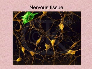
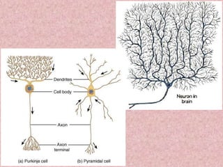
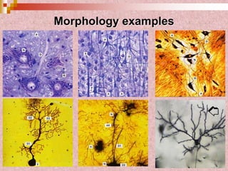
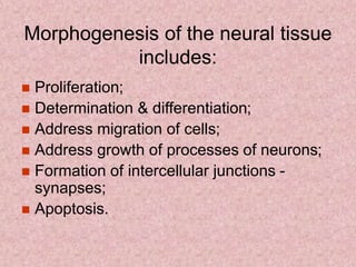
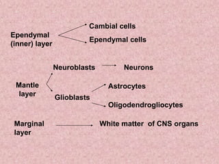
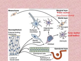
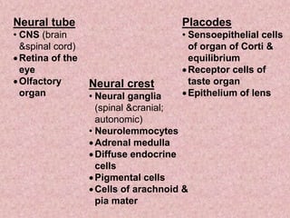
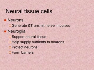
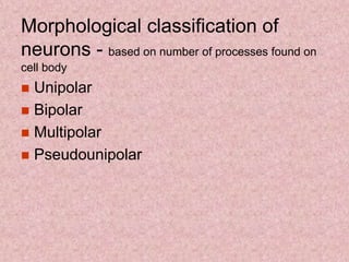
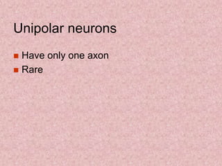
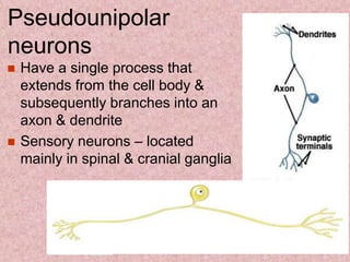
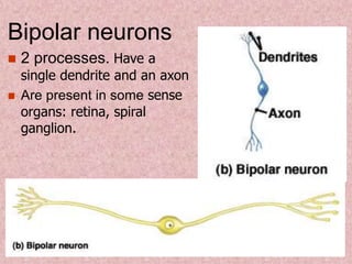
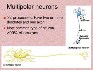
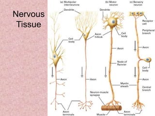
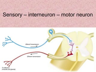
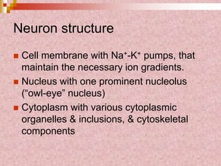
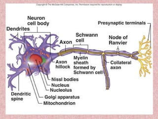
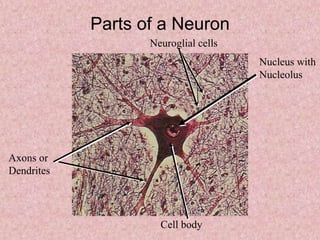
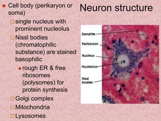
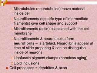
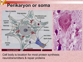
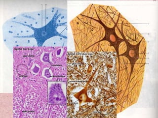
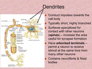
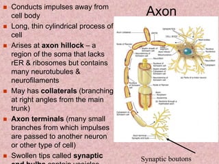
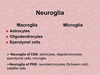
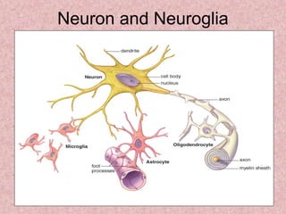
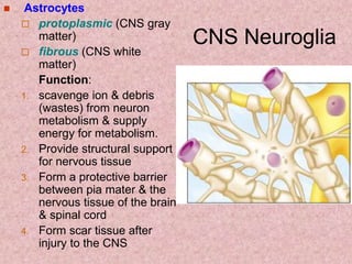
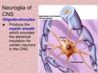
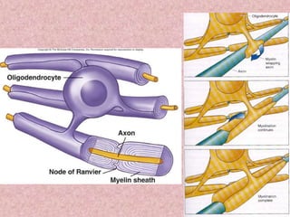
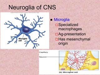
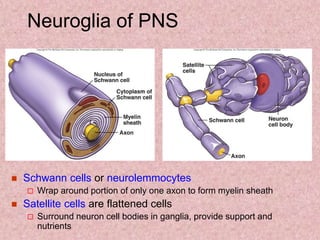
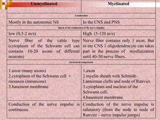
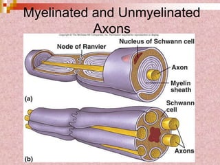
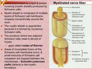
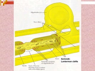
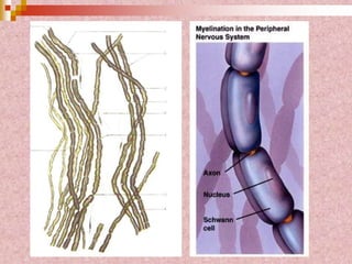
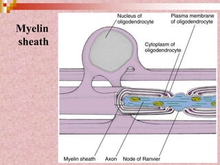
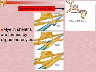
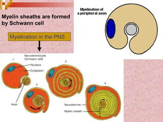
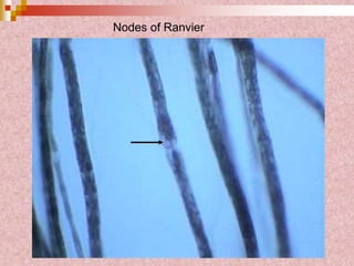
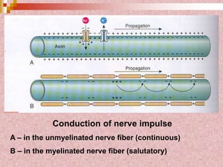
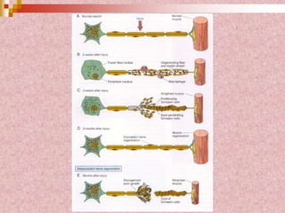
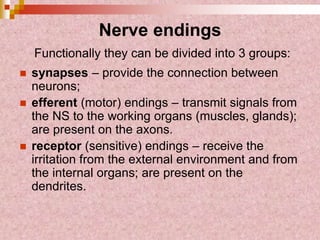
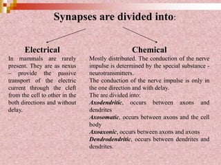
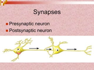
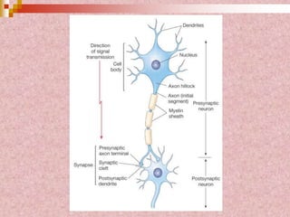
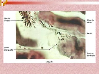
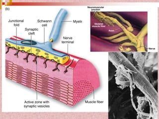
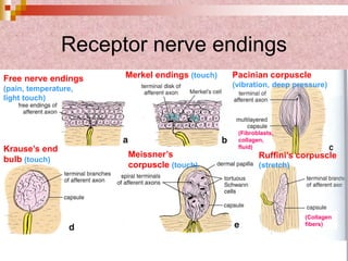
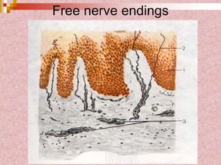
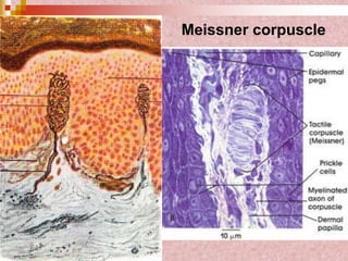
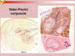
Nervous tissue
Morphology examples
 Conducts electrical impulses (signals) to the
CNS & transmits impulses from the CNS to
various structures of the body
 Conveys information from one area to
another
Neural tissue
Morphogenesis of the neural tissue
includes:
 Proliferation;
 Determination & differentiation;
 Address migration of cells;
 Address growth of processes of neurons;
 Formation of intercellular junctions -
synapses;
 Apoptosis.
Neurulation
Cells in the neural tube
Ependymal
(inner) layer
Cambial cells
Ependymal cells
Mantle
layer
Neuroblasts
Glioblasts
Neurons
Astrocytes
Oligodendrogliocytes
Marginal
layer
White matter of CNS organs
Gray matter
(cell bodies)
White matter
(myelinated axons)
Neural tube
• CNS (brain
&spinal cord)
Retina of the
eye
Olfactory
organ
Neural crest
• Neural ganglia
(spinal &cranial;
autonomic)
• Neurolemmocytes
Adrenal medulla
Diffuse endocrine
cells
Pigmental cells
Cells of arachnoid &
pia mater
Placodes
• Sensoepithelial cells
of organ of Corti &
equilibrium
Receptor cells of
taste organ
Epithelium of lens
 Neurons
Generate &Transmit nerve impulses
 Neuroglia
Support neural tissue
Help supply nutrients to neurons
Protect neurons
Form barriers
Neural tissue cells
Morphological classification of
neurons - based on number of processes found on
cell body
 Unipolar
 Bipolar
 Multipolar
 Pseudounipolar
Unipolar neurons
 Have only one axon
 Rare
Pseudounipolar
neurons
 Have a single process that
extends from the cell body &
subsequently branches into an
axon & dendrite
 Sensory neurons – located
mainly in spinal & cranial ganglia
Bipolar neurons
 2 processes. Have a
single dendrite and an axon
 Are present in some sense
organs: retina, spiral
ganglion.
Multipolar neurons
 >2 processes. Have two or more
dendrites and one axon
 Most common type of neuron.
>99% of neurons
Functional classification of neurons
 motor neurons - efferent (conduct impulses from CNS
to other neurons, muscles or glands);
 sensory neurons - afferent (receive stimuli from the
internal & external environment). Conduct nerve
impulses to the CNS.
 interneurons act as connectors of neurons in chain.
They most commonly connect sensory & motor neurons.
 secretory neurons
- neurons of hypothalamus: supraoptic & paraventricular
nuclei – neurons produce hormones: vasopressin &
oxytocin);
- all neurons produce neurotransmitters of synapses.
Nervous
Tissue
Sensory – interneuron – motor neuron
Neurons
 Functional unit of nervous system
 Special neuronal characteristics
Convey APs (excitable)
Longevity
Do not divide
High metabolic rate
Neuron structure
 Cell membrane with Na+-K+ pumps, that
maintain the necessary ion gradients.
 Nucleus with one prominent nucleolus
(“owl-eye” nucleus)
 Cytoplasm with various cytoplasmic
organelles & inclusions, & cytoskeletal
components
Neuron has
 Cell body (perikaryon, soma)
 Processes:
 Only one axon
 One & more dendrites
Nucleus with
Nucleolus
Parts of a Neuron
Axons or
Dendrites
Cell body
Neuroglial cells
Neuron structure Cell body (perikaryon or
soma)
 single nucleus with
prominent nucleolus
 Nissl bodies
(chromatophilic
substance) are stained
basophilic
 rough ER & free
ribosomes
(polysomes) for
protein synthesis
 Golgi complex
 Mitochondria
 Lysosomes
Microtubules (neurotubules) move material
inside cell
Neurofilaments (specific type of intermediate
filaments) give cell shape and support
Microfilaments (actin) associated with the cell
membrane
Neurofilaments & neurotubules form
neurofibrils – is artefact. Neurofibrils appear at
time of slide preparing & can be distinguish
inside of neurons
Lipofuscin pigment clumps (harmless aging)
Lipid inclusions
 Cell processes = dendrites & axon
Perikaryon or soma
Cell body is location for most protein synthesis
neurotransmitters & repair proteins
Dendrites
 Conduct impulses towards the
cell body
 Typically short, highly branched
 Surfaces specialized for
contact with other neurons
(spines) – increase the area
useful for synapse formation
 Have arborized terminals –
permit a neuron to receive
stimuli at the same time from
many other neurons
 Contains neurofibrils & Nissl
bodies
Axon Conducts impulses away from
cell body
 Long, thin cylindrical process of
cell
 Arises at axon hillock – a
region of the soma that lacks
rER & ribosomes but contains
many neurotubules &
neurofilaments
 May has collaterals (branching
at right angles from the main
trunk)
 Axon terminals (many small
branches from which impulses
are passed to another neuron
or other type of cell)
 Swollen tips called synaptic
Transport
 Dendritic – the movement of
substances & organelles through the
dindrites
 Axonal - the movement of
substances & organelles through the
axon
Axonal Transport
 Axonal transport system moves substances
slow axonal flow
 movement in one direction only -- away from cell body
 movement at 1-5 mm per day
fast axonal flow
 transports in either direction
 at 100-500 mm per day
 moves organelles & materials along surface of
microtubules
 for use or for recycling in cell body
•Anterograde transport – carries material away from the soma
•Retrograde transport – carries material toward the soma for
reutilization, recycling, or degradation
Neuroglia
Macroglia
 Astrocytes
 Oligodendrocytes
 Ependymal cells
Microglia
 Neuroglia of CNS: astrocytes, oligodendrocytes,
ependymal cells, microglia
Neuroglia of PNS: neurolemmocytes (Schwann cell),
satellite cells
Neuron and Neuroglia
CNS Neuroglia
 Astrocytes
 protoplasmic (CNS gray
matter)
 fibrous (CNS white
matter)
Function:
1. scavenge ion & debris
(wastes) from neuron
metabolism & supply
energy for metabolism.
2. Provide structural support
for nervous tissue
3. Form a protective barrier
between pia mater & the
nervous tissue of the brain
& spinal cord
4. Form scar tissue after
injury to the CNS
Neuroglia of CNS
 Astrocytes
 Promote tight junctions
to form blood-brain
barrier
1. Endothelium of the
capillary (between
endothelial cells there
are tight junctions)
2. basement membrane
of endothelium
3. perivascular
membrane – is formed
by foot processes of
astrocytes
Blood-brain barrier
1 – endothelium; 2- basement membrane; 3 – astrocyte’s body, 4 –
foot processes of astrocyte; 5 – neuron, 6 – neuron’s processes; 7-
oligodendroglial cell
Neuroglia of CNS
 Ependymal Cells
 Line ventricles of the
brain, spinal cord
central canal, choroid
plexus
 Secrete & move liquor
 Form blood-liquor
barrier:
1. Endothelium of
capillary
2. Basement membrane
of endothelium
3. Loose connective
tissue
4. Basement membrane
of ependymal cells
5. Ependymal cells
Oligodendrocytes
 Produce the
myelin sheath
which provides
the electrical
insulation for
certain neurons
in the CNS
Neuroglia of
CNS
Neuroglia of CNS
 Microglia
Specialized
macrophages
Ag-presentation
Has mesenchymal
origin
Supporting cells in the PNS
 Satellite cells
 Schwann cells / neurolemmocytes
Neuroglia of PNS
 Schwann cells or neurolemmocytes
 Wrap around portion of only one axon to form myelin sheath
 Satellite cells are flattened cells
 Surround neuron cell bodies in ganglia, provide support and
nutrients
Satellite Cells
The End
Nerve fibers
 Myelinated fibers
 Unmyelinated fibers
Unmyelinated Myelinated
Localization
Mostly in the autonomic NS In the CNS and PNS
Speed of the conduction of the nerve impulse
low (0,5-2 m/s) High (5-120 m/s)
Nerve fiber of the cable type
(cytoplasm of the Schwann cell can
contains 10-20 axons of different
neurons)
Nerve fiber contains only 1 axon. But
in tne CNS 1 oligodendrocyte can takes
part in the process of myelinization
until 40-50 nerve fibers.
Structural components
1.axon (many axons)
2.cytoplasm of the Schwann cell +
mesaxon (mesaxons)
3.basement membrane
1.axon
2.myelin sheath with Schmidt-
Lanterman clefts and node of Ranvier.
3.cytoplasm and nucleus of the
Schwann cell.
4.basement membrane.
Conduction of the nerve impulse is
continuous.
Conduction of the nerve impulse is
salutatory (from the node to node of
Ranvier – nerve impulse jumps)
Myelinated and Unmyelinated
Axons
Unmyelinated nerve fibers
 Axons surrounded by a lipid & protein
covering (myelin sheath) produced by
Schwann cells.
 Myelin sheath is composed of multiple
layers of Schwann cell membrane
wrapped concentrically around the
axon.
 The myelin sheath is segmented
because it is formed by numerous
Schwann cells.
 The junctions where two adjacent
Schwann cells meet is devoid of
myelin.
 gaps called nodes of Ranvier
 Areas of incomplete fusion of the
Schwann cell membrane occur, &
small amounts of Schwann cell
cytoplasm are trapped between the
membranes – Schmidt-Lanterman
clefts (defects in the myelin
formation)
Myelinated nerve fiber
Schmidt-
Lanterman clefts
Myelin
sheath
Myelination in the CNS
Myelin sheaths
are formed by
oligodendrocytes
Myelination in the PNS
Myelin sheaths are formed
by Schwann cell
Myelin Sheath
 Whitish, fatty (protein-lipid), segmented
sheath around most long axons
 It functions in:
Protection of the axon
Electrically insulating fibers from one another
Increasing the speed of nerve impulse
transmission
Myelin Sheaths
Nodes of Ranvier
 Gaps in the myelin sheath between
adjacent Schwann cells
 They are the sites where collaterals can
arise
Nodes of Ranvier
Conduction of nerve impulse
A – in the unmyelinated nerve fiber (continuous)
B – in the myelinated nerve fiber (salutatory)
Nerve endings
Functionally they can be divided into 3 groups:
 synapses – provide the connection between
neurons;
 efferent (motor) endings – transmit signals from
the NS to the working organs (muscles, glands);
are present on the axons.
 receptor (sensitive) endings – receive the
irritation from the external environment and from
the internal organs; are present on the
dendrites.
Electrical
In mammals are rarely
present. They are as nexus
– provide the passive
transport of the electric
current through the cleft
from the cell to other in the
both directions and without
delay.
Chemical
Mostly distributed. The conduction of the nerve
impulse is determined by the special substance -
neurotransmitters.
The conduction of the nerve impulse is only in
the one direction and with delay.
The are divided into:
Axodendritic, occurs between axons and
dendrites
Axosomatic, occurs between axons and the cell
body
Axoaxonic, occurs between axons and axons
Dendrodendritic, occurs between dendrites and
dendrites.
Synapses are divided into:
Synapses
 Presynaptic neuron
 Postsynaptic neuron
Synapses
 Axodendritic synapses
 Axosomatic synapses
 Axoaxonic synapses
 Dendrodendritic synapses
Synapse structure
 Presynaptic element
Axon terminal
Synaptic vesicles
Neurotransmitters
Mitochondria
 Synaptic cleft
 Postsynaptic
elements
NT receptors
May generate AP
Synaptic
Transmission
 An AP reaches the
axon terminal of the
presynaptic cell and
causes V-gated Ca2+
channels to open.
 Ca2+ rushes in, binds
to regulatory proteins &
initiates NT exocytosis.
 NTs diffuse across the
synaptic cleft and then
bind to receptors on
the postsynaptic
membrane and initiate
some sort of response
on the postsynaptic
cell.
Efferent nerve endings
Motor
Are present in the striated and
smooth muscles.
By structure they are like synapses,
but there are some features: nearly
to the muscle fiber the axon loses
the myelin sheath and gives some
small branches. They are covered
by the Schwann cells and
basement membrane.
The transmission of the excitation
is provided by the neurotransmitter
- acetylcholine.
Secretory
Are present in the glands
Can make next influences:
- hydrokinetic (mobilization of
the water);
- proteokinetic (secretion of the
proteins);
- synthetic (to increase the
synthesis);
- trophic (to maintain the normal
structure and function).
 Motor unit
 One neuron
 Muscle cells stimulated
by that neuron
• Neuromuscular
junctions – association
site of nerve and
muscle
Receptor nerve endings
 exteroreceptors (receive the signals from
the external environment). They are: visual,
auditory, olfactory, taste, tactile receptors.
 interoreceptors. They are divided into
visceroreceptors – receive signals from
the inner organs; and proprioreceptors –
receptors of the locomotor system.
Physiological classification of the
receptor nerve endings
 mechanoreceptors (pressure, vibration)
 chemoreceptors (taste, smell)
 thermoreceptors (cold, warm)
 pain receptors
Morphological classification
Receptor nerve endings
Free(simple)
They are consists of
terminal branches of the
dendrites of the sensory
neuron.
They provide the
perception of the pain,
cold, warm, tactile
signals.
They are present inside of
the epithelium and in the
loose connective tissue,
which is located beneath.
It is consists of only of the
dendrite.
Restricted (compound)
Encapsulated
They are surrounded by the
connective tissue capsule.
Structure:
branches of the dendrite
surrounding Schwann cells
connective tissue capsule.
Examples:
Vater-Pacini corpuscles
Meissner’s tactile corpuscles
Ruffini’s curpuscles
Bulb of Krause
Neuromuscular spindles
Tendon organ of Golgi
Unencapsulated
They are consist of the
branches of the
dendrites that are
surrounded by the
Schwann cells.
They are present in
the dermis of the skin
and in the lamina
propria of the tunica
mucosa.
Free nerve endings
(pain, temperature,
light touch)
Merkel endings (touch) Pacinian corpuscle
(vibration, deep pressure)
Krause’s end
bulb (touch)
Meissner’s
corpuscle (touch)
Ruffini’s corpuscle
(stretch)
(Fibroblasts,
collagen,
fluid)
(Collagen
fibers)
Receptor nerve endings
Free nerve endings
Meissner corpuscle
Vater-Pacini
corpuscle
Pacinian corpuscle
Neuromuscular spindle
Vater-Pacini corpuscles – are present on the connective
tissue of the skin and inner organs. They are responsible for the
sensation of the pressure and vibration.
Meissner’s tactile corpuscles - are located in the papillary
layer of the dermis in skin, mostly: tips of fingers, lips, nipple
and area which is around.
Ruffini’s curpuscles – are located in the connective tissue of
the skin and in the capsules of the articulations. Take in
pressure.
Bulb of Krause – is present in the papillary layer of the dermis,
lamina propria of the tunica mucosa in the oral cavity. It is
mechanoreceptor.
Neuromuscular spindle – receptors of the sprain of the
muscle fibers. It has motor and sensory innervations.
Tendon organ of Golgi – receptor of the sprain. It is located in
the places where the skeletal muscle fibers join to the tendon.
Good bye! ☻

Nervous tissue 1

  • 2.
  • 4.
  • 5.
     Conducts electricalimpulses (signals) to the CNS & transmits impulses from the CNS to various structures of the body  Conveys information from one area to another Neural tissue
  • 6.
    Morphogenesis of theneural tissue includes:  Proliferation;  Determination & differentiation;  Address migration of cells;  Address growth of processes of neurons;  Formation of intercellular junctions - synapses;  Apoptosis.
  • 7.
  • 8.
    Cells in theneural tube
  • 9.
    Ependymal (inner) layer Cambial cells Ependymalcells Mantle layer Neuroblasts Glioblasts Neurons Astrocytes Oligodendrogliocytes Marginal layer White matter of CNS organs
  • 10.
    Gray matter (cell bodies) Whitematter (myelinated axons)
  • 11.
    Neural tube • CNS(brain &spinal cord) Retina of the eye Olfactory organ Neural crest • Neural ganglia (spinal &cranial; autonomic) • Neurolemmocytes Adrenal medulla Diffuse endocrine cells Pigmental cells Cells of arachnoid & pia mater Placodes • Sensoepithelial cells of organ of Corti & equilibrium Receptor cells of taste organ Epithelium of lens
  • 12.
     Neurons Generate &Transmitnerve impulses  Neuroglia Support neural tissue Help supply nutrients to neurons Protect neurons Form barriers Neural tissue cells
  • 13.
    Morphological classification of neurons- based on number of processes found on cell body  Unipolar  Bipolar  Multipolar  Pseudounipolar
  • 14.
    Unipolar neurons  Haveonly one axon  Rare
  • 15.
    Pseudounipolar neurons  Have asingle process that extends from the cell body & subsequently branches into an axon & dendrite  Sensory neurons – located mainly in spinal & cranial ganglia
  • 16.
    Bipolar neurons  2processes. Have a single dendrite and an axon  Are present in some sense organs: retina, spiral ganglion.
  • 17.
    Multipolar neurons  >2processes. Have two or more dendrites and one axon  Most common type of neuron. >99% of neurons
  • 18.
    Functional classification ofneurons  motor neurons - efferent (conduct impulses from CNS to other neurons, muscles or glands);  sensory neurons - afferent (receive stimuli from the internal & external environment). Conduct nerve impulses to the CNS.  interneurons act as connectors of neurons in chain. They most commonly connect sensory & motor neurons.  secretory neurons - neurons of hypothalamus: supraoptic & paraventricular nuclei – neurons produce hormones: vasopressin & oxytocin); - all neurons produce neurotransmitters of synapses.
  • 19.
  • 20.
    Sensory – interneuron– motor neuron
  • 21.
    Neurons  Functional unitof nervous system  Special neuronal characteristics Convey APs (excitable) Longevity Do not divide High metabolic rate
  • 22.
    Neuron structure  Cellmembrane with Na+-K+ pumps, that maintain the necessary ion gradients.  Nucleus with one prominent nucleolus (“owl-eye” nucleus)  Cytoplasm with various cytoplasmic organelles & inclusions, & cytoskeletal components
  • 23.
    Neuron has  Cellbody (perikaryon, soma)  Processes:  Only one axon  One & more dendrites
  • 25.
    Nucleus with Nucleolus Parts ofa Neuron Axons or Dendrites Cell body Neuroglial cells
  • 26.
    Neuron structure Cellbody (perikaryon or soma)  single nucleus with prominent nucleolus  Nissl bodies (chromatophilic substance) are stained basophilic  rough ER & free ribosomes (polysomes) for protein synthesis  Golgi complex  Mitochondria  Lysosomes
  • 27.
    Microtubules (neurotubules) movematerial inside cell Neurofilaments (specific type of intermediate filaments) give cell shape and support Microfilaments (actin) associated with the cell membrane Neurofilaments & neurotubules form neurofibrils – is artefact. Neurofibrils appear at time of slide preparing & can be distinguish inside of neurons Lipofuscin pigment clumps (harmless aging) Lipid inclusions  Cell processes = dendrites & axon
  • 28.
    Perikaryon or soma Cellbody is location for most protein synthesis neurotransmitters & repair proteins
  • 30.
    Dendrites  Conduct impulsestowards the cell body  Typically short, highly branched  Surfaces specialized for contact with other neurons (spines) – increase the area useful for synapse formation  Have arborized terminals – permit a neuron to receive stimuli at the same time from many other neurons  Contains neurofibrils & Nissl bodies
  • 31.
    Axon Conducts impulsesaway from cell body  Long, thin cylindrical process of cell  Arises at axon hillock – a region of the soma that lacks rER & ribosomes but contains many neurotubules & neurofilaments  May has collaterals (branching at right angles from the main trunk)  Axon terminals (many small branches from which impulses are passed to another neuron or other type of cell)  Swollen tips called synaptic
  • 32.
    Transport  Dendritic –the movement of substances & organelles through the dindrites  Axonal - the movement of substances & organelles through the axon
  • 33.
    Axonal Transport  Axonaltransport system moves substances slow axonal flow  movement in one direction only -- away from cell body  movement at 1-5 mm per day fast axonal flow  transports in either direction  at 100-500 mm per day  moves organelles & materials along surface of microtubules  for use or for recycling in cell body •Anterograde transport – carries material away from the soma •Retrograde transport – carries material toward the soma for reutilization, recycling, or degradation
  • 34.
    Neuroglia Macroglia  Astrocytes  Oligodendrocytes Ependymal cells Microglia  Neuroglia of CNS: astrocytes, oligodendrocytes, ependymal cells, microglia Neuroglia of PNS: neurolemmocytes (Schwann cell), satellite cells
  • 35.
  • 36.
    CNS Neuroglia  Astrocytes protoplasmic (CNS gray matter)  fibrous (CNS white matter) Function: 1. scavenge ion & debris (wastes) from neuron metabolism & supply energy for metabolism. 2. Provide structural support for nervous tissue 3. Form a protective barrier between pia mater & the nervous tissue of the brain & spinal cord 4. Form scar tissue after injury to the CNS
  • 38.
    Neuroglia of CNS Astrocytes  Promote tight junctions to form blood-brain barrier 1. Endothelium of the capillary (between endothelial cells there are tight junctions) 2. basement membrane of endothelium 3. perivascular membrane – is formed by foot processes of astrocytes
  • 39.
    Blood-brain barrier 1 –endothelium; 2- basement membrane; 3 – astrocyte’s body, 4 – foot processes of astrocyte; 5 – neuron, 6 – neuron’s processes; 7- oligodendroglial cell
  • 41.
    Neuroglia of CNS Ependymal Cells  Line ventricles of the brain, spinal cord central canal, choroid plexus  Secrete & move liquor  Form blood-liquor barrier: 1. Endothelium of capillary 2. Basement membrane of endothelium 3. Loose connective tissue 4. Basement membrane of ependymal cells 5. Ependymal cells
  • 43.
    Oligodendrocytes  Produce the myelinsheath which provides the electrical insulation for certain neurons in the CNS Neuroglia of CNS
  • 45.
    Neuroglia of CNS Microglia Specialized macrophages Ag-presentation Has mesenchymal origin
  • 46.
    Supporting cells inthe PNS  Satellite cells  Schwann cells / neurolemmocytes
  • 47.
    Neuroglia of PNS Schwann cells or neurolemmocytes  Wrap around portion of only one axon to form myelin sheath  Satellite cells are flattened cells  Surround neuron cell bodies in ganglia, provide support and nutrients
  • 48.
  • 50.
  • 51.
    Nerve fibers  Myelinatedfibers  Unmyelinated fibers
  • 52.
    Unmyelinated Myelinated Localization Mostly inthe autonomic NS In the CNS and PNS Speed of the conduction of the nerve impulse low (0,5-2 m/s) High (5-120 m/s) Nerve fiber of the cable type (cytoplasm of the Schwann cell can contains 10-20 axons of different neurons) Nerve fiber contains only 1 axon. But in tne CNS 1 oligodendrocyte can takes part in the process of myelinization until 40-50 nerve fibers. Structural components 1.axon (many axons) 2.cytoplasm of the Schwann cell + mesaxon (mesaxons) 3.basement membrane 1.axon 2.myelin sheath with Schmidt- Lanterman clefts and node of Ranvier. 3.cytoplasm and nucleus of the Schwann cell. 4.basement membrane. Conduction of the nerve impulse is continuous. Conduction of the nerve impulse is salutatory (from the node to node of Ranvier – nerve impulse jumps)
  • 53.
  • 54.
  • 55.
     Axons surroundedby a lipid & protein covering (myelin sheath) produced by Schwann cells.  Myelin sheath is composed of multiple layers of Schwann cell membrane wrapped concentrically around the axon.  The myelin sheath is segmented because it is formed by numerous Schwann cells.  The junctions where two adjacent Schwann cells meet is devoid of myelin.  gaps called nodes of Ranvier  Areas of incomplete fusion of the Schwann cell membrane occur, & small amounts of Schwann cell cytoplasm are trapped between the membranes – Schmidt-Lanterman clefts (defects in the myelin formation) Myelinated nerve fiber
  • 56.
  • 58.
  • 59.
    Myelination in theCNS Myelin sheaths are formed by oligodendrocytes
  • 60.
    Myelination in thePNS Myelin sheaths are formed by Schwann cell
  • 61.
    Myelin Sheath  Whitish,fatty (protein-lipid), segmented sheath around most long axons  It functions in: Protection of the axon Electrically insulating fibers from one another Increasing the speed of nerve impulse transmission
  • 62.
  • 63.
    Nodes of Ranvier Gaps in the myelin sheath between adjacent Schwann cells  They are the sites where collaterals can arise
  • 64.
  • 65.
    Conduction of nerveimpulse A – in the unmyelinated nerve fiber (continuous) B – in the myelinated nerve fiber (salutatory)
  • 67.
    Nerve endings Functionally theycan be divided into 3 groups:  synapses – provide the connection between neurons;  efferent (motor) endings – transmit signals from the NS to the working organs (muscles, glands); are present on the axons.  receptor (sensitive) endings – receive the irritation from the external environment and from the internal organs; are present on the dendrites.
  • 68.
    Electrical In mammals arerarely present. They are as nexus – provide the passive transport of the electric current through the cleft from the cell to other in the both directions and without delay. Chemical Mostly distributed. The conduction of the nerve impulse is determined by the special substance - neurotransmitters. The conduction of the nerve impulse is only in the one direction and with delay. The are divided into: Axodendritic, occurs between axons and dendrites Axosomatic, occurs between axons and the cell body Axoaxonic, occurs between axons and axons Dendrodendritic, occurs between dendrites and dendrites. Synapses are divided into:
  • 69.
  • 71.
    Synapses  Axodendritic synapses Axosomatic synapses  Axoaxonic synapses  Dendrodendritic synapses
  • 73.
    Synapse structure  Presynapticelement Axon terminal Synaptic vesicles Neurotransmitters Mitochondria  Synaptic cleft  Postsynaptic elements NT receptors May generate AP
  • 75.
    Synaptic Transmission  An APreaches the axon terminal of the presynaptic cell and causes V-gated Ca2+ channels to open.  Ca2+ rushes in, binds to regulatory proteins & initiates NT exocytosis.  NTs diffuse across the synaptic cleft and then bind to receptors on the postsynaptic membrane and initiate some sort of response on the postsynaptic cell.
  • 76.
    Efferent nerve endings Motor Arepresent in the striated and smooth muscles. By structure they are like synapses, but there are some features: nearly to the muscle fiber the axon loses the myelin sheath and gives some small branches. They are covered by the Schwann cells and basement membrane. The transmission of the excitation is provided by the neurotransmitter - acetylcholine. Secretory Are present in the glands Can make next influences: - hydrokinetic (mobilization of the water); - proteokinetic (secretion of the proteins); - synthetic (to increase the synthesis); - trophic (to maintain the normal structure and function).
  • 77.
     Motor unit One neuron  Muscle cells stimulated by that neuron • Neuromuscular junctions – association site of nerve and muscle
  • 80.
    Receptor nerve endings exteroreceptors (receive the signals from the external environment). They are: visual, auditory, olfactory, taste, tactile receptors.  interoreceptors. They are divided into visceroreceptors – receive signals from the inner organs; and proprioreceptors – receptors of the locomotor system.
  • 81.
    Physiological classification ofthe receptor nerve endings  mechanoreceptors (pressure, vibration)  chemoreceptors (taste, smell)  thermoreceptors (cold, warm)  pain receptors
  • 82.
    Morphological classification Receptor nerveendings Free(simple) They are consists of terminal branches of the dendrites of the sensory neuron. They provide the perception of the pain, cold, warm, tactile signals. They are present inside of the epithelium and in the loose connective tissue, which is located beneath. It is consists of only of the dendrite. Restricted (compound) Encapsulated They are surrounded by the connective tissue capsule. Structure: branches of the dendrite surrounding Schwann cells connective tissue capsule. Examples: Vater-Pacini corpuscles Meissner’s tactile corpuscles Ruffini’s curpuscles Bulb of Krause Neuromuscular spindles Tendon organ of Golgi Unencapsulated They are consist of the branches of the dendrites that are surrounded by the Schwann cells. They are present in the dermis of the skin and in the lamina propria of the tunica mucosa.
  • 83.
    Free nerve endings (pain,temperature, light touch) Merkel endings (touch) Pacinian corpuscle (vibration, deep pressure) Krause’s end bulb (touch) Meissner’s corpuscle (touch) Ruffini’s corpuscle (stretch) (Fibroblasts, collagen, fluid) (Collagen fibers) Receptor nerve endings
  • 84.
  • 85.
  • 86.
  • 87.
  • 88.
  • 89.
    Vater-Pacini corpuscles –are present on the connective tissue of the skin and inner organs. They are responsible for the sensation of the pressure and vibration. Meissner’s tactile corpuscles - are located in the papillary layer of the dermis in skin, mostly: tips of fingers, lips, nipple and area which is around. Ruffini’s curpuscles – are located in the connective tissue of the skin and in the capsules of the articulations. Take in pressure. Bulb of Krause – is present in the papillary layer of the dermis, lamina propria of the tunica mucosa in the oral cavity. It is mechanoreceptor. Neuromuscular spindle – receptors of the sprain of the muscle fibers. It has motor and sensory innervations. Tendon organ of Golgi – receptor of the sprain. It is located in the places where the skeletal muscle fibers join to the tendon.
  • 90.