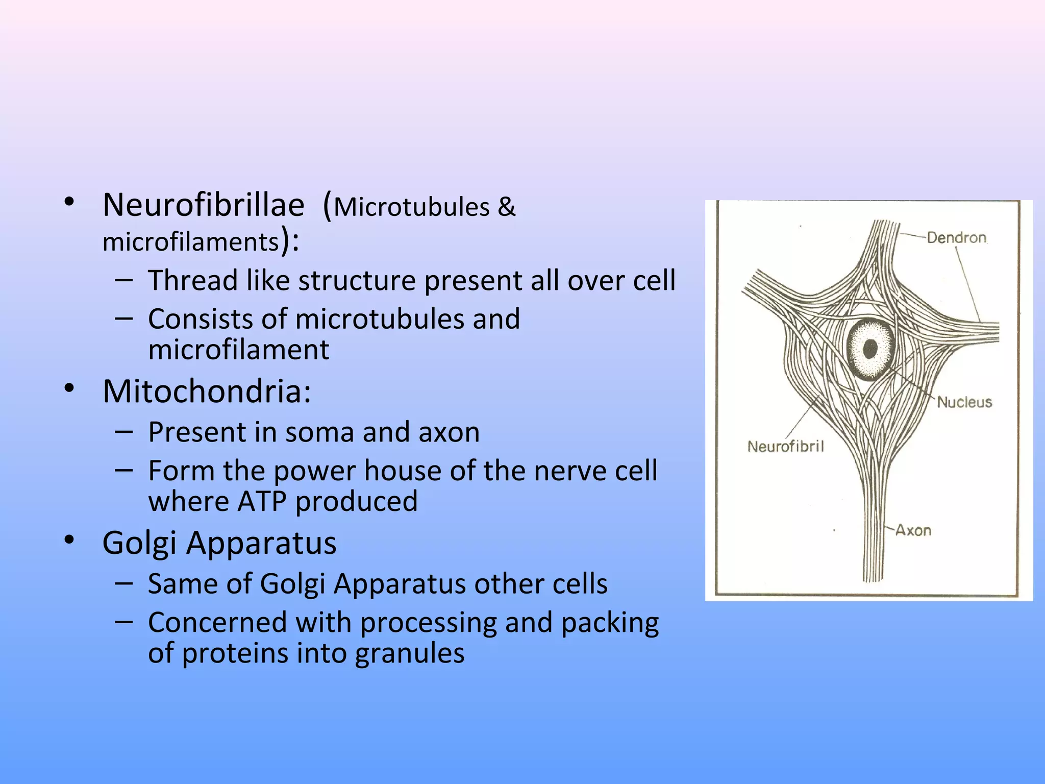
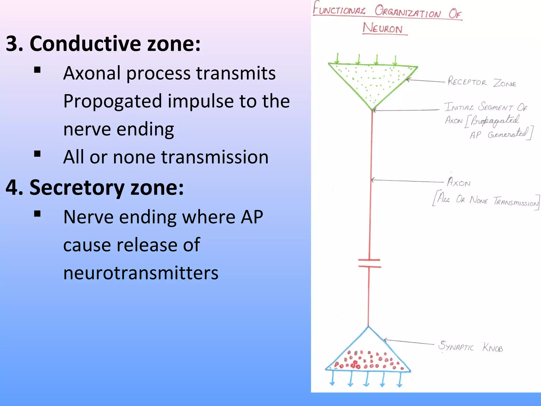
NERVE MUSCLE PHYSIOLOGY
Dr. Sonia Bajaj
H.o.D
Department of Zoology
• INTRODUCTION
• STRUCTURE & FUNCTION OF NEURON
• MYELINOGENESIS
Introduction
• Human CNS contain >100 billion neurons
• 50-100 times this number glial cells
• About 40% human genes participating its
formation
• Specialized function of muscle – contraction
• Specialized function of neurons – integration &
transmission of nerve impulse
• Along with endocrine, nervous system forms the
major control system for body functions
NERVOUS SYSTEM
CENTRAL NERVOUS SYSTEM PERIPHERAL NERVOUS
SYSTEM
Brain Spinal Cord Autonomic nervous
system
Somatic nervous
system
Sympathetic
nervous system
Parasympathetic
nervous system
NeuroN
• Structural and functional unit of nervous system
• Similar to other cell in body having nucleus and
most organelles in cytoplasm
• Different from other cells:
I. Neurons has branches or processes- dendrites and
Axon
II. Have nissl granules and neurofibrillae
III. No centrosome- loss power of division
IV. Contain and secrete neurotransmitter
Classification of Neuron
1. Depending upon the number of poles
2. Depending upon the function
3. Depending upon the length of axon
1. Depending upon the number of
poles
a. Unipolar:
• Having only one pole
• From single pole both axon and dendrites arise
• Present in embryonic stage in human being
b. Bipolar:
• Having two poles
• Axon arises one pole and dendrites other pole
c. Multipolar:
• Nucleus having multipoles
• Axon arise one pole & all other pole give rise dendrites
2. Depending upon the functions
• Motor or efferent neurons:
– Carry impulses from CNS to peripheral effector
organs e.g., muscles/glands/blood vessels
– Generally each motor neurons has long axon and
short dendrites
• Sensory or afferent neurons:
– Carry impulses from periphery to CNS
– Generally each neuron has short axon a long
dendrites
3. Depending upon the length of axon
• Golgi Type I neurons:
– Have long axons
– Cell body situated in CNS and their axon reaches
remote peripheral organs
• Golgi type II neurons:
– Have short axons
– Present in cerebral cortex and spinal cord
Structure of Neuron
• Structural and functional unit of nervous system
• Consists of nerve cell body with all its processes axon
and dendrites
• All neurons contain one and only one axon
• But dendrites may be absent one or many
• Axon carries impulses from the soma towards a
centrifugal directions (away from soma)
• Dendrites brings impulse from distance centripetally
(towards the soma)
• Nerve cell means a neuron where as nerve cell body
means soma
• Neuron can be divided in to:
– Cell body (nerve cell body)
– Dendrites
– Axon
– Nerve terminals
i. Nerve cell body:
• AKA soma, perikaryon
• Various size and forms – stellate, round, pyramidal
• It maintains the functional and anatomical integrity of axon
cut part distal to cut degenerate
• Cytoplasm contains:
– Nucleus
– Nissl bodies or Nissl granules
– Neurofibrillae
– Mitochondria
– Golgi apparatus
• Nissl granules and neurofibrillae found only in nerve cell
not in other cells
• Soma are present in :
- Grey matter of CNS
- Nuclei of brain e.g., cranial N. Nuclei/Basal
ganglia/Ganglia of CNS
- All neurons contain soma
- All processes do not survive without soma
CONTI
i. Nucleus:
– Each neuron has centrally placed one nucleus in
soma
– Prominent nucleoli which contains ribose nucleic
acid
– No centrosome – loss power of division
ii. Nissl granules or bodies:
– Named after discoverer FRANZ NISSL in 19th
century
– Also called tigroid substance (spotted appearance
when properly stained)
– Small basophilic granules or membrane bound
cavities found in clusters or clumps in soma
– Present in cell body and dendrites but absent in
axon and axon hillock
– Composed of ribonucleoprotein (RNA + Protein)
Ribosome= RNA+ protein
– Synthesize proteins of neurons which transported to axon
by axonal flow
– When demand of protein synthesis great nissl granules
over work and may altogether disappear (chromatolysis)
e.g, fatigue, anoxic, injured
– Reappear following recovery of neurons from fatigue or
after regeneration
• Neurofibrillae (Microtubules &
microfilaments):
– Thread like structure present all over cell
– Consists of microtubules and
microfilament
• Mitochondria:
– Present in soma and axon
– Form the power house of the nerve cell
where ATP produced
• Golgi Apparatus
– Same of Golgi Apparatus other cells
– Concerned with processing and packing
of proteins into granules
DENDRITES:
• Tapering and branching extension of soma
• Dendrites of cerebral cortex and cerebellar cortex show
knobby projections called dendritic spine
• May be absent if present may be one or many in number
• Conduct impulses towards the cell body
• Generate local potential not action potential as well as
integrate activity
• Has Nissl granules and neurofibrils
• Dendrites and soma constitute input zone
AxoN
• Each axon has only one axon
• Arises from axon hillock of soma
• Carry impulses away from cell body
• Cannot synthesize own protein depends upon
soma
• Branched only at its terminal end called
synaptic knobe, terminal button, axon
telodendria
• Axon may be medullary or non medullary
• Synaptic knobs Terminal buttons or Axon
Telodendria
• Axon divides into terminal branches and each
ending in numbers of synaptic knobs
• Contain granules or vesicles which contain
synaptic transmittors
• Specialized to convert electrical signal (AP) to
chemical signal
Axis cylinder
• Has long central core of cytoplasm- axoplasm
• Axoplasm covered by membrane – axolemma
continuation of cell membrane of soma
• Axoplasm along with axolemma- axis cylinder
• Contain mitochondria, neurofibrils and axoplasm,
vesicles
• Axis cylinder covered by neurilemma in non
myelinated nerve fiber
• Nerve fiber insulated by myelin sheath –
myelinated nerve fiber
MyElIN ShEATh
• Concentric layers of protein alternating with lipid
• Nerve fiber insulated by myelin sheath- myelinated nerve
fiber
• Protein lipid complex wrapped around axon >100 times
• Outside the CNS (peripheral nerve) myelin produced by
Schwann cells
• Inside the CNS myelin sheath produced by
oligodendrogliocytes
• Myelin is compacted when extracellular membrane
protein (Po) locked extracellular portion of Po
apposing membrane
• Not continuous sheath absent at regular intervals
• Where sheath absent – node of Ranvier (1µm)
• Segment between two node- internode (1mm)
Myelinogenesis
• Formation of myelin sheath around the axon
• Peripheral nerve started 4th
month of IUL and
completed few years after birth
• Pyramidal tract remain unmyelinated at birth and
completed around end of 2nd
year of life
• Outside its CNS myelin sheath formed by
Schwann cells
• Before myelinogenesis Schwann cells (Double
layer) close to axolemma as in non myelinated
nerve fiber
• Membrane of Schwann cells wrappe up and rotate
around the axon many concentric layer but not
cytoplasm
• These concentric layer compacted – produce myelin
sheath
• Cytoplasm of cell not deposited in myelin sheath
• Nucleus of cell remain in between myelin sheath and
neurilemma
• Myelinogenosis in CNS occurs by
oligodendrogeocytes
non Myelinated nerve
• No myelin sheath formation
• Nerve fiber simply covered by Schwann cells, no
wrapping
• No internode and node of Ranvier
• Neurilemma and axis cylinder close to each other
• In CNS no neurilemma
• Myelinogenosis in CNS by oligodenogliocytes not
by Schwann cells
iMportance and Myelin
sheath
• Propagation of AP very fast d/t saltatory conduction
(possible only in myelinated nerve fiber)
• Myelination results quicker mobility in higher animals
• Have high insulating capacity so prevents cross stimulation
neurileMMa
• AKA sheath of schwann
• This membrane which surrounds axis cylinder
• Contain schwann cells which have flattend and
elongated nuclei
• One nucleus is present in each internode of axon
• Nucleus situated between myelin sheath and
neurilemma
• Non myelinated nerve fiber neurilemma surrounds
axolemma continuously
• At node of ranvier neurilemma invaginates upto
axolemma
Functions oF
neurileMMa
• Non myelinated nerve fiber – serve as covering
membrane
• In myelinated nerve fiber necessory for
myelinogenesis
• Neurilemma absent in CNS
• Oligodendrogliocytes are responsible for
myelinogenesis in CNS
Functional division oF
neuron
• Divided in to four zone:
1. Receptor or dendritic zone:
– Multiple local potential generated by synaptic
connection are integrated
2. Origin of conducted impulse:
– Propagated action potential generated (Initial of
segment of spinal motor neuron)
– Initial node of Ranvier in sensory neuron
3. Conductive zone:
 Axonal process transmits
Propogated impulse to the
nerve ending
 All or none transmission
4. Secretory zone:
 Nerve ending where AP
cause release of
neurotransmitters
Axonal Transport
• Transport subs
from soma to synaptic ending
• Fast axonal transport- Membrane bound organelles
& mitochondria ( 400 mm/ day)
• Slow axonal transport- Subs
dissolved in cytoplasm-
Proteins (1mm/ day)
• Requires ATP/ Ca++
& microtubules for guide
• Occurs in both direction anterograde & retrograde
• Anterograde transport- Synaptic vesicles/ proteins
• Retrograde transport- Neurotrophins /viruses
Neurotrophins – Neurotrophic Factors
• Protein substances
• Play important role in growth and functioning
of nervous tissue
• Secreted by many tissue in body e.g., muscles/
neurons/ astrocytes
• Functions:
– Facilitate initial growth and development of nerve
cells in CNS & PNS
• Promote survival and repair of nerve cell
• Maintenance of nerve tissue and neural
transmission
• Recently – neurotrophins capable of making
damaged neuron regrow
• Used reversing devastating symptoms of nervous
disorders like Parkinson disease, Alzheimer's
disease,
• Commercial preparation for treatment of some
neural diseases
Type
• 1st
protein identified as neurotrophin- nerve
growth factor (NGF)
• Now many numbers neurotrophin identified
1. Nerve growth factors (NGF)
– Promote early growth and development in
neurons
– Major action on sympathetic and sensory neurons
especially neurons concerned with pain
– AKA sympathetic NGF (major actions on
sympathetic neurons
• Commercial preparation of NGF extracted from
snake venoms and submaxillary gland of male mouse
• Used in sympathetic neurons disease as well as many
neurons disorders:
– Alzeimer’s disease
– Neuron degeneration in aging
Other Neurotrophins:
2. Brain derived neurotrophic growth factor
(BDGF)
• 1st
discovered in pig brain now found in human
brain and sperm
• Promotes survival of sensory and motor neurons
• Enhances growth of cholinergic, dopaminergic
neurons and optic nerve
• Also regulate synaptic transmission
• Commercial preparation used to treat motor
neuron disease
CONTI
3. Ciliary Neurotrophic factor (CNTF)
• Found in neurological cells/ astrocytes and
Schwann cells
• Potent protective action on dopaminergic
neurons
• Used for treatment of Parkinson’s disease
• 4. Fibroblast growth factors:
• Promoting fibroblastic growth
5. Glial cell line- derived neurotrophic factor
(GDNF)
– Maintains mid bran dopaminergic neurons
6. Leukemia inhibitory factors (LIF)
– Enhances the growth of neurons
7. Insulin like growth factor I (IGF-I)
8. Transforming growth factor
9. Fibroblast growth factor
10. Platelet – derived growth factors
NeuroTrophiN 3 (NT-3)
• Act on motor sympathetic and sensory organs
neurons
• Regulates release of neurotransmitter from NMJ
• Useful for treatment of motor neuron
neuropathy and diabetic neuropathy
• Others sub belong to neurotrophin family NT-4,
NT-5 α leukemia inhibitory factors
• NT-4 & NT-5 act on sympathetic and sensory and
motor neurons
Neuroglia
• Neuroglia or glia (glia- glue) supporting cells of
nervous system
• Non excitable and do not transmit nerve
impulse
• 10-50 time as many glial cells as neurons
• Capable of multiplying by mitosis
• Schwann cells invest axon also glial cells
ClassifiCaTioN of
Neuroglial Cells
1. Central Neuroglial cells
– Astrocytes:
– Fibrous astrocytes
– Protoplasmic astrocytes
– Microglia
– Oligodendrocytes
2. Peripheral neuroglial cells
– Schwann cells
– Satellite cells
ClassifiCaTioN of Nerve
fibers
1. Depending upon structure
– Myelinated nerve fibers
– Non myelinated nerve fibers
2. Depending upon distribution
– Somatic nerve fibers (supply skeletal muscles)
– Visceral or autonomic (supply internal organs)
• Depending upon origin
– Cranial nerve (arising from brain)
– Spinal nerve (arising from spinal cord)
Thank You

Nervous system

  • 1.
    NERVE MUSCLE PHYSIOLOGY Dr.Sonia Bajaj H.o.D Department of Zoology
  • 2.
    • INTRODUCTION • STRUCTURE& FUNCTION OF NEURON • MYELINOGENESIS
  • 3.
    Introduction • Human CNScontain >100 billion neurons • 50-100 times this number glial cells • About 40% human genes participating its formation • Specialized function of muscle – contraction • Specialized function of neurons – integration & transmission of nerve impulse • Along with endocrine, nervous system forms the major control system for body functions
  • 4.
    NERVOUS SYSTEM CENTRAL NERVOUSSYSTEM PERIPHERAL NERVOUS SYSTEM Brain Spinal Cord Autonomic nervous system Somatic nervous system Sympathetic nervous system Parasympathetic nervous system
  • 5.
    NeuroN • Structural andfunctional unit of nervous system • Similar to other cell in body having nucleus and most organelles in cytoplasm • Different from other cells: I. Neurons has branches or processes- dendrites and Axon II. Have nissl granules and neurofibrillae III. No centrosome- loss power of division IV. Contain and secrete neurotransmitter
  • 6.
    Classification of Neuron 1.Depending upon the number of poles 2. Depending upon the function 3. Depending upon the length of axon
  • 7.
    1. Depending uponthe number of poles a. Unipolar: • Having only one pole • From single pole both axon and dendrites arise • Present in embryonic stage in human being b. Bipolar: • Having two poles • Axon arises one pole and dendrites other pole c. Multipolar: • Nucleus having multipoles • Axon arise one pole & all other pole give rise dendrites
  • 9.
    2. Depending uponthe functions • Motor or efferent neurons: – Carry impulses from CNS to peripheral effector organs e.g., muscles/glands/blood vessels – Generally each motor neurons has long axon and short dendrites • Sensory or afferent neurons: – Carry impulses from periphery to CNS – Generally each neuron has short axon a long dendrites
  • 10.
    3. Depending uponthe length of axon • Golgi Type I neurons: – Have long axons – Cell body situated in CNS and their axon reaches remote peripheral organs • Golgi type II neurons: – Have short axons – Present in cerebral cortex and spinal cord
  • 11.
    Structure of Neuron •Structural and functional unit of nervous system • Consists of nerve cell body with all its processes axon and dendrites • All neurons contain one and only one axon • But dendrites may be absent one or many • Axon carries impulses from the soma towards a centrifugal directions (away from soma) • Dendrites brings impulse from distance centripetally (towards the soma) • Nerve cell means a neuron where as nerve cell body means soma
  • 13.
    • Neuron canbe divided in to: – Cell body (nerve cell body) – Dendrites – Axon – Nerve terminals
  • 14.
    i. Nerve cellbody: • AKA soma, perikaryon • Various size and forms – stellate, round, pyramidal • It maintains the functional and anatomical integrity of axon cut part distal to cut degenerate • Cytoplasm contains: – Nucleus – Nissl bodies or Nissl granules – Neurofibrillae – Mitochondria – Golgi apparatus • Nissl granules and neurofibrillae found only in nerve cell not in other cells
  • 15.
    • Soma arepresent in : - Grey matter of CNS - Nuclei of brain e.g., cranial N. Nuclei/Basal ganglia/Ganglia of CNS - All neurons contain soma - All processes do not survive without soma
  • 16.
    CONTI i. Nucleus: – Eachneuron has centrally placed one nucleus in soma – Prominent nucleoli which contains ribose nucleic acid – No centrosome – loss power of division
  • 17.
    ii. Nissl granulesor bodies: – Named after discoverer FRANZ NISSL in 19th century – Also called tigroid substance (spotted appearance when properly stained) – Small basophilic granules or membrane bound cavities found in clusters or clumps in soma – Present in cell body and dendrites but absent in axon and axon hillock
  • 18.
    – Composed ofribonucleoprotein (RNA + Protein) Ribosome= RNA+ protein – Synthesize proteins of neurons which transported to axon by axonal flow – When demand of protein synthesis great nissl granules over work and may altogether disappear (chromatolysis) e.g, fatigue, anoxic, injured – Reappear following recovery of neurons from fatigue or after regeneration
  • 19.
    • Neurofibrillae (Microtubules& microfilaments): – Thread like structure present all over cell – Consists of microtubules and microfilament • Mitochondria: – Present in soma and axon – Form the power house of the nerve cell where ATP produced • Golgi Apparatus – Same of Golgi Apparatus other cells – Concerned with processing and packing of proteins into granules
  • 20.
    DENDRITES: • Tapering andbranching extension of soma • Dendrites of cerebral cortex and cerebellar cortex show knobby projections called dendritic spine • May be absent if present may be one or many in number • Conduct impulses towards the cell body • Generate local potential not action potential as well as integrate activity • Has Nissl granules and neurofibrils • Dendrites and soma constitute input zone
  • 21.
    AxoN • Each axonhas only one axon • Arises from axon hillock of soma • Carry impulses away from cell body • Cannot synthesize own protein depends upon soma • Branched only at its terminal end called synaptic knobe, terminal button, axon telodendria
  • 22.
    • Axon maybe medullary or non medullary • Synaptic knobs Terminal buttons or Axon Telodendria • Axon divides into terminal branches and each ending in numbers of synaptic knobs • Contain granules or vesicles which contain synaptic transmittors • Specialized to convert electrical signal (AP) to chemical signal
  • 23.
    Axis cylinder • Haslong central core of cytoplasm- axoplasm • Axoplasm covered by membrane – axolemma continuation of cell membrane of soma • Axoplasm along with axolemma- axis cylinder • Contain mitochondria, neurofibrils and axoplasm, vesicles • Axis cylinder covered by neurilemma in non myelinated nerve fiber • Nerve fiber insulated by myelin sheath – myelinated nerve fiber
  • 24.
    MyElIN ShEATh • Concentriclayers of protein alternating with lipid • Nerve fiber insulated by myelin sheath- myelinated nerve fiber • Protein lipid complex wrapped around axon >100 times • Outside the CNS (peripheral nerve) myelin produced by Schwann cells • Inside the CNS myelin sheath produced by oligodendrogliocytes
  • 25.
    • Myelin iscompacted when extracellular membrane protein (Po) locked extracellular portion of Po apposing membrane • Not continuous sheath absent at regular intervals • Where sheath absent – node of Ranvier (1µm) • Segment between two node- internode (1mm)
  • 26.
    Myelinogenesis • Formation ofmyelin sheath around the axon • Peripheral nerve started 4th month of IUL and completed few years after birth • Pyramidal tract remain unmyelinated at birth and completed around end of 2nd year of life • Outside its CNS myelin sheath formed by Schwann cells • Before myelinogenesis Schwann cells (Double layer) close to axolemma as in non myelinated nerve fiber
  • 27.
    • Membrane ofSchwann cells wrappe up and rotate around the axon many concentric layer but not cytoplasm • These concentric layer compacted – produce myelin sheath • Cytoplasm of cell not deposited in myelin sheath • Nucleus of cell remain in between myelin sheath and neurilemma • Myelinogenosis in CNS occurs by oligodendrogeocytes
  • 29.
    non Myelinated nerve •No myelin sheath formation • Nerve fiber simply covered by Schwann cells, no wrapping • No internode and node of Ranvier • Neurilemma and axis cylinder close to each other • In CNS no neurilemma • Myelinogenosis in CNS by oligodenogliocytes not by Schwann cells
  • 31.
    iMportance and Myelin sheath •Propagation of AP very fast d/t saltatory conduction (possible only in myelinated nerve fiber) • Myelination results quicker mobility in higher animals • Have high insulating capacity so prevents cross stimulation
  • 32.
    neurileMMa • AKA sheathof schwann • This membrane which surrounds axis cylinder • Contain schwann cells which have flattend and elongated nuclei • One nucleus is present in each internode of axon • Nucleus situated between myelin sheath and neurilemma • Non myelinated nerve fiber neurilemma surrounds axolemma continuously • At node of ranvier neurilemma invaginates upto axolemma
  • 33.
    Functions oF neurileMMa • Nonmyelinated nerve fiber – serve as covering membrane • In myelinated nerve fiber necessory for myelinogenesis • Neurilemma absent in CNS • Oligodendrogliocytes are responsible for myelinogenesis in CNS
  • 34.
    Functional division oF neuron •Divided in to four zone: 1. Receptor or dendritic zone: – Multiple local potential generated by synaptic connection are integrated 2. Origin of conducted impulse: – Propagated action potential generated (Initial of segment of spinal motor neuron) – Initial node of Ranvier in sensory neuron
  • 35.
    3. Conductive zone: Axonal process transmits Propogated impulse to the nerve ending  All or none transmission 4. Secretory zone:  Nerve ending where AP cause release of neurotransmitters
  • 36.
    Axonal Transport • Transportsubs from soma to synaptic ending • Fast axonal transport- Membrane bound organelles & mitochondria ( 400 mm/ day) • Slow axonal transport- Subs dissolved in cytoplasm- Proteins (1mm/ day) • Requires ATP/ Ca++ & microtubules for guide • Occurs in both direction anterograde & retrograde • Anterograde transport- Synaptic vesicles/ proteins • Retrograde transport- Neurotrophins /viruses
  • 37.
    Neurotrophins – NeurotrophicFactors • Protein substances • Play important role in growth and functioning of nervous tissue • Secreted by many tissue in body e.g., muscles/ neurons/ astrocytes • Functions: – Facilitate initial growth and development of nerve cells in CNS & PNS
  • 38.
    • Promote survivaland repair of nerve cell • Maintenance of nerve tissue and neural transmission • Recently – neurotrophins capable of making damaged neuron regrow • Used reversing devastating symptoms of nervous disorders like Parkinson disease, Alzheimer's disease, • Commercial preparation for treatment of some neural diseases
  • 39.
    Type • 1st protein identifiedas neurotrophin- nerve growth factor (NGF) • Now many numbers neurotrophin identified 1. Nerve growth factors (NGF) – Promote early growth and development in neurons – Major action on sympathetic and sensory neurons especially neurons concerned with pain – AKA sympathetic NGF (major actions on sympathetic neurons
  • 40.
    • Commercial preparationof NGF extracted from snake venoms and submaxillary gland of male mouse • Used in sympathetic neurons disease as well as many neurons disorders: – Alzeimer’s disease – Neuron degeneration in aging
  • 41.
    Other Neurotrophins: 2. Brainderived neurotrophic growth factor (BDGF) • 1st discovered in pig brain now found in human brain and sperm • Promotes survival of sensory and motor neurons • Enhances growth of cholinergic, dopaminergic neurons and optic nerve • Also regulate synaptic transmission • Commercial preparation used to treat motor neuron disease
  • 42.
    CONTI 3. Ciliary Neurotrophicfactor (CNTF) • Found in neurological cells/ astrocytes and Schwann cells • Potent protective action on dopaminergic neurons • Used for treatment of Parkinson’s disease • 4. Fibroblast growth factors: • Promoting fibroblastic growth
  • 43.
    5. Glial cellline- derived neurotrophic factor (GDNF) – Maintains mid bran dopaminergic neurons 6. Leukemia inhibitory factors (LIF) – Enhances the growth of neurons 7. Insulin like growth factor I (IGF-I) 8. Transforming growth factor 9. Fibroblast growth factor 10. Platelet – derived growth factors
  • 44.
    NeuroTrophiN 3 (NT-3) •Act on motor sympathetic and sensory organs neurons • Regulates release of neurotransmitter from NMJ • Useful for treatment of motor neuron neuropathy and diabetic neuropathy • Others sub belong to neurotrophin family NT-4, NT-5 α leukemia inhibitory factors • NT-4 & NT-5 act on sympathetic and sensory and motor neurons
  • 45.
    Neuroglia • Neuroglia orglia (glia- glue) supporting cells of nervous system • Non excitable and do not transmit nerve impulse • 10-50 time as many glial cells as neurons • Capable of multiplying by mitosis • Schwann cells invest axon also glial cells
  • 46.
    ClassifiCaTioN of Neuroglial Cells 1.Central Neuroglial cells – Astrocytes: – Fibrous astrocytes – Protoplasmic astrocytes – Microglia – Oligodendrocytes 2. Peripheral neuroglial cells – Schwann cells – Satellite cells
  • 47.
    ClassifiCaTioN of Nerve fibers 1.Depending upon structure – Myelinated nerve fibers – Non myelinated nerve fibers 2. Depending upon distribution – Somatic nerve fibers (supply skeletal muscles) – Visceral or autonomic (supply internal organs) • Depending upon origin – Cranial nerve (arising from brain) – Spinal nerve (arising from spinal cord)
  • 48.