Download to read offline

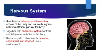
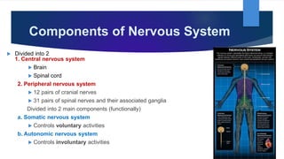
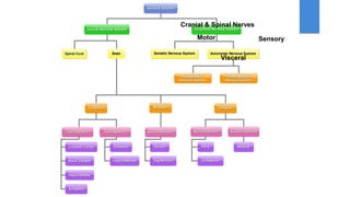
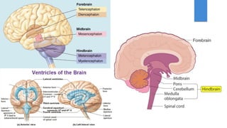
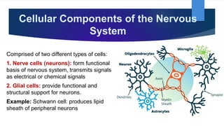
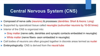
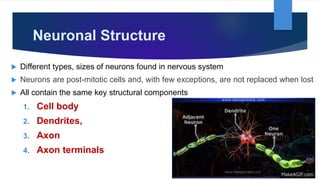


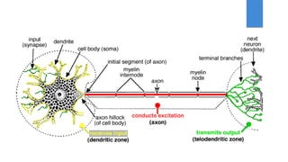
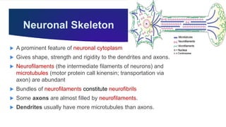
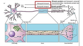
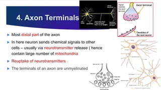

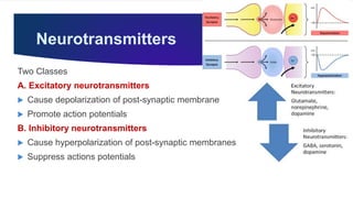
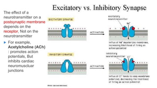
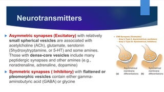
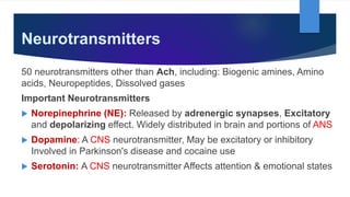

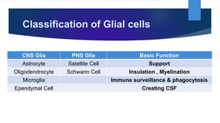
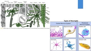

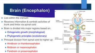
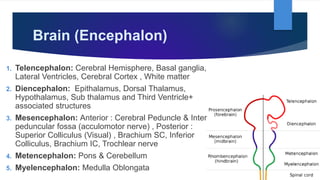
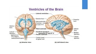
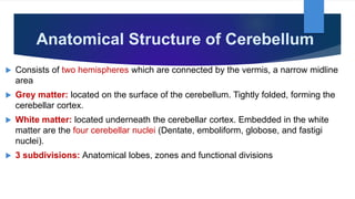
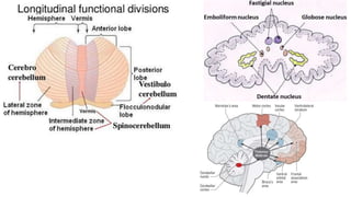
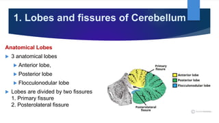
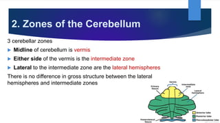

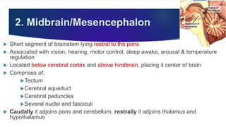
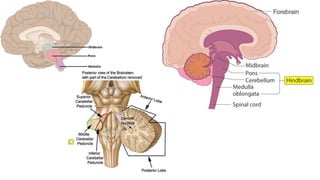


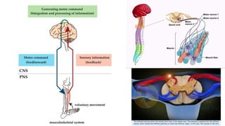
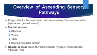
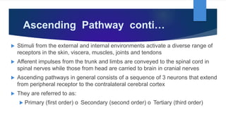
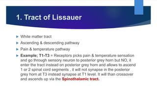
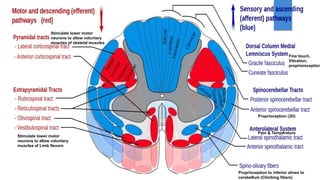

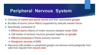

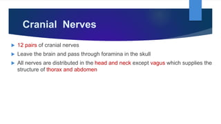
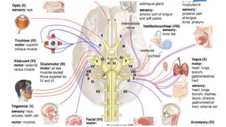


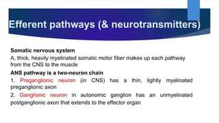

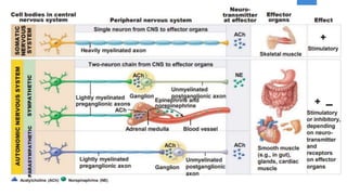

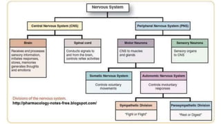


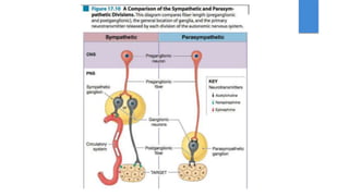
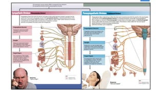
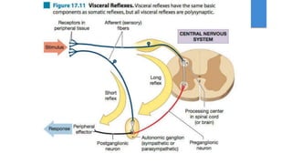


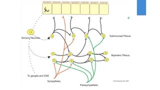


The document provides a comprehensive overview of the nervous system, detailing its structure, functions, and components, including the central and peripheral systems. It describes the different types of neurons, their classifications, cellular components, and functional aspects, such as neurotransmitters and synapses. Furthermore, it covers the brain's anatomical regions and roles, particularly highlighting the cerebellum and its subdivisions.


