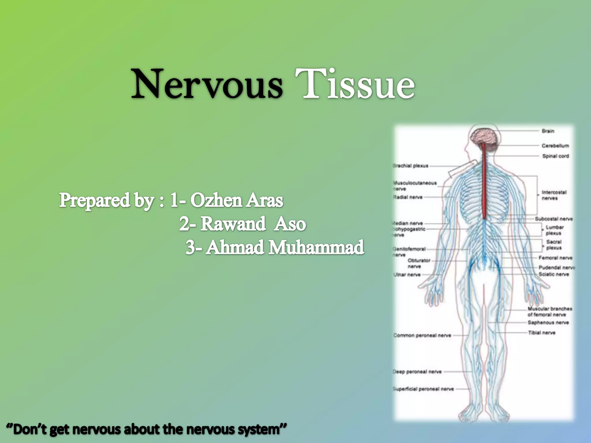
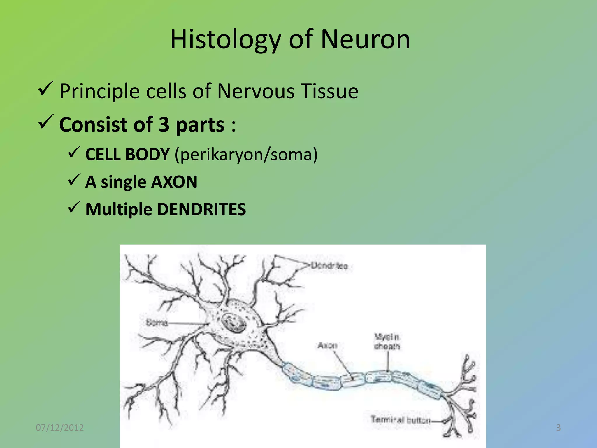
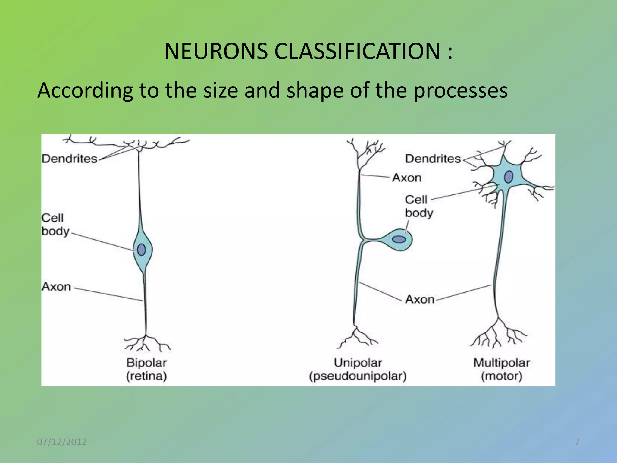
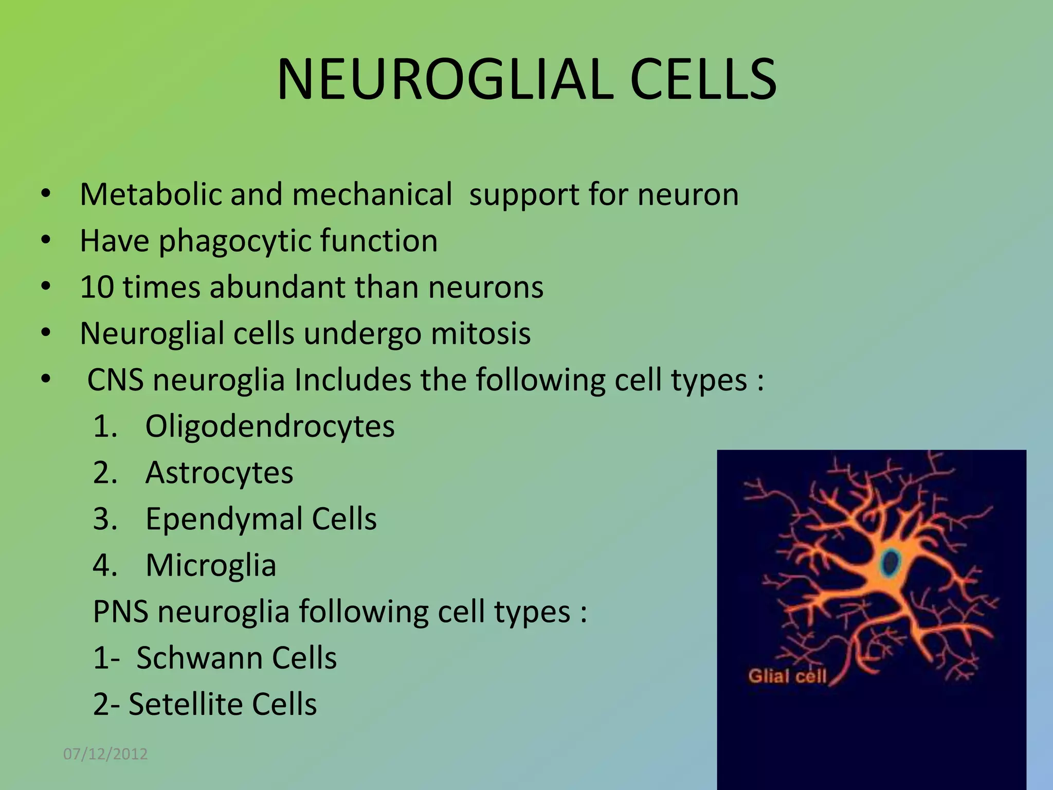
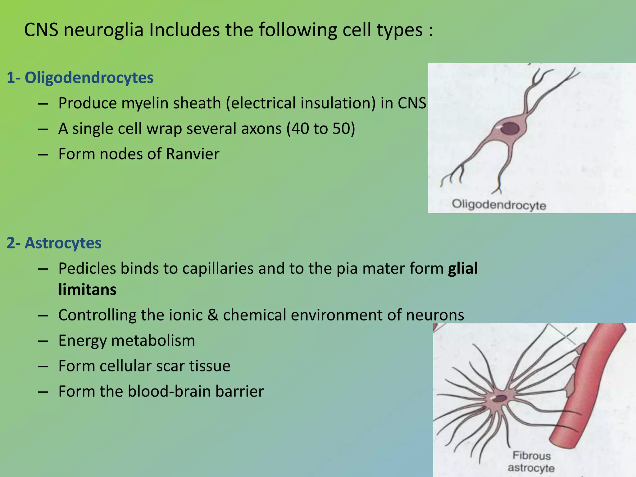
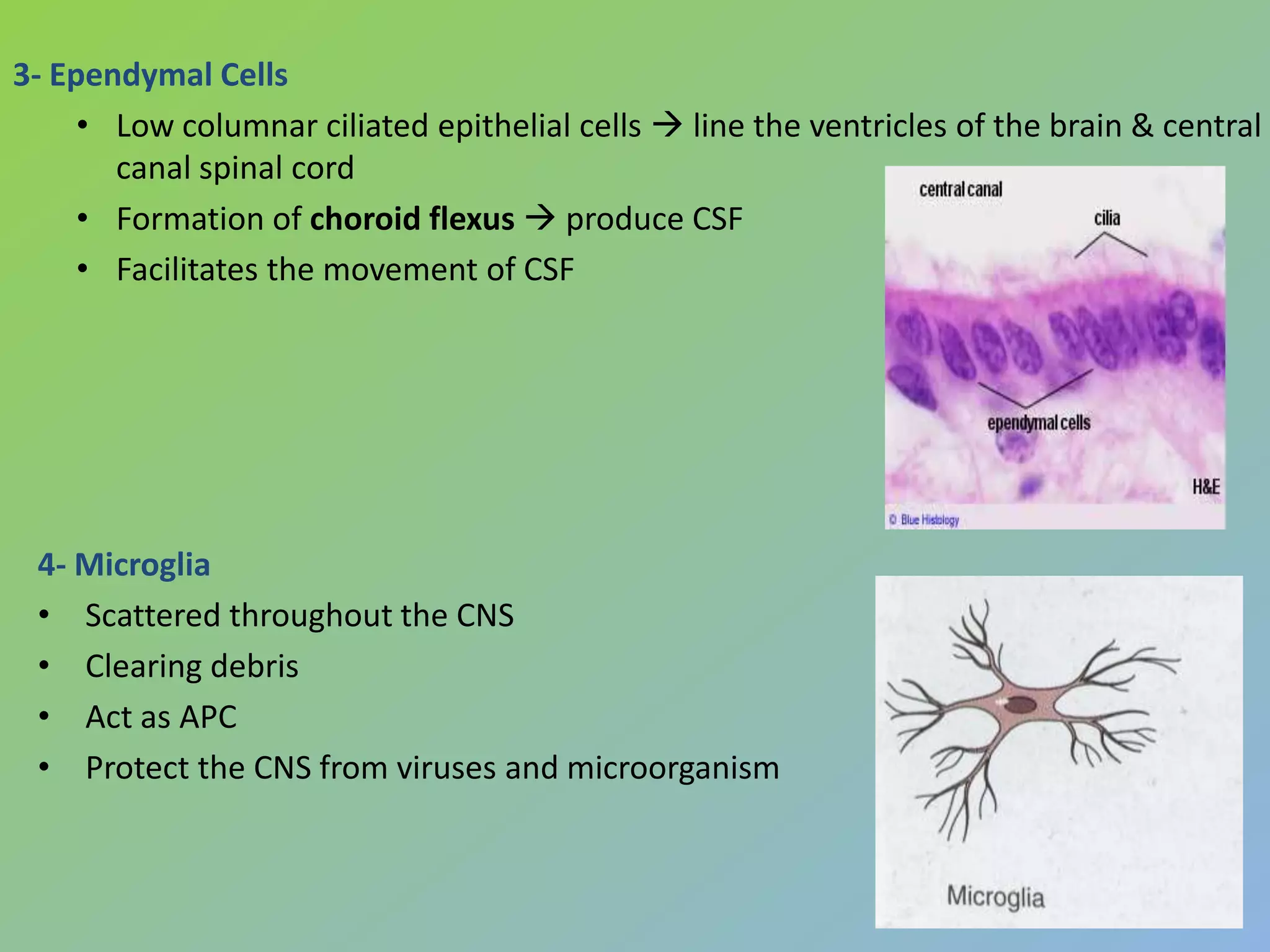
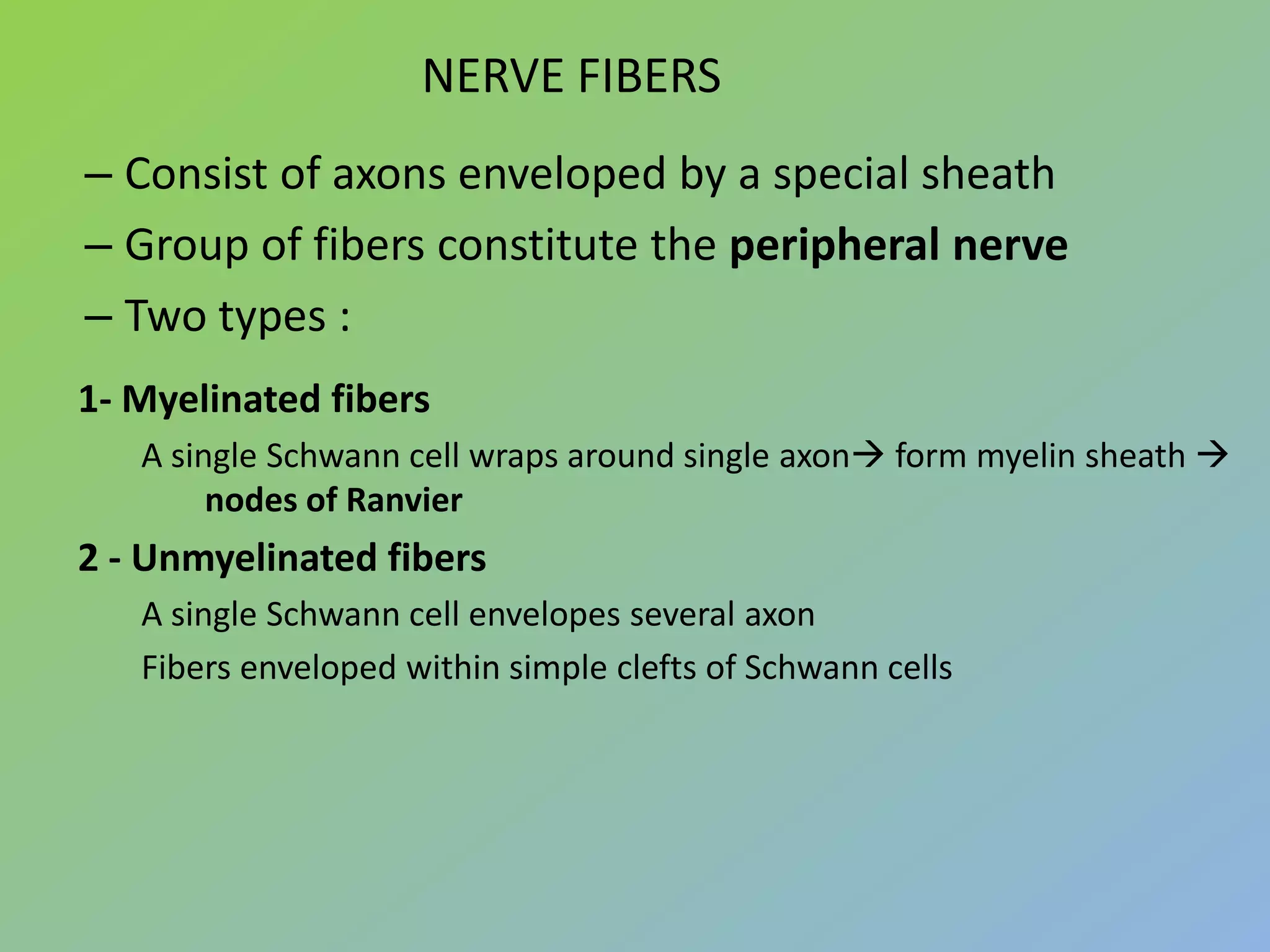
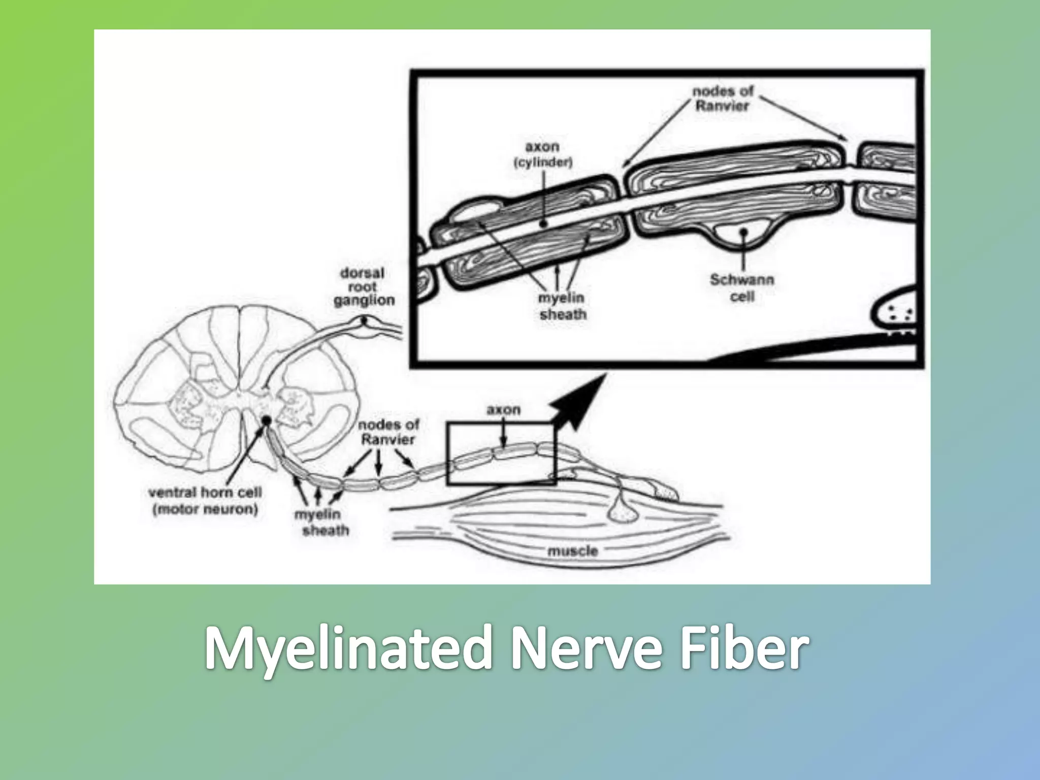
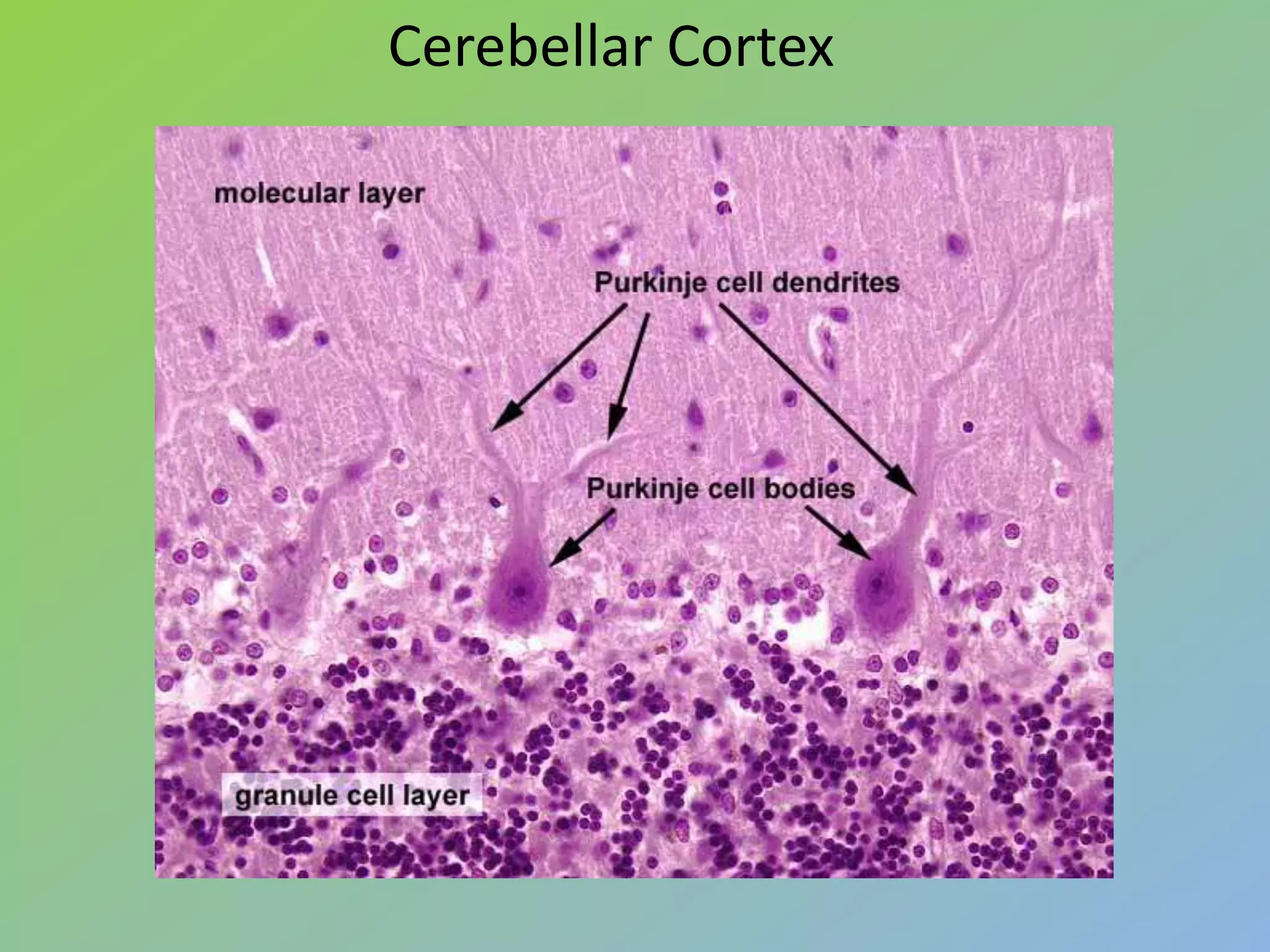
07/12/2012   2
Histology of Neuron
 Principle cells of Nervous Tissue
 Consist of 3 parts :
       CELL BODY (perikaryon/soma)
       A single AXON
       Multiple DENDRITES


07/12/2012                             3
CELL BODY (PERIKARYON)
•   Central portion of the cell
•   Generally are polygonal
•   Different shape and size  characteristic regions of nervous system
•   Contain : Nucleus and Perinuclear cytoplasm
   Nucleus :
•   large, spherical to ovoid and centraly located
•   a single prominent nucleolus
•    finely dispersed chromatin
 Cytoplasm : contains :
• R.E.R and S.E.R.
• Polyribosomes
• Basic dyes (a+b) Nissl Bodies
• Golgi complex

     07/12/2012                                                           4
CELL BODY (PERIKARYON)
•   Multiple elongated processes
•   Cytoplasmic~perikaryon (devoid golgi complex)
•   Receiving stimuli


•Single process up to 100 cm
•Originate from axon hillock
•Devoid ribosome
•conducting impulse away from the soma
•It maintains Axonal transport
NEURONS CLASSIFICATION :
According to the size and shape of the processes


07/12/2012                                         7
NEUROGLIAL CELLS
•    Metabolic and mechanical support for neuron
•    Have phagocytic function
•    10 times abundant than neurons
•    Neuroglial cells undergo mitosis
•    CNS neuroglia Includes the following cell types :
      1. Oligodendrocytes
      2. Astrocytes
      3. Ependymal Cells
      4. Microglia
      PNS neuroglia following cell types :
      1- Schwann Cells
      2- Setellite Cells
    07/12/2012                                           8
CNS neuroglia Includes the following cell types :

1- Oligodendrocytes
    – Produce myelin sheath (electrical insulation) in CNS
    – A single cell wrap several axons (40 to 50)
    – Form nodes of Ranvier


2- Astrocytes
    – Pedicles binds to capillaries and to the pia mater form glial
       limitans
    – Controlling the ionic & chemical environment of neurons
    – Energy metabolism
    – Form cellular scar tissue
    – Form the blood-brain barrier
3- Ependymal Cells
     • Low columnar ciliated epithelial cells  line the ventricles of the brain & central
       canal spinal cord
     • Formation of choroid flexus  produce CSF
     • Facilitates the movement of CSF


 4- Microglia
 • Scattered throughout the CNS
 • Clearing debris
 • Act as APC
 • Protect the CNS from viruses and microorganism
PNS neuroglia Includes the following cell types :
1- Schwann cells
• surround all axons of neurons in the PNS creating a neurilemma around them.
Neurilemma allows for potential regeneration of damaged axons
• creates myelin sheath around most axons of PNS


2 - Satellite cells
• support groups of cell bodies of neurons within ganglia of the PNS
•Grey matter consists of
neuronal cell bodies
 neuropil (dendrites and unmyelinated axons)
 glial cells (astroglia and oligodendrocytes)

•Grey matter contains neural cell bodies, in contrast to white matter,
which does not and mostly contains myelinated axon tracts.

•The color difference arises mainly from the whiteness of myelin


•White matter is composed of bundles of myelinated nerve cell processes.

•consists mostly of glial cells and myelinated axons that transmit signals from one region of
the cerebrum to another and between the cerebrum and lower brain centers.

•Its white color is due to its usual preservation in formaldehyde.
NERVE FIBERS
– Consist of axons enveloped by a special sheath
– Group of fibers constitute the peripheral nerve
– Two types :
1- Myelinated fibers
   A single Schwann cell wraps around single axon form myelin sheath 
        nodes of Ranvier
2 - Unmyelinated fibers
   A single Schwann cell envelopes several axon
   Fibers enveloped within simple clefts of Schwann cells
CONNECTIVE TISSUE INVESTMENTS OF NERVES
                     • Epineureum
                       – Dense collagenous Con. Tissue
                         with thick elastic fiber
                       – Prevent damage by
                         overstreching
                     • Perineureum
                       – Dense con. Tissue
                       – Layers of epithelioids
                       – Isolates neural environment
                         (blood-nerve barrier)
                     • Endoneureum
                       – Loose con. Tissue
                       – Regulation of
                         microenvironment of nerve
                         fiber
                                                       16
07/12/2012
GANGLIA
• Ovoid structure containing neuronal cell bodies, glial cells
  supported by connective tissue
• Function : Relay stations to transmit impulses
• Types :
   1- Sensory Ganglia (cell bodies of sensory neuron)
       • Unipolar cell bodies enveloped by cuboidal capsule cells
                – Cranial ganglia : Associated with the cranial nerve
                – Spinal ganglia : Associated with the spinal nerve

   2 -Autonomic Ganglia (cell bodies of postganglionic autonomic nerves)
       • Multipolar neuron enveloped by satellite cells
       • Some are located within certain organ (intramural)


   07/12/2012                                                              17
Sensory Ganglia   Autonomic Ganglia
MENINGES
                  • The dura mater
                     – dense, collagenous connective
                       tissue
                  • The arachnoid layer:
                     – fibroblasts, collagen & elastic
                       fibers
                     – Layer in contact with dura mater
                       & a system of trabeculae
                     – Form arachnoid villi
                  • The pia mater
                     – Loose con. Tissue with blood
                       vessel
                     – There is a physical barrier
                       between pia mater & neuron


07/12/2012                                            19
Cerebellar Cortex
 •Surface of cerebellum show transverse fissures which divide the cerebellum into a
 number of lobules
 .
•Section of cerebellar cortex shows three layers, from out to inside are:

1 - Molecular Layer. :
• Outer most layer mainly consists of cell processes.
•These cell process may be dendrites or unmyelinated axon.
•In this layer make the profuse synapses

2 - Purkinje Cell layer :
•This layer contains the cell bodies of large multipolar neurons called Purkinje cells.
•The purkinje cell consists large flask shaped body.
•cytoplasm contains large number of Nissl granules.

3- Granular Layer :
•This layer consists of thickly populated small cells called granule cells.
•This layer shows irregular scattered lighter staining area called as glomeruli or island.
Cerebellar Cortex
Nervous tissue (Histology)

Nervous tissue (Histology)

  • 2.
  • 3.
    Histology of Neuron Principle cells of Nervous Tissue  Consist of 3 parts :  CELL BODY (perikaryon/soma)  A single AXON  Multiple DENDRITES 07/12/2012 3
  • 4.
    CELL BODY (PERIKARYON) • Central portion of the cell • Generally are polygonal • Different shape and size  characteristic regions of nervous system • Contain : Nucleus and Perinuclear cytoplasm  Nucleus : • large, spherical to ovoid and centraly located • a single prominent nucleolus • finely dispersed chromatin  Cytoplasm : contains : • R.E.R and S.E.R. • Polyribosomes • Basic dyes (a+b) Nissl Bodies • Golgi complex 07/12/2012 4
  • 5.
  • 6.
    Multiple elongated processes • Cytoplasmic~perikaryon (devoid golgi complex) • Receiving stimuli •Single process up to 100 cm •Originate from axon hillock •Devoid ribosome •conducting impulse away from the soma •It maintains Axonal transport
  • 7.
    NEURONS CLASSIFICATION : Accordingto the size and shape of the processes 07/12/2012 7
  • 8.
    NEUROGLIAL CELLS • Metabolic and mechanical support for neuron • Have phagocytic function • 10 times abundant than neurons • Neuroglial cells undergo mitosis • CNS neuroglia Includes the following cell types : 1. Oligodendrocytes 2. Astrocytes 3. Ependymal Cells 4. Microglia PNS neuroglia following cell types : 1- Schwann Cells 2- Setellite Cells 07/12/2012 8
  • 9.
    CNS neuroglia Includesthe following cell types : 1- Oligodendrocytes – Produce myelin sheath (electrical insulation) in CNS – A single cell wrap several axons (40 to 50) – Form nodes of Ranvier 2- Astrocytes – Pedicles binds to capillaries and to the pia mater form glial limitans – Controlling the ionic & chemical environment of neurons – Energy metabolism – Form cellular scar tissue – Form the blood-brain barrier
  • 10.
    3- Ependymal Cells • Low columnar ciliated epithelial cells  line the ventricles of the brain & central canal spinal cord • Formation of choroid flexus  produce CSF • Facilitates the movement of CSF 4- Microglia • Scattered throughout the CNS • Clearing debris • Act as APC • Protect the CNS from viruses and microorganism
  • 11.
    PNS neuroglia Includesthe following cell types : 1- Schwann cells • surround all axons of neurons in the PNS creating a neurilemma around them. Neurilemma allows for potential regeneration of damaged axons • creates myelin sheath around most axons of PNS 2 - Satellite cells • support groups of cell bodies of neurons within ganglia of the PNS
  • 12.
    •Grey matter consistsof neuronal cell bodies  neuropil (dendrites and unmyelinated axons)  glial cells (astroglia and oligodendrocytes) •Grey matter contains neural cell bodies, in contrast to white matter, which does not and mostly contains myelinated axon tracts. •The color difference arises mainly from the whiteness of myelin •White matter is composed of bundles of myelinated nerve cell processes. •consists mostly of glial cells and myelinated axons that transmit signals from one region of the cerebrum to another and between the cerebrum and lower brain centers. •Its white color is due to its usual preservation in formaldehyde.
  • 14.
    NERVE FIBERS – Consistof axons enveloped by a special sheath – Group of fibers constitute the peripheral nerve – Two types : 1- Myelinated fibers A single Schwann cell wraps around single axon form myelin sheath  nodes of Ranvier 2 - Unmyelinated fibers A single Schwann cell envelopes several axon Fibers enveloped within simple clefts of Schwann cells
  • 16.
    CONNECTIVE TISSUE INVESTMENTSOF NERVES • Epineureum – Dense collagenous Con. Tissue with thick elastic fiber – Prevent damage by overstreching • Perineureum – Dense con. Tissue – Layers of epithelioids – Isolates neural environment (blood-nerve barrier) • Endoneureum – Loose con. Tissue – Regulation of microenvironment of nerve fiber 16 07/12/2012
  • 17.
    GANGLIA • Ovoid structurecontaining neuronal cell bodies, glial cells supported by connective tissue • Function : Relay stations to transmit impulses • Types : 1- Sensory Ganglia (cell bodies of sensory neuron) • Unipolar cell bodies enveloped by cuboidal capsule cells – Cranial ganglia : Associated with the cranial nerve – Spinal ganglia : Associated with the spinal nerve 2 -Autonomic Ganglia (cell bodies of postganglionic autonomic nerves) • Multipolar neuron enveloped by satellite cells • Some are located within certain organ (intramural) 07/12/2012 17
  • 18.
    Sensory Ganglia Autonomic Ganglia
  • 19.
    MENINGES • The dura mater – dense, collagenous connective tissue • The arachnoid layer: – fibroblasts, collagen & elastic fibers – Layer in contact with dura mater & a system of trabeculae – Form arachnoid villi • The pia mater – Loose con. Tissue with blood vessel – There is a physical barrier between pia mater & neuron 07/12/2012 19
  • 20.
    Cerebellar Cortex •Surfaceof cerebellum show transverse fissures which divide the cerebellum into a number of lobules . •Section of cerebellar cortex shows three layers, from out to inside are: 1 - Molecular Layer. : • Outer most layer mainly consists of cell processes. •These cell process may be dendrites or unmyelinated axon. •In this layer make the profuse synapses 2 - Purkinje Cell layer : •This layer contains the cell bodies of large multipolar neurons called Purkinje cells. •The purkinje cell consists large flask shaped body. •cytoplasm contains large number of Nissl granules. 3- Granular Layer : •This layer consists of thickly populated small cells called granule cells. •This layer shows irregular scattered lighter staining area called as glomeruli or island.
  • 21.