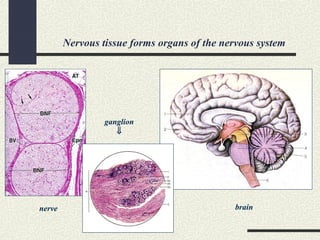
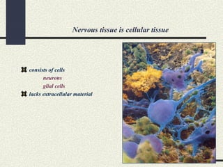
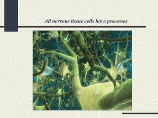
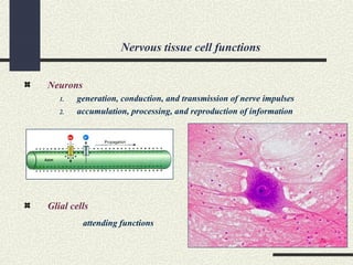
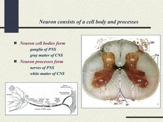
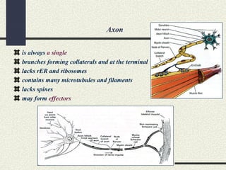
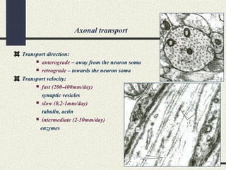
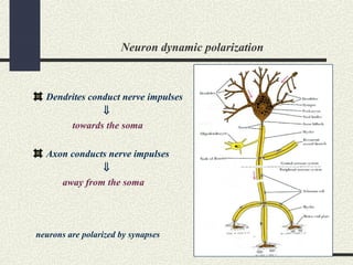
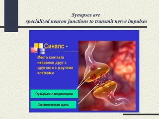
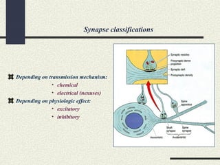
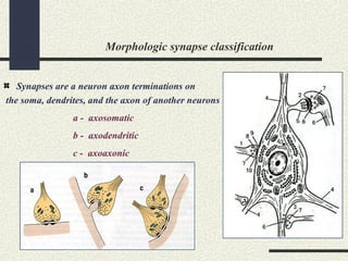
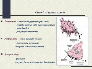
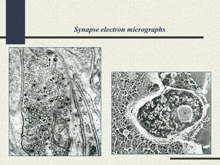
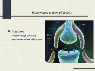
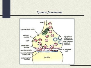
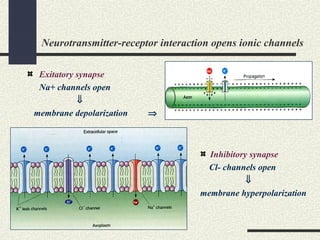
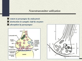
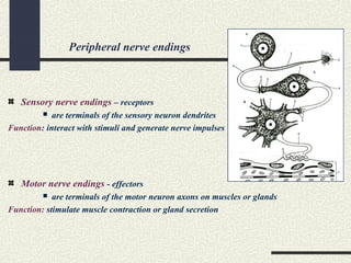
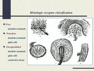
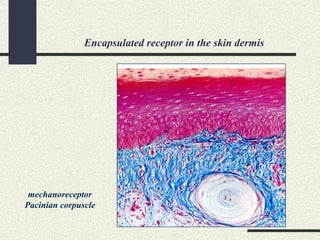
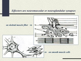
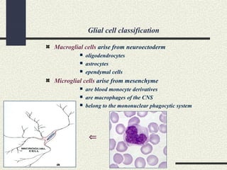
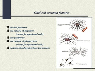
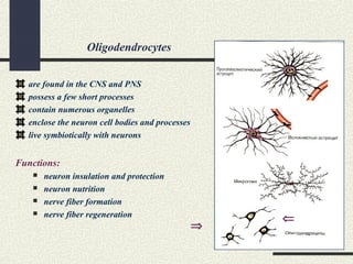
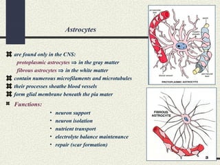
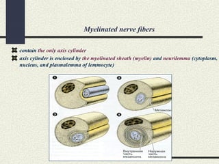
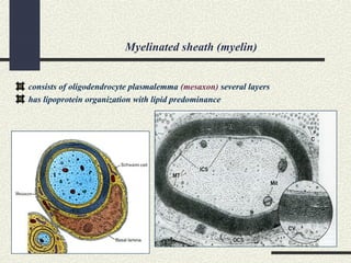
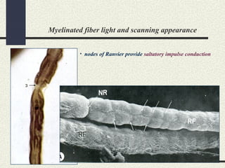
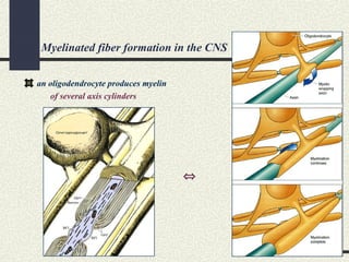
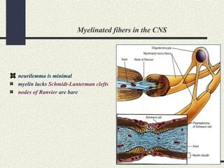
Nervous Tissue
Lecturer – professor Boronikhina Tatiana Vladimirovna
Nervous tissue forms organs of the nervous system
nerve
ganglion
⇓
brain
Nervous tissue is cellular tissue
consists of cells
neurons
glial cells
lacks extracellular material
All nervous tissue cells have processes
Nervous tissue cell functions
Neurons
1. generation, conduction, and transmission of nerve impulses
2. accumulation, processing, and reproduction of information
Glial cells
attending functions
Nervous tissue embryonic origins
Neural tube ⇒
neurons and glial cells of CNC
Neural crest ⇒
neurons and glial cells of PNS
Mesenchyme ⇒
microglial cells of CNS
Neuron consists of a cell body and processes
Neuron cell bodies form
ganglia of PNS
gray matter of CNS
Neuron processes form
nerves of PNS
white matter of CNS
Morphologic neuron classification
Bipolar
Multipolar
Pseudounipolar
Neuron cytology
Nucleus is large and euchromatic
with well-developed nucleoli
Cytoplasm is rich in organelles
 rER
 mitochondria
 Golgi apparatus
 microtubules and filaments
 sER
 lysosomes
Neuron cell body (soma, perikaryon)
Neurons are obligatory aerobic cells
contain numerous mitochondria
use O2 - 25% in adults
50% in children
blood glucose is a substratum of respiration
Time of revivification in clinical death is limited
5 – 6 minutes
15 – 16 minutes in hypothermia or pharmacologic defence
Basophilic clumps or Nissl bodies
rER
Nissl bodies
Neuron rER synthesizes membrane proteins
proteins are necessary for
synaptic vesicle formation
neuron growth and differentiation
intracellular regeneration
Golgi apparatus takes part in synaptic vesicle formation
Golgi apparatus in neuron cell bodies
synaptic vesicles
“Neurofibrils” are aggregated microtubules and filaments
Silver impregnation
⇑
electron micrograph
of axon cytoplasm
Neuron processes
Dendrites
are numerous or single
branch dichotomically directly from the soma (arborization)
contain all organelles
possess spines to increase the synaptic surface ⇒
may form receptors
Axon
is always a single
branches forming collaterals and at the terminal
lacks rER and ribosomes
contains many microtubules and filaments
lacks spines
may form effectors
Axonal transport
Transport direction:
 anterograde – away from the neuron soma
 retrograde – towards the neuron soma
Transport velocity:
 fast (200-400mm/day)
synaptic vesicles
 slow (0,2-1mm/day)
tubulin, actin
 intermediate (2-50mm/day)
enzymes
Neuron dynamic polarization
Dendrites conduct nerve impulses
⇓
towards the soma
Axon conducts nerve impulses
⇓
away from the soma
neurons are polarized by synapses
Synapses are
specialized neuron junctions to transmit nerve impulses
Synapse classifications
Depending on transmission mechanism:
• chemical
• electrical (nexuses)
Depending on physiologic effect:
• excitatory
• inhibitory
Morphologic synapse classification
Synapses are a neuron axon terminations on
the soma, dendrites, and the axon of another neurons
a - axosomatic
b - axodendritic
c - axoaxonic
Chemical synapse parts
Presynapse – axon ending (presynaptic knob)
synaptic vesicles with neurotransmitters
mitochondria
presynaptic membrane
Postsynapse – soma, dendrite, or axon
postsynaptic membrane
receptors to neurotransmitters
Synaptic cleft
filaments
enzymes for neurotransmitter inactivation
Synapse electron micrographs
Parasynapse is from glial cells
functions
synaptic cleft isolation
neurotransmitter utilization
Synapse functioning
Neurotransmitter-receptor interaction opens ionic channels
Exitatory synapse
Na+ channels open
⇓
membrane depolarization ⇒
Inhibitory synapse
Cl- channels open
⇓
membrane hyperpolarization
Neurotransmitter utilization
return to presynapse by endocytosis
destruction in synaptic cleft by enzymes
absorption by parasynapse
Peripheral nerve endings
Sensory nerve endings – receptors
 are terminals of the sensory neuron dendrites
Function: interact with stimuli and generate nerve impulses
Motor nerve endings - effectors
 are terminals of the motor neuron axons on muscles or glands
Function: stimulate muscle contraction or gland secretion
Histologic receptor classification
Free
dendrite terminals
Non-free
dendrite terminals
glial cells
Encapsulated
dendrite terminals
glial cells
connective tissue
Encapsulated receptor in the skin dermis
mechanoreceptor
Pacinian corpuscle
Effectors are neuromuscular or neuroglandular synapses
⇐ on smooth muscle cells
on skeletal muscle fiber ⇒
Motor end plate
Presynapse – motorneuron axon
Postsynapse – sarcolemma
Neurotransmitter – acetylcholine
Cleft enzyme - acetylcholinesterase
Parasynapse - lemmocytes
Functional neuron classification
Sensory neurons
pseudounipolar
bipolar
multipolar (rarely)
Motor neurons
multipolar
Associative neurons
multipolar or bipolar
Neuron capacity for regeneration
Physiologic regeneration
intracellular level (static population)
Reparative regeneration
soma damage ⇒ cell death
process damage ⇒ processes are repaired
Nervous stem cells
be discovered in the brain
⇒
Glial cell classification
Macroglial cells arise from neuroectoderm
 oligodendrocytes
 astrocytes
 ependymal cells
Microglial cells arise from mesenchyme
 are blood monocyte derivatives
 are macrophages of the CNS
 belong to the mononuclear phagocytic system
⇐
Glial cell common features
possess processes
are capable of migration
(except for ependymal cells)
can proliferate
are capable of phagocytosis
(except for ependymal cells)
perform attending functions for neurons
Oligodendrocytes
are found in the CNS and PNS
possess a few short processes
contain numerous organelles
enclose the neuron cell bodies and processes
live symbiotically with neurons
Functions:
 neuron insulation and protection
 neuron nutrition
 nerve fiber formation
 nerve fiber regeneration
⇐
⇒
Astrocytes
are found only in the CNS:
protoplasmic astrocytes ⇒ in the gray matter
fibrous astrocytes ⇒ in the white matter
contain numerous microfilaments and microtubules
their processes sheathe blood vessels
form glial membrane beneath the pia mater
Functions:
• neuron support
• neuron isolation
• nutrient transport
• electrolyte balance maintenance
• repair (scar formation)
Ependymal cells
are found in the CNS:
line the spinal canal and brain ventricles
form epithelium-like layer
are cylindrical or cuboidal in shape
apical cell pole possesses cilia
basal cell pole form process
Functions:
• production of cerebrospinal fluid (CSF)
• exchange between CSF and nervous tissue
• neuron support
⇒
Ependymal cells line the spinal canal
Nerve fibers are neuron processes enveloped by
oligodendrocyte sheaths
Unmyelinated nerve fibers
contain dendrites (except for ANS motor neuron axon)
untreated fibers are grey in colour
are slow - nerve impulse conduction velocity is 8-10 m/sec
Myelinated nerve fibers
contain axons (except for sensory neuron dendrites)
untreated fibers are white in colour
are fast - nerve impulse conduction velocity is 80-120 m/sec
Function of sheaths – electric insulation of neuron process
Unmyelinated nerve fibers
contains several (15-20) axis cylinders or neuron processes
the so-called “cable-type fibers”
axis cylinders are enclosed by cytoplasm and plasmalemma of oligodendrocytes
(lemmocytes or Schwann cells in the PNS)
Myelinated nerve fibers
contain the only axis cylinder
axis cylinder is enclosed by the myelinated sheath (myelin) and neurilemma (cytoplasm,
nucleus, and plasmalemma of lemmocyte)
Myelinated sheath (myelin)
consists of oligodendrocyte plasmalemma (mesaxon) several layers
has lipoprotein organization with lipid predominance
Myelinated sheath structures
Node of Ranvier
sites between adjacent lemmocytes
lacked myelin
Schmidt-Lanterman clefts (only in PNS)
sites where the Schwann cell cytoplasm
is preserved to function like nutritious canals
Myelinated fiber light and scanning appearance
• nodes of Ranvier provide saltatory impulse conduction
PNS nerve fibers are enclosed by endoneurium
Includes
basal lamina
collagen fibers
Is produced
by lemmocytes
Myelinated fiber formation in the CNS
an oligodendrocyte produces myelin
of several axis cylinders
⇔
Myelinated fibers in the CNS
neurilemma is minimal
myelin lacks Schmidt-Lanterman clefts
nodes of Ranvier are bare
Demyelinization
Nerve fiber regeneration
Thank you for attention!

Nervous tissue

  • 1.
    Nervous Tissue Lecturer –professor Boronikhina Tatiana Vladimirovna
  • 2.
    Nervous tissue formsorgans of the nervous system nerve ganglion ⇓ brain
  • 3.
    Nervous tissue iscellular tissue consists of cells neurons glial cells lacks extracellular material
  • 4.
    All nervous tissuecells have processes
  • 5.
    Nervous tissue cellfunctions Neurons 1. generation, conduction, and transmission of nerve impulses 2. accumulation, processing, and reproduction of information Glial cells attending functions
  • 6.
    Nervous tissue embryonicorigins Neural tube ⇒ neurons and glial cells of CNC Neural crest ⇒ neurons and glial cells of PNS Mesenchyme ⇒ microglial cells of CNS
  • 7.
    Neuron consists ofa cell body and processes Neuron cell bodies form ganglia of PNS gray matter of CNS Neuron processes form nerves of PNS white matter of CNS
  • 8.
  • 9.
    Neuron cytology Nucleus islarge and euchromatic with well-developed nucleoli Cytoplasm is rich in organelles  rER  mitochondria  Golgi apparatus  microtubules and filaments  sER  lysosomes
  • 10.
    Neuron cell body(soma, perikaryon)
  • 11.
    Neurons are obligatoryaerobic cells contain numerous mitochondria use O2 - 25% in adults 50% in children blood glucose is a substratum of respiration
  • 12.
    Time of revivificationin clinical death is limited 5 – 6 minutes 15 – 16 minutes in hypothermia or pharmacologic defence
  • 13.
    Basophilic clumps orNissl bodies rER Nissl bodies
  • 14.
    Neuron rER synthesizesmembrane proteins proteins are necessary for synaptic vesicle formation neuron growth and differentiation intracellular regeneration
  • 15.
    Golgi apparatus takespart in synaptic vesicle formation Golgi apparatus in neuron cell bodies synaptic vesicles
  • 16.
    “Neurofibrils” are aggregatedmicrotubules and filaments Silver impregnation ⇑ electron micrograph of axon cytoplasm
  • 17.
  • 18.
    Dendrites are numerous orsingle branch dichotomically directly from the soma (arborization) contain all organelles possess spines to increase the synaptic surface ⇒ may form receptors
  • 19.
    Axon is always asingle branches forming collaterals and at the terminal lacks rER and ribosomes contains many microtubules and filaments lacks spines may form effectors
  • 20.
    Axonal transport Transport direction: anterograde – away from the neuron soma  retrograde – towards the neuron soma Transport velocity:  fast (200-400mm/day) synaptic vesicles  slow (0,2-1mm/day) tubulin, actin  intermediate (2-50mm/day) enzymes
  • 21.
    Neuron dynamic polarization Dendritesconduct nerve impulses ⇓ towards the soma Axon conducts nerve impulses ⇓ away from the soma neurons are polarized by synapses
  • 22.
    Synapses are specialized neuronjunctions to transmit nerve impulses
  • 23.
    Synapse classifications Depending ontransmission mechanism: • chemical • electrical (nexuses) Depending on physiologic effect: • excitatory • inhibitory
  • 24.
    Morphologic synapse classification Synapsesare a neuron axon terminations on the soma, dendrites, and the axon of another neurons a - axosomatic b - axodendritic c - axoaxonic
  • 25.
    Chemical synapse parts Presynapse– axon ending (presynaptic knob) synaptic vesicles with neurotransmitters mitochondria presynaptic membrane Postsynapse – soma, dendrite, or axon postsynaptic membrane receptors to neurotransmitters Synaptic cleft filaments enzymes for neurotransmitter inactivation
  • 26.
  • 27.
    Parasynapse is fromglial cells functions synaptic cleft isolation neurotransmitter utilization
  • 28.
  • 29.
    Neurotransmitter-receptor interaction opensionic channels Exitatory synapse Na+ channels open ⇓ membrane depolarization ⇒ Inhibitory synapse Cl- channels open ⇓ membrane hyperpolarization
  • 30.
    Neurotransmitter utilization return topresynapse by endocytosis destruction in synaptic cleft by enzymes absorption by parasynapse
  • 31.
    Peripheral nerve endings Sensorynerve endings – receptors  are terminals of the sensory neuron dendrites Function: interact with stimuli and generate nerve impulses Motor nerve endings - effectors  are terminals of the motor neuron axons on muscles or glands Function: stimulate muscle contraction or gland secretion
  • 32.
    Histologic receptor classification Free dendriteterminals Non-free dendrite terminals glial cells Encapsulated dendrite terminals glial cells connective tissue
  • 33.
    Encapsulated receptor inthe skin dermis mechanoreceptor Pacinian corpuscle
  • 34.
    Effectors are neuromuscularor neuroglandular synapses ⇐ on smooth muscle cells on skeletal muscle fiber ⇒
  • 35.
    Motor end plate Presynapse– motorneuron axon Postsynapse – sarcolemma Neurotransmitter – acetylcholine Cleft enzyme - acetylcholinesterase Parasynapse - lemmocytes
  • 36.
    Functional neuron classification Sensoryneurons pseudounipolar bipolar multipolar (rarely) Motor neurons multipolar Associative neurons multipolar or bipolar
  • 37.
    Neuron capacity forregeneration Physiologic regeneration intracellular level (static population) Reparative regeneration soma damage ⇒ cell death process damage ⇒ processes are repaired
  • 38.
    Nervous stem cells bediscovered in the brain ⇒
  • 39.
    Glial cell classification Macroglialcells arise from neuroectoderm  oligodendrocytes  astrocytes  ependymal cells Microglial cells arise from mesenchyme  are blood monocyte derivatives  are macrophages of the CNS  belong to the mononuclear phagocytic system ⇐
  • 40.
    Glial cell commonfeatures possess processes are capable of migration (except for ependymal cells) can proliferate are capable of phagocytosis (except for ependymal cells) perform attending functions for neurons
  • 41.
    Oligodendrocytes are found inthe CNS and PNS possess a few short processes contain numerous organelles enclose the neuron cell bodies and processes live symbiotically with neurons Functions:  neuron insulation and protection  neuron nutrition  nerve fiber formation  nerve fiber regeneration ⇐ ⇒
  • 42.
    Astrocytes are found onlyin the CNS: protoplasmic astrocytes ⇒ in the gray matter fibrous astrocytes ⇒ in the white matter contain numerous microfilaments and microtubules their processes sheathe blood vessels form glial membrane beneath the pia mater Functions: • neuron support • neuron isolation • nutrient transport • electrolyte balance maintenance • repair (scar formation)
  • 43.
    Ependymal cells are foundin the CNS: line the spinal canal and brain ventricles form epithelium-like layer are cylindrical or cuboidal in shape apical cell pole possesses cilia basal cell pole form process Functions: • production of cerebrospinal fluid (CSF) • exchange between CSF and nervous tissue • neuron support ⇒
  • 44.
    Ependymal cells linethe spinal canal
  • 45.
    Nerve fibers areneuron processes enveloped by oligodendrocyte sheaths Unmyelinated nerve fibers contain dendrites (except for ANS motor neuron axon) untreated fibers are grey in colour are slow - nerve impulse conduction velocity is 8-10 m/sec Myelinated nerve fibers contain axons (except for sensory neuron dendrites) untreated fibers are white in colour are fast - nerve impulse conduction velocity is 80-120 m/sec Function of sheaths – electric insulation of neuron process
  • 46.
    Unmyelinated nerve fibers containsseveral (15-20) axis cylinders or neuron processes the so-called “cable-type fibers” axis cylinders are enclosed by cytoplasm and plasmalemma of oligodendrocytes (lemmocytes or Schwann cells in the PNS)
  • 47.
    Myelinated nerve fibers containthe only axis cylinder axis cylinder is enclosed by the myelinated sheath (myelin) and neurilemma (cytoplasm, nucleus, and plasmalemma of lemmocyte)
  • 48.
    Myelinated sheath (myelin) consistsof oligodendrocyte plasmalemma (mesaxon) several layers has lipoprotein organization with lipid predominance
  • 49.
    Myelinated sheath structures Nodeof Ranvier sites between adjacent lemmocytes lacked myelin Schmidt-Lanterman clefts (only in PNS) sites where the Schwann cell cytoplasm is preserved to function like nutritious canals
  • 50.
    Myelinated fiber lightand scanning appearance • nodes of Ranvier provide saltatory impulse conduction
  • 51.
    PNS nerve fibersare enclosed by endoneurium Includes basal lamina collagen fibers Is produced by lemmocytes
  • 52.
    Myelinated fiber formationin the CNS an oligodendrocyte produces myelin of several axis cylinders ⇔
  • 53.
    Myelinated fibers inthe CNS neurilemma is minimal myelin lacks Schmidt-Lanterman clefts nodes of Ranvier are bare
  • 54.
  • 55.
  • 56.
    Thank you forattention!