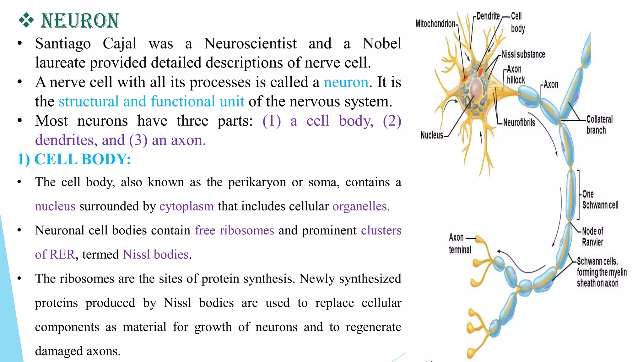
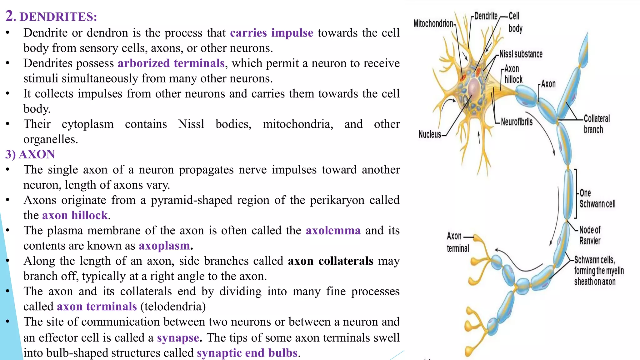
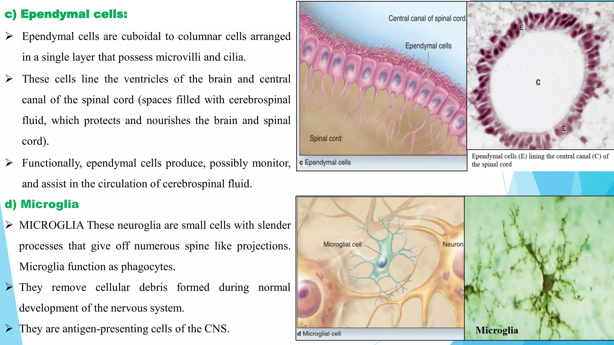
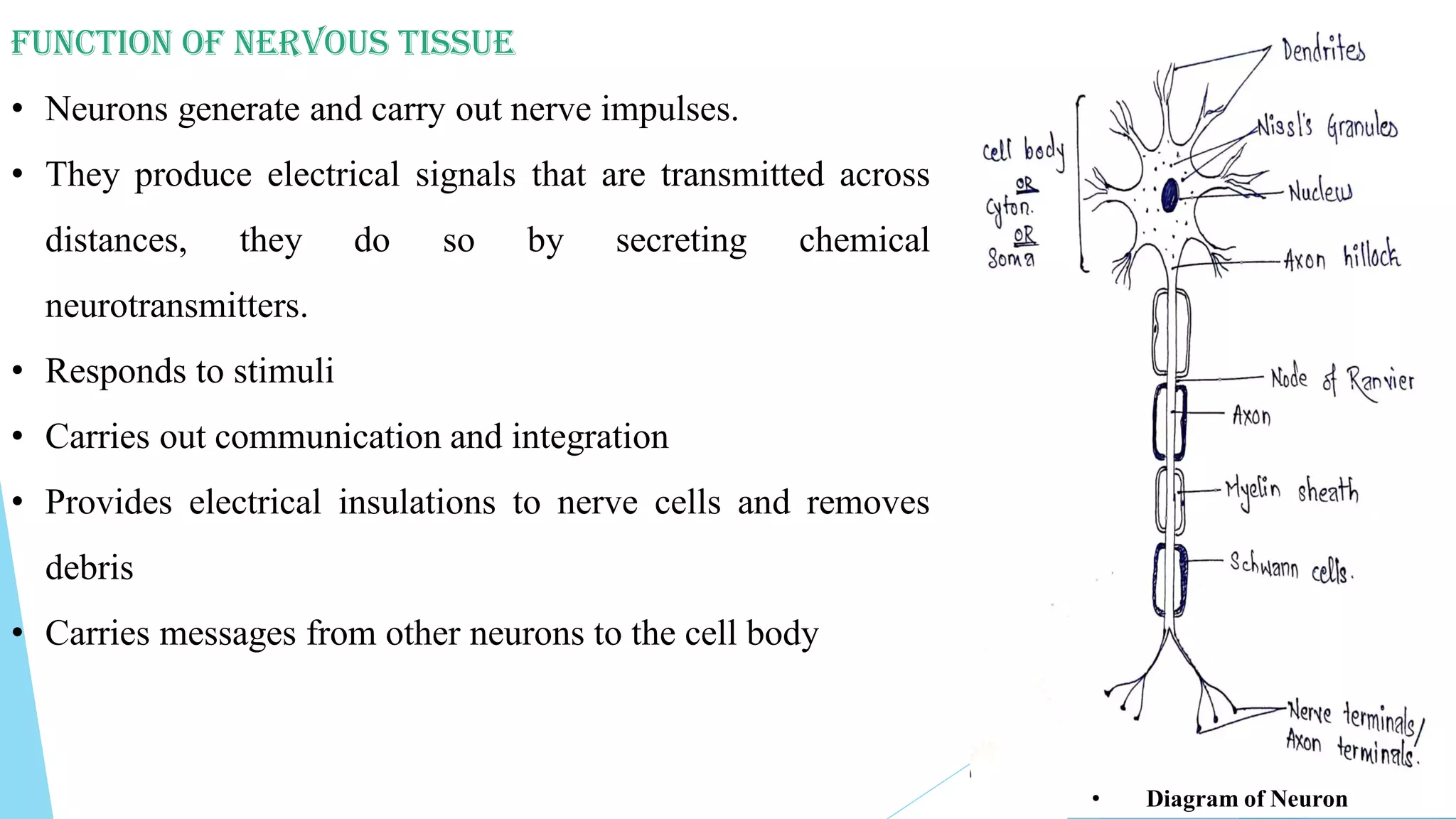
This document summarizes nervous tissue, including its cells and classification. It describes the two main cell types: neurons, which transmit electrical signals, and neuroglia cells, which support and protect neurons. Neurons are composed of a cell body, dendrites that receive signals, and an axon that transmits signals. There are different types of neurons classified by their structure. Neuroglia include oligodendrocytes, astrocytes, ependymal cells, microglia, Schwann cells, and satellite cells that each have distinct functions in supporting the nervous system. Together, neurons and glia allow the nervous system to coordinate voluntary and involuntary body functions through electrical signaling.
9953330565 Low Rate Call Girls In Rohini Delhi NCR
Nervous tissue
1. Chapter: 2
The Tissues
Part 2: Nervous tissue
By: Bithika Baidya
Content:
1. Introduction to nervous tissue
2. Cells of nervous tissue
-Neuron
-Neuroglia cell and types of neuroglia cells
3. Classification of neurons
4. Function of nervous tissue
2. ❖ Introduction to nervous tissue
• Nervous tissue is the term for group of organized
cells in nervous system (consist of CNS and PNS).
• Nervous system coordinates all the voluntary and
involuntary action of body by sending and carrying
signals to and from the different parts of the body.
• The signal sent by the nervous system are electrical
signal called nerve impulse and they are transmitted
by specialized nervous system cells called the neuron
or nerve cells.
https://www.google.com/search?q=neuron+animation&source=lnms&tbm=isch&
sa=X&ved=2ahUKEwjxqTQ2LXrAhWzwzgGHQOMDEoQ_AUoAnoECA4QBA&biw=139
6&bih=686#imgrc=BqVkPTYUPPRpeM&imgdii=qOpqWKqkDQNkFM
3. ❑ CELLS OF NERVOUS SYSTEM :
❖ Nervous tissue consists of two types of cells:
a) Neurons:
Neuron provide most of the unique functions of the
nervous system, such as sensing, controlling muscle
activity, regulating glandular secretions, etc.
a) Neuroglia:
Neuroglia support, nourish, and protect the neurons.
4. ❖ Neuron
• Santiago Cajal was a Neuroscientist and a Nobel
laureate provided detailed descriptions of nerve cell.
• A nerve cell with all its processes is called a neuron. It is
the structural and functional unit of the nervous system.
• Most neurons have three parts: (1) a cell body, (2)
dendrites, and (3) an axon.
1) CELL BODY:
• The cell body, also known as the perikaryon or soma, contains a
nucleus surrounded by cytoplasm that includes cellular organelles.
• Neuronal cell bodies contain free ribosomes and prominent clusters
of RER, termed Nissl bodies.
• The ribosomes are the sites of protein synthesis. Newly synthesized
proteins produced by Nissl bodies are used to replace cellular
components as material for growth of neurons and to regenerate
damaged axons.
5. 2. DENDRITES:
• Dendrite or dendron is the process that carries impulse towards the cell
body from sensory cells, axons, or other neurons.
• Dendrites possess arborized terminals, which permit a neuron to receive
stimuli simultaneously from many other neurons.
• It collects impulses from other neurons and carries them towards the cell
body.
• Their cytoplasm contains Nissl bodies, mitochondria, and other
organelles.
3) AXON
• The single axon of a neuron propagates nerve impulses toward another
neuron, length of axons vary.
• Axons originate from a pyramid-shaped region of the perikaryon called
the axon hillock.
• The plasma membrane of the axon is often called the axolemma and its
contents are known as axoplasm.
• Along the length of an axon, side branches called axon collaterals may
branch off, typically at a right angle to the axon.
• The axon and its collaterals end by dividing into many fine processes
called axon terminals (telodendria)
• The site of communication between two neurons or between a neuron and
an effector cell is called a synapse. The tips of some axon terminals swell
into bulb-shaped structures called synaptic end bulbs.
6. ❖ Neuroglia:
• Neuroglial cells are special type of interstitial cells that out number
neurons by approximately 10 to 1.
• These cells are embedded in a web of tissue composed of modified
ectodermal elements; the entire supporting structure is termed the
neuroglia.
• They function to support and protect neurons, but they do not
conduct impulses
• There are six types of glial cells
a) Oligodendrocytes
b) Astrocytes- ii) Protoplasmic astrocytes iii) Fibrous astrocytes
c) Ependymal cells
d) Microglia
e) Schwann cells
f) Satellite cells
7. a) Oligodendrocytes
Oligodendrocytes (Gr. oligos, small, few + dendron, tree + kytos,
cell) produce the myelin sheaths, the myelin sheath is a
multilayered lipid and protein covering around some axons that
insulates them and increases the speed of nerve impulse conduction.
Such axons are said to be myelinated.
b) Astrocytes:
▪ Astrocytes (Gr. astron, star + kytos) have a large number of
radiating processes. Astrocytes are by far the most numerous
glial cells of the CNS, as well as the most diverse structurally
and functionally.
▪ Those with relatively few, long processes are called fibrous
astrocytes and are typical in white matter; those with many
shorter, branched processes are called protoplasmic astrocytes
and predominate in the gray matter.
8. c) Ependymal cells:
➢ Ependymal cells are cuboidal to columnar cells arranged
in a single layer that possess microvilli and cilia.
➢ These cells line the ventricles of the brain and central
canal of the spinal cord (spaces filled with cerebrospinal
fluid, which protects and nourishes the brain and spinal
cord).
➢ Functionally, ependymal cells produce, possibly monitor,
and assist in the circulation of cerebrospinal fluid.
d) Microglia
➢ MICROGLIA These neuroglia are small cells with slender
processes that give off numerous spine like projections.
Microglia function as phagocytes.
➢ They remove cellular debris formed during normal
development of the nervous system.
➢ They are antigen-presenting cells of the CNS.
9. e) Schwann Cells
➢ These cells encircle PNS axons.
➢ Like oligodendrocytes, they form the myelin sheath around
axons.
➢ However, a single oligodendrocyte myelinates several
axons, but each Schwann cell myelinates a single axon.
➢ Schwann cells participate in axon regeneration, which is
more easily accomplished in the PNS than in the CNS
f) Satellite cells
➢ These flat cells surround the cell bodies of neurons of PNS
ganglia.
➢ Besides providing structural support, satellite cells regulate
the exchanges of materials between neuronal cell bodies and
interstitial fluid
11. • Classification of Neurons
Structurally, neurons are classified according to the
number of processes extending from the cell body
1. Multipolar neurons usually have several dendrites
and one axon. Most neurons in the brain and spinal
cord are of this type.
2. Bipolar neurons have one main dendrite and one
axon.They are found in the retina of the eye, in the
inner ear, and in the olfactory area of the brain.
3. Unipolar neurons have dendrites and one axon
that are fused together to form a continuous process
that emerges from the cell body. These neurons are
also known as pseudounipolar neurons because
they begin in the embryo as bipolar neurons.
During development, the dendrites and axon fuse
together and become a single process.
4. Anaxonic neuron: It is a type of neuron where the
differentiation of axon from the dendrites cannot be
done. Such anaxonic neurons are present in brain
and retina.
12. Function Of Nervous Tissue
• Neurons generate and carry out nerve impulses.
• They produce electrical signals that are transmitted across
distances, they do so by secreting chemical
neurotransmitters.
• Responds to stimuli
• Carries out communication and integration
• Provides electrical insulations to nerve cells and removes
debris
• Carries messages from other neurons to the cell body
• Diagram of Neuron
13. Question:
1. Draw a well-labeled diagram of Neuron. Describe its structural component.
2. Write a note on neuroglia cells.
3. Classification of neurons
4. Define: Axon, Nissl bodies, Myelin sheath, Axolemma, Axoplasm, Node of Ranvier, Axon Hillock, Saltatory conduction,
Neuroglia cells, anaxonic neurons
References:
• Principles of Anatomy and Physiology 12th Edition by Gerard J. Tortora and Bryan Derrickson Valencia
• CC Chatterjee' s Human Physiology 12th edition
• Cell biology and histology / Leslie P. Gartner, James L. Hiatt, Judy M. Strum. — 6th ed
• Junqueira’s Basic Histology Text and atlas 13th Edition, Anthony L. Mescher
Thank You….